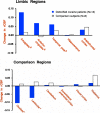
Limbic activation during cue-induced cocaine craving
- PMID: 9892292
- PMCID: PMC2820826
- DOI: 10.1176/ajp.156.1.11
Limbic activation during cue-induced cocaine craving
Abstract
Objective: Since signals for cocaine induce limbic brain activation in animals and cocaine craving in humans, the objective of this study was to test whether limbic activation occurs during cue-induced craving in humans.
Method: Using positron emission tomography, the researchers measured relative regional cerebral blood flow (CBF) in limbic and comparison brain regions of 14 detoxified male cocaine users and six cocaine-naive comparison subjects during exposure to both non-drug-related and cocaine-related videos and during resting baseline conditions.
Results: During the cocaine video, the cocaine users experienced craving and showed a pattern of increases in limbic (amygdala and anterior cingulate) CBF and decreases in basal ganglia CBF relative to their responses to the non-drug video. This pattern did not occur in the cocaine-naive comparison subjects, and the two groups did not differ in their responses in the comparison regions (i.e., the dorsolateral prefrontal cortex, cerebellum, thalamus, and visual cortex).
Conclusions: These findings indicate that limbic activation is one component of cue-induced cocaine craving. Limbic activation may be similarly involved in appetitive craving for other drugs and for natural rewards.
Figures


Similar articles
-
Neural activity related to drug craving in cocaine addiction.Arch Gen Psychiatry. 2001 Apr;58(4):334-41. doi: 10.1001/archpsyc.58.4.334. Arch Gen Psychiatry. 2001. PMID: 11296093
-
Changes in regional cerebral blood flow elicited by craving memories in abstinent opiate-dependent subjects.Am J Psychiatry. 2001 Oct;158(10):1680-6. doi: 10.1176/appi.ajp.158.10.1680. Am J Psychiatry. 2001. PMID: 11579002
-
Cue-induced cocaine craving: neuroanatomical specificity for drug users and drug stimuli.Am J Psychiatry. 2000 Nov;157(11):1789-98. doi: 10.1176/appi.ajp.157.11.1789. Am J Psychiatry. 2000. PMID: 11058476
-
Functional neuroimaging studies of alcohol cue reactivity: a quantitative meta-analysis and systematic review.Addict Biol. 2013 Jan;18(1):121-33. doi: 10.1111/j.1369-1600.2012.00464.x. Epub 2012 May 10. Addict Biol. 2013. PMID: 22574861 Free PMC article. Review.
-
Functional imaging of craving.Alcohol Res Health. 1999;23(3):187-96. Alcohol Res Health. 1999. PMID: 10890814 Free PMC article. Review.
Cited by
-
Modulating reward and aversion: Insights into addiction from the paraventricular nucleus.CNS Neurosci Ther. 2024 Sep;30(9):e70046. doi: 10.1111/cns.70046. CNS Neurosci Ther. 2024. PMID: 39295107 Free PMC article. Review.
-
Memory Reconsolidation Updating in Substance Addiction: Applications, Mechanisms, and Future Prospects for Clinical Therapeutics.Neurosci Bull. 2024 Sep 12. doi: 10.1007/s12264-024-01294-z. Online ahead of print. Neurosci Bull. 2024. PMID: 39264570 Review.
-
Perineuronal Nets in the Rat Medial Prefrontal Cortex Alter Hippocampal-Prefrontal Oscillations and Reshape Cocaine Self-Administration Memories.J Neurosci. 2024 Aug 21;44(34):e0468242024. doi: 10.1523/JNEUROSCI.0468-24.2024. J Neurosci. 2024. PMID: 38991791
-
Mapping the Neural Substrates of Cocaine Craving: A Systematic Review.Brain Sci. 2024 Mar 29;14(4):329. doi: 10.3390/brainsci14040329. Brain Sci. 2024. PMID: 38671981 Free PMC article. Review.
-
EcoHIV Infection Modulates the Effects of Cocaine Exposure Pattern and Abstinence on Cocaine Seeking and Neuroimmune Protein Expression in Male Mice.bioRxiv [Preprint]. 2024 Apr 19:2024.04.15.589615. doi: 10.1101/2024.04.15.589615. bioRxiv. 2024. PMID: 38659915 Free PMC article. Preprint.
References
-
- Childress AR, Ehrman RN, Rohsenow D, Robbins SJ, O'Brien CP. Classically conditioned factors in drug dependence. In: Lowinson J, Ruiz P, Millman R, editors. Comprehensive Textbook of Substance Abuse. Williams & Wilkins; Baltimore: 1993. pp. 56–69.
-
- Rothman R, Glowa JR. A review of the effects of dopamimetic agents on humans, animals and drug-seeking behavior, and its implications for medication development: focus on GBR12909. Mol Neurobiol. 1995;11:1–19. - PubMed
-
- Kosten TT, McCance E. A review of pharmacotherapies for substance abuse. Am J Addict. 1996;5:58–65.
-
- Gawin FH, Kleber HD. Abstinence symptomatology and psychiatric diagnosis in cocaine abusers. Arch Gen Psychiatry. 1986;43:107–113. - PubMed
-
- Jaffe JJ, Cascella NG, Kumor KM, Sherer MA. Cocaine-induced cocaine craving. Psychopharmacology (Berl) 1989;97:59–64. - PubMed
Publication types
MeSH terms
Substances
Grants and funding
LinkOut - more resources
Full Text Sources
Other Literature Sources
Medical

