Comparative Study
. 1998 Dec 1;12(23):3715-27.
doi: 10.1101/gad.12.23.3715.

A novel class of evolutionarily conserved genes defined by piwi are essential for stem cell self-renewal

Affiliations
Comparative Study

A novel class of evolutionarily conserved genes defined by piwi are essential for stem cell self-renewal

D N Cox et al. Genes Dev. .

Abstract

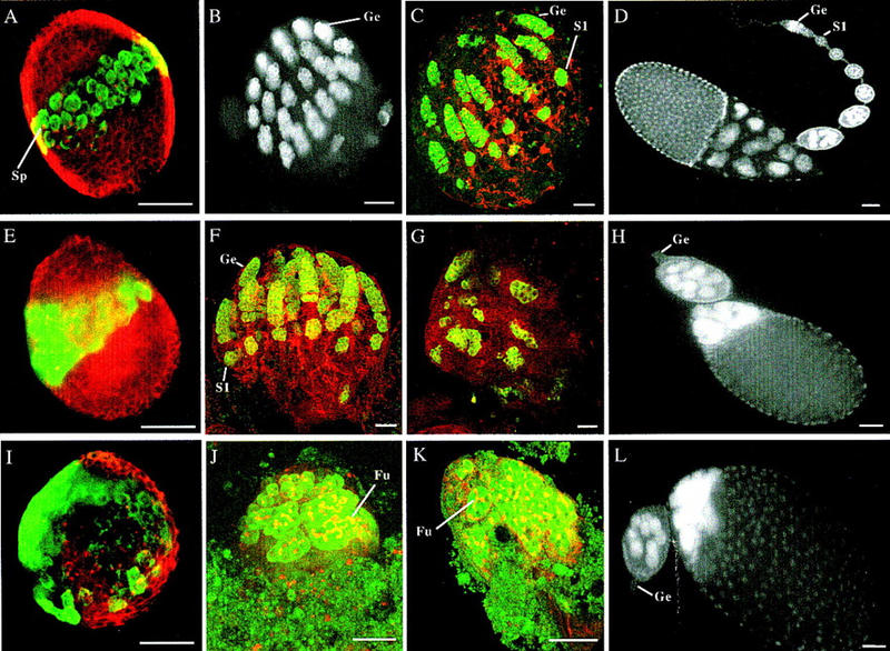
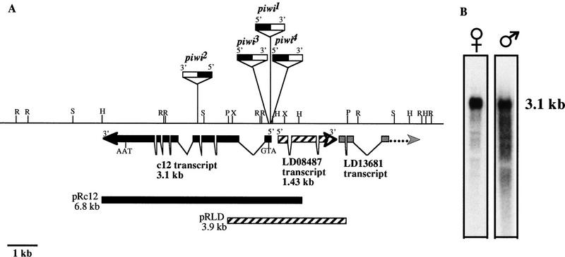
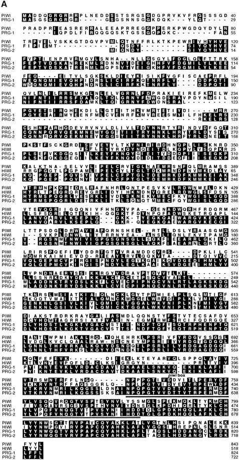
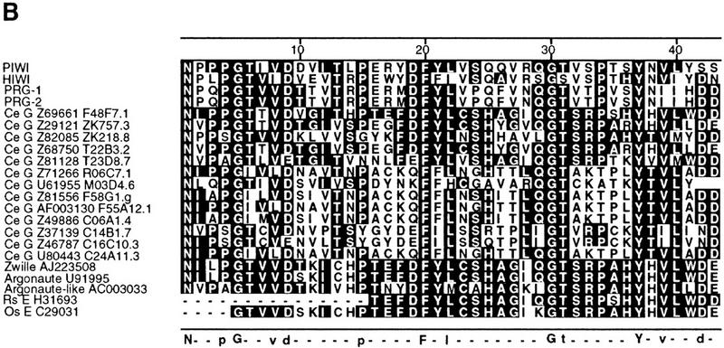
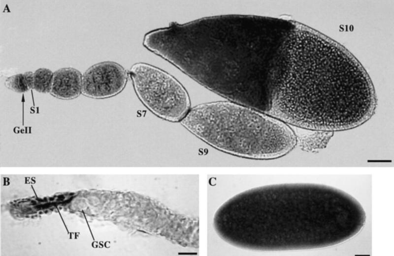
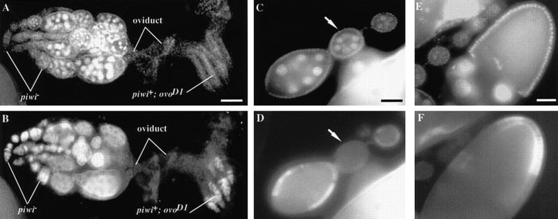
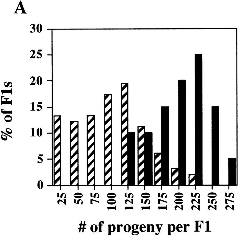
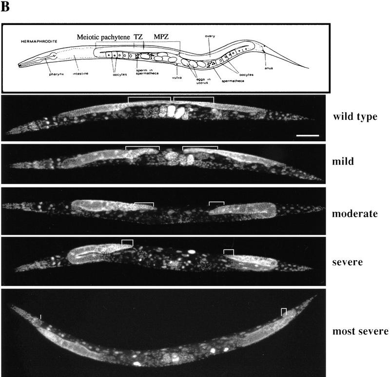
Germ-line stem cells (GSCs) serve as the source for gametogenesis in diverse organisms. We cloned and characterized the Drosophila piwi gene and showed that it is required for the asymmetric division of GSCs to produce and maintain a daughter GSC but is not essential for the further differentiation of the committed daughter cell. Genetic mosaic and RNA in situ analyses suggest that piwi expression in adjacent somatic cells regulates GSC division. piwi encodes a highly basic novel protein, well conserved during evolution. We isolated piwi homologs in Caenorhabditis elegans and humans and also identified Arabidopsis piwi-like genes known to be required for meristem cell maintenance. Decreasing C. elegans piwi expression reduces the proliferation of GSC-equivalent cells. Thus, piwi represents a novel class of genes required for GSC division in diverse organisms.


Figures

Figure 1
Figure 1
piwi is required for the self-renewing division of GSCs during oogenesis. (A,E,I) Confocal images showing third instar larval ovaries from wild-type, piwi2, and piwi1 mutant flies, respectively, stained for a germ-line-specific marker VASA in green and spectrin in red to mark somatic cells and spectrosomes/fusomes. GSCs are positioned medially along the anterioposterior axis in the wild-type (A) and piwi2 ovary (E); however, GSCs are mispositioned in the piwi1 mutant ovary (I). Spectrosomes (Sp) appear as red dots in germ cells. (B,J) Confocal images of 24-hr pupal ovaries from wild-type and piwi1 mutant flies, respectively, with B stained solely for VASA and J is stained for both VASA in green and spectrin in red. At this stage, the wild-type ovary has partitioned into individual germaria (Ge), which contain GSCs and early germ-line cysts. In piwi1 mutant ovaries, germ cells still appear to have been partitioned, although the partition is distorted by the highly differentiated large germ-line cysts containing elaborate fusomes (Fu). The number of cysts does not exceed the number of GSCs. (C,F,K) Confocal images of 48-hr pupal ovaries from wild-type, piwi2 and piwi1 mutant flies, respectively, stained with VASA in green and spectrin in red. By this stage, in both wild-type (C) and piwi2 mutant ovaries (F), germaria have developed fully and stage 1 egg chambers (S1) have budded off the germarium in synchrony because of continued GSC division. However, in piwi1 mutant ovaries (K), the number of germ-line cysts remains unchanged despite their continued differentiation. (G) Confocal image of a 72-hr pupal ovary from piwi2 mutant flies stained with VASA in green and spectrin stained in red. At this stage, GSCs fail to self-renew and some existing germ-line cysts begin to degenerate. (D,H,L), DAPI images of 0-to-1-day old adult ovarioles from wild-type, piwi2, and piwi1 mutant flies, respectively. Both piwi1 (L) and piwi2 (H) mutant ovarioles typically contain two normal or abnormal egg chambers connected to a germaria lacking germ lines (Ge). In contrast, wild-type ovarioles contain a long string of developing egg chambers produced continuously by the germarium (D). piwi3 phenotype is indistinguishable from that of piwi2 (data not shown). Bars in all panels, 50 μm.
Figure 2
Figure 2
Molecular map of the piwi locus and piwi Northern analyses. (A) Molecular map of the piwi region. Restriction enzyme map for 15 kb of genomic DNA surrounding the piwi mutations is shown. Three transcripts are represented by thick arrows indicating their relative positions, sizes, splice sites, and direction of transcription. The relative insertion sites of the P-element alleles are indicated above the restriction map with orientation of insertion indicated by black (5′) and white (3′) boxes. The P elements are not shown according to scale, with piwi2 insertion partially mapped. (R) EcoRI; (H) HindIII; (P) PstI; (S) SalI; (X) XhoI. (B) piwi RNA is present in piwi1/CyO adult males and females. The female and male lanes contain ∼10 and 1.5 μg of poly(A)+ RNA, respectively. They are exposed for 3.5 and 12 hr, respectively.
Figure 3
Figure 3
Alignment of predicted PIWI protein sequence with its homologs. (A) Sequence alignment of predicted PIWI proteins from Drosophila (PIWI), C. elegans (PRG-1 and PRG-2) and human (HIWI). The full-length amino acid sequence of PRG-1 and PRG-2 was deduced from the nucleotide sequence of two independent cDNA clones. Partial amino acid sequence of HIWI was deduced from the nucleotide sequence of a partial cDNA clone from a human testes library. The alignments were produced with CLUSTALW software (DNAStar). Identical residues to a consensus are highlighted. (B) The piwi box domain in 22 different proteins. The alignments were generated by Block Maker analysis (Henikoff et al. 1995) against amino acids 740–782 of the predicted piwi sequence. The amino acid sequences were deduced from genomic DNA sequences for C. elegans (Ce G) or from cDNA (EST) sequences (E) for Rattus sp. (Rs) and Oryza sativa (Os). GenBank accession numbers and cosmid designations (Ce) are indicated for each sequence. Residues identical to the consensus are highlighted. Shown below the alignments: Residues that are absolutely conserved are shown as uppercase letters and residues that are 80% or more conserved are shown as lowercase letters; dashes indicate spacing.
Figure 3
Figure 3
Alignment of predicted PIWI protein sequence with its homologs. (A) Sequence alignment of predicted PIWI proteins from Drosophila (PIWI), C. elegans (PRG-1 and PRG-2) and human (HIWI). The full-length amino acid sequence of PRG-1 and PRG-2 was deduced from the nucleotide sequence of two independent cDNA clones. Partial amino acid sequence of HIWI was deduced from the nucleotide sequence of a partial cDNA clone from a human testes library. The alignments were produced with CLUSTALW software (DNAStar). Identical residues to a consensus are highlighted. (B) The piwi box domain in 22 different proteins. The alignments were generated by Block Maker analysis (Henikoff et al. 1995) against amino acids 740–782 of the predicted piwi sequence. The amino acid sequences were deduced from genomic DNA sequences for C. elegans (Ce G) or from cDNA (EST) sequences (E) for Rattus sp. (Rs) and Oryza sativa (Os). GenBank accession numbers and cosmid designations (Ce) are indicated for each sequence. Residues identical to the consensus are highlighted. Shown below the alignments: Residues that are absolutely conserved are shown as uppercase letters and residues that are 80% or more conserved are shown as lowercase letters; dashes indicate spacing.
Figure 4
Figure 4
piwi mRNA expression in Drosophila ovaries. (A) A complete Ore-R ovariole showing germ-line piwi expression in region II of the germarium (GeII), and in early stage egg chambers up to stage 10 (S10). (B) In the germarium, the piwi RNA is also detected in the terminal filament cells (TF) and epithelial sheath cells (ES) apical to GSCs. (C) The piwi RNA is uniformly present in early embryos. Bars, 50, 10, 50 μm for A, B, and C, respectively.
Figure 5
Figure 5
piwi function is dispensable in the germ-line and differentiated follicle cells for GSC division. (A,B) piwi is dispensable in the germ line. A pair of ovaries containing ovarioles with homozygous piwi germ-line clones and ovarioles with piwi+ ovoD1 germ line stained for DAPI to mark DNA (A) and for VASA to mark germ cells (B). The piwi+ ovoD1 germ-line cells are arrested at the beginning of oogenesis in the germarium because of the ovoD1 mutation. In contrast, in ovarioles in the left ovary containing piwi germ-line clones, GSCs have divided normally, giving rise to a progression of morphologically wild-type egg chambers that eventually develop into mature eggs. This indicates that piwi is not required in the germ line for GSC division and subsequent steps of oogenesis. Bar in A, 100 μm. (C–F) piwi clones in differentiated follicle cells do not effect normal GSC division and egg chamber development. Egg chambers were stained with DAPI (C,E) and a monoclonal anti-MYC antibody (D,F). Absence of Myc staining is indicative of homozygous piwi clones. Induction of somatic clones around an entire early stage egg chamber such as the ones in C and D (see arrow) had no effect on GSC division and oogenesis. Similarly, removal of piwi function from some follicle cells of later stage egg chambers also had no effect on oogenesis (E,F). Therefore, piwi function is dispensable in differentiated follicle cells for oogenesis including GSC division. Bars in C and E, 50 μm.
Figure 6
Figure 6
prg–RNAi significantly reduces germ-line proliferation in C. elegans. (A) Reducing prg activity affects fertility. The graph indicates the number of progeny produced by a single F1 worm from an RNAi prg-1 or pBST-injected mother vs. the percentage of F1 animals that produced that given number of progeny. Comparison of prg–RNAi (hatched bars) with pBST RNAi (solid bars) animals reveals two distinctive distributions. (B) Reducing prg activity results in reductions in both the MPZ and the mitotic index. Variable defects in both the size of the MPZ and the mitotic index were observed by DAPI staining. The MPZ in each micrograph is denoted in white brackets. In moderate to severely defective animals, a 50% reduction in the number of mitotic nuclei and a concomitant fivefold reduction in the mitotic index were typically observed. Bar, 100 μm for all panels.
Figure 6
Figure 6
prg–RNAi significantly reduces germ-line proliferation in C. elegans. (A) Reducing prg activity affects fertility. The graph indicates the number of progeny produced by a single F1 worm from an RNAi prg-1 or pBST-injected mother vs. the percentage of F1 animals that produced that given number of progeny. Comparison of prg–RNAi (hatched bars) with pBST RNAi (solid bars) animals reveals two distinctive distributions. (B) Reducing prg activity results in reductions in both the MPZ and the mitotic index. Variable defects in both the size of the MPZ and the mitotic index were observed by DAPI staining. The MPZ in each micrograph is denoted in white brackets. In moderate to severely defective animals, a 50% reduction in the number of mitotic nuclei and a concomitant fivefold reduction in the mitotic index were typically observed. Bar, 100 μm for all panels.

Similar articles

Cited by

References

    1. Altschul SF, Gish W, Miller W, Myers EW, Lipman DJ. Basic local alignment search tool. J Mol Biol. 1990;215:403–410. - PubMed
    1. Bohmert K, Camus I, Bellini C, Bouchez D, Caboche M, Benning C. AGO1 defines a novel locus of Arabidopsis controlling leaf development. EMBO J. 1998;17:170–180. - PMC - PubMed
    1. Brown EH, King RC. Oogonial and spermatogonial differentiation within a mosaic gonad of Drosophila melanogaster. Growth. 1962;26:53–70. - PubMed
    1. ————— Studies on the events resulting in the formation of an egg chamber in Drosophila melanogaster. Growth. 1964;28:41–81. - PubMed
    1. Brown NH, Kafatos FC. Functional libraries from Drosophila embryos. J Mol Biol. 1988;203:425–437. - PubMed

Publication types

MeSH terms

Associated data