. 1998 Aug;9(8):2185-200.
doi: 10.1091/mbc.9.8.2185.

Activation of both MAP kinase and phosphatidylinositide 3-kinase by Ras is required for hepatocyte growth factor/scatter factor-induced adherens junction disassembly

Affiliations
Free PMC article

Activation of both MAP kinase and phosphatidylinositide 3-kinase by Ras is required for hepatocyte growth factor/scatter factor-induced adherens junction disassembly

S Potempa et al. Mol Biol Cell. 1998 Aug.
Free PMC article

Abstract

Hepatocyte growth factor/scatter factor (HGF/SF) stimulates the motility of epithelial cells, initially inducing centrifugal spreading of colonies followed by disruption of cell-cell junctions and subsequent cell scattering. In Madin-Darby canine kidney cells, HGF/SF-induced motility involves actin reorganization mediated by Ras, but whether Ras and downstream signals regulate the breakdown of intercellular adhesions has not been established. Both HGF/SF and V12Ras induced the loss of the adherens junction proteins E-cadherin and beta-catenin from intercellular junctions during cell spreading, and the HGF/SF response was blocked by dominant-negative N17Ras. Desmosomes and tight junctions were regulated separately from adherens junctions, because they were not disrupted by V12Ras. MAP kinase, phosphatidylinositide 3-kinase (PI 3-kinase), and Rac were required downstream of Ras, because loss of adherens junctions was blocked by the inhibitors PD098059 and LY294002 or by dominant-inhibitory mutants of MAP kinase kinase 1 or Rac1. All of these inhibitors also prevented HGF/SF-induced cell scattering. Interestingly, activated Raf or the activated p110alpha subunit of PI 3-kinase alone did not induce disruption of adherens junctions. These results indicate that activation of both MAP kinase and PI 3-kinase by Ras is required for adherens junction disassembly and that this is essential for the motile response to HGF/SF.


Figures

Figure 1
Figure 1
HGF/SF-induced scattering is blocked by the MAPKK1/2 inhibitor PD098059. Phase-contrast pictures of a colony of MDCK cells are shown before the addition of HGF/SF (a) or at 16 h (b) after addition of HGF/SF. Cells in c were preincubated with 50 μM PD098059 before addition of HGF/SF for 16 h. The bar in c represents 50 μm and applies also to a and b.
Figure 2
Figure 2
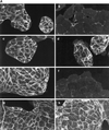
Disruption of adherens junctions by HGF/SF is blocked by the MAPKK1/2 inhibitor PD098059 and the PI 3-kinase inhibitor LY294002. (A) MDCK cells were stimulated with HGF/SF for 4 h (b and f) or were preincubated with 50 μM PD098059 (c and g) or 20 μM LY294002 (d and h) before addition of HGF/SF for 4 h. MDCK cells were then fixed and stained for β-catenin (a–d) and E-cadherin (e–h) localization. Bar, 10 μm. (B) MDCK cells were treated as indicated with 20 μM LY294002 (LY) or 50 μM PD098059 (PD) before addition of HGF/SF (HGF) for 4 h. Disrupted junctions in control cells (cont) and stimulated cells were defined as junctions where no E-cadherin and β-catenin could be detected at the sites of intercellular contact and were subsequently counted. The numbers represent the mean of 300 cells, which were counted in three independent experiments.
Figure 2
Figure 2
Disruption of adherens junctions by HGF/SF is blocked by the MAPKK1/2 inhibitor PD098059 and the PI 3-kinase inhibitor LY294002. (A) MDCK cells were stimulated with HGF/SF for 4 h (b and f) or were preincubated with 50 μM PD098059 (c and g) or 20 μM LY294002 (d and h) before addition of HGF/SF for 4 h. MDCK cells were then fixed and stained for β-catenin (a–d) and E-cadherin (e–h) localization. Bar, 10 μm. (B) MDCK cells were treated as indicated with 20 μM LY294002 (LY) or 50 μM PD098059 (PD) before addition of HGF/SF (HGF) for 4 h. Disrupted junctions in control cells (cont) and stimulated cells were defined as junctions where no E-cadherin and β-catenin could be detected at the sites of intercellular contact and were subsequently counted. The numbers represent the mean of 300 cells, which were counted in three independent experiments.
Figure 3
Figure 3
E-cadherin and β-catenin are redistributed from the NP-40-insoluble fraction to the soluble fraction in response to HGF/SF. Cell lysates of unstimulated control cells (C) or cells incubated with HGF/SF for 4 h (HGF) or cells treated with 20 μM LY 294002 for 1 h before addition of HGF/SF (HGF/LY) or 50 μM PD98059 (HGF/PD) were fractionated into a NP-40-soluble (S) and NP-40-insoluble (I) fraction. Equal sample volumes were loaded and probed for E-cadherin (top blot) and β-catenin (bottom blot).
Figure 4
Figure 4
HGF/SF induces sustained activation of p42/p44 MAPK. Cells were stimulated with HGF/SF (HGF) for 10 min or 4 or 16 h with (+) or without (−) preincubation with 50 μM PD098059. The cells were subsequently lysed, and the lysates were electrophoresed on an SDS-gel. The top panel shows Western blotting for the activated, phosphorylated form of p42/p44 MAPK (pMAPK). The bottom panel shows the blot reprobed with an anti-ERK1 antibody against total p42/p44 MAPK (ERK1/ERK2).
Figure 5
Figure 5
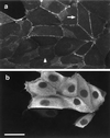
Dominant negative mutants of MAPKK1 block disruption of adherens junctions. MDCK cells were microinjected with a construct expressing a dominant negative mutant of MAPKK1, AFG MAPKK1 (100 ng/μl). After microinjection cells were incubated for 6 h before addition of HGF/SF for 2 h. Cells were stained for β-catenin (a) and for expression of the mutant MAPKK1 using the rabbit anti-MAPKK1 antibody (b). The arrow in a shows β-catenin localizing to adherens junctions in injected cells, and the arrowhead indicates disrupted adherens junctions in uninjected cells. Bar, 10 μm.
Figure 6
Figure 6
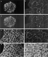
Adherens junctions are disrupted before tight and desmosomal junctions in response to HGF/SF. Unstimulated MDCK cells were costained for β-catenin (a) and ZO-1 (c) or for β-catenin (e) and desmoplakin (g). Costaining of cells treated with HGF/SF for 4 h with β-catenin (b) and ZO-1 (d) or with β-catenin (f) and desmoplakin (h) indicates that β-catenin is lost from intercellular junctions (e.g., arrow in b and arrowhead in f) where ZO-1 (d, arrow) and desmoplakin (h, arrowhead) are still present. Bar, 10 μm.
Figure 7
Figure 7
Ras disrupts adherens junctions through p42/p44 MAPK and PI 3-kinase but does not disrupt desmosomes or tight junctions. (A) Cells were microinjected with V12Ras (35 ng/μl), fixed after 1 h, and stained for β-catenin (a and c) or ZO-1 (e). Microinjected cells were detected by coinjection of TRITC–dextran (b, d, and f). The arrow in a shows the dispersion of β-catenin in injected cells, and the arrowhead indicates the localization of β-catenin at adherens junctions in uninjected cells. Cells were preincubated with 50 μM PD098059 in c and d before microinjection. Bar, 10 μm. (B) Cells microinjected with V12Ras (ras) were preincubated with or without LY294002 (LY) or PD098059 (PD) for 1 h before injection, as indicated. Cells were fixed 1 h after injection and stained for β-catenin (bc) and E-cadherin (ec) or incubated for 16 h and stained for ZO-1 (zo-1). The numbers represent the mean of at least three independent experiments (the error bars represent the mean of 5 experiments) where 100 cells were injected in each experiment or where 100 uninjected cells (control) were counted.
Figure 7
Figure 7
Ras disrupts adherens junctions through p42/p44 MAPK and PI 3-kinase but does not disrupt desmosomes or tight junctions. (A) Cells were microinjected with V12Ras (35 ng/μl), fixed after 1 h, and stained for β-catenin (a and c) or ZO-1 (e). Microinjected cells were detected by coinjection of TRITC–dextran (b, d, and f). The arrow in a shows the dispersion of β-catenin in injected cells, and the arrowhead indicates the localization of β-catenin at adherens junctions in uninjected cells. Cells were preincubated with 50 μM PD098059 in c and d before microinjection. Bar, 10 μm. (B) Cells microinjected with V12Ras (ras) were preincubated with or without LY294002 (LY) or PD098059 (PD) for 1 h before injection, as indicated. Cells were fixed 1 h after injection and stained for β-catenin (bc) and E-cadherin (ec) or incubated for 16 h and stained for ZO-1 (zo-1). The numbers represent the mean of at least three independent experiments (the error bars represent the mean of 5 experiments) where 100 cells were injected in each experiment or where 100 uninjected cells (control) were counted.
Figure 8
Figure 8
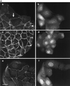
Ras and Rac mediate the disruption of adherens junctions. In a, cells were microinjected with a construct expressing N17Ras (100 ng/μl). After microinjection cells were incubated for 6 h before addition of HGF/SF for 2 h and stained for β-catenin. In c, cells were microinjected with N17Rac1 protein (150 ng/μl) and incubated for 2 h with HGF/SF before staining for β-catenin. Cells were coinjected with V12Ras (100 ng/μl) and N17Rac1 (100 ng/μl), incubated for 2 h, and subsequently stained for β-catenin in e. The arrowheads show β-catenin localization at intercellular junctions in injected cells, and the arrows indicate dispersed adherens junctions in uninjected cells (a and c). Microinjected cells were detected by either staining with a rat anti-Ras antibody (b) or coinjection of TRITC–dextran (d and f). Bar, 10 μm.
Figure 9
Figure 9
Expression of membrane-targeted p110α and Raf does not disrupt adherens junctions. Cells were microinjected with plasmids encoding membrane-targeted p110α (pSG5-5-Myc-p110α-WT-3-K-Ras; 100 ng/μl; (a and b), Raf (pEFHm-RAFCAAX-Myc; c and d), or p110α and Raf (e and f), incubated for 4 h, and then stained for β-catenin (a, c, and e) and for expression of p110α with a rabbit anti-p110α antibody (b and f). Cells injected with the Raf plasmid were visualized by coinjection of TRITC–dextran (d). Control experiments showed that in cells coinjected with TRITC–dextran and the Raf plasmid, exogenous Raf (myc tagged) was expressed in 100% of injected cells. The arrows indicate the presence of β-catenin at adherens junctions of injected cells. Bar, 10 μm.

Similar articles

Cited by

References

    1. Alessi DR, Cuenda A, Cohen P, Dudley DT, Saltiel AR. PD 098059 is a specific inhibitor of the activation of mitogen-activated protein kinase in vitro and in vivo. J Biol Chem. 1995;270:27489–27494. - PubMed
    1. Alessi DR, Saito Y, Campbell DG, Cohen P, Sithanandam G, Rapp U, Ashworth A, Marshall CJ, Cowley SJ. Identification of the sites of MAP kinase kinase-1 phosphorylated by p74raf-1. EMBO J. 1994;13:1610–1619. - PMC - PubMed
    1. Anderson JM, Stevenson BR, Jesaitis LA, Goodenough DA, Mooseker MS. Characterization of ZO-1, a protein component of tight junction from mouse liver and Madin-Darby canine kidney cells. J Cell Biol. 1988;106:1141–1149. - PMC - PubMed
    1. Avruch J, Zhang X-F, Kyriakis JM. Raf meets Ras: completing the framework of a signal transduction pathway. Trends Biochem Sci. 1994;19:279–283. - PubMed
    1. Balda MS, Whitney JA, Flores C, Gonzáles S, Cereijdo M, Matter K. Functional dissociation of paracellular permeability and transepithelial electrical resistance and disruption of the apical-basolateral intramembrane diffusion barrier by expression of a mutant tight junction membrane protein. J Cell Biol. 1996;134:1031–1049. - PMC - PubMed

MeSH terms