Use of differential display analysis to assess the effect of human cytomegalovirus infection on the accumulation of cellular RNAs: induction of interferon-responsive RNAs
- PMID: 9391139
- PMCID: PMC28419
- DOI: 10.1073/pnas.94.25.13985
Use of differential display analysis to assess the effect of human cytomegalovirus infection on the accumulation of cellular RNAs: induction of interferon-responsive RNAs
Abstract
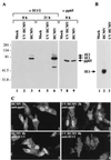
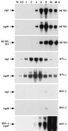
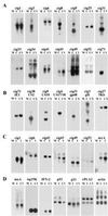
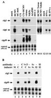
We used differential display analysis to identify mRNAs that accumulate to enhanced levels in human cytomegalovirus-infected cells as compared with mock-infected cells. RNAs were compared at 8 hr after infection of primary human fibroblasts. Fifty-seven partial cDNA clones were isolated, representing about 26 differentially expressed mRNAs. Eleven of the mRNAs were virus-coded, and 15 were of cellular origin. Six of the partial cDNA sequences have not been reported previously. All of the cellular mRNAs identified in the screen are induced by interferon alpha. The induction in virus-infected cells, however, does not involve the action of interferon or other small signaling molecules. Neutralizing antibodies that block virus infection also block the induction. These RNAs accumulate after infection with virus that has been inactivated by treatment with UV light, indicating that the inducer is present in virions. We conclude that human cytomegalovirus induces interferon-responsive mRNAs.
Figures


Similar articles
-
Altered cellular mRNA levels in human cytomegalovirus-infected fibroblasts: viral block to the accumulation of antiviral mRNAs.J Virol. 2001 Dec;75(24):12319-30. doi: 10.1128/JVI.75.24.12319-12330.2001. J Virol. 2001. PMID: 11711622 Free PMC article.
-
Human cytomegalovirus latent infection alters the expression of cellular and viral microRNA.Gene. 2014 Feb 25;536(2):272-8. doi: 10.1016/j.gene.2013.12.012. Epub 2013 Dec 18. Gene. 2014. PMID: 24361963
-
Rhesus cytomegalovirus particles prevent activation of interferon regulatory factor 3.J Virol. 2005 May;79(10):6419-31. doi: 10.1128/JVI.79.10.6419-6431.2005. J Virol. 2005. PMID: 15858025 Free PMC article.
-
Human cytomegalovirus: a review of developments between 1970 and 1976. Part II. Experimental developments.Biomedicine. 1977 Apr;26(2):86-97. Biomedicine. 1977. PMID: 194635 Review.
-
Alterations in TLRs as new molecular markers of congenital infections with Human cytomegalovirus?Pathog Dis. 2014 Feb;70(1):3-16. doi: 10.1111/2049-632X.12083. Epub 2013 Sep 10. Pathog Dis. 2014. PMID: 23929630 Review.
Cited by
-
Viperin: An ancient radical SAM enzyme finds its place in modern cellular metabolism and innate immunity.J Biol Chem. 2020 Aug 14;295(33):11513-11528. doi: 10.1074/jbc.REV120.012784. Epub 2020 Jun 16. J Biol Chem. 2020. PMID: 32546482 Free PMC article. Review.
-
Cytomegalovirus Restructures Lipid Rafts via a US28/CDC42-Mediated Pathway, Enhancing Cholesterol Efflux from Host Cells.Cell Rep. 2016 Jun 28;16(1):186-200. doi: 10.1016/j.celrep.2016.05.070. Epub 2016 Jun 16. Cell Rep. 2016. PMID: 27320924 Free PMC article.
-
Transcriptome signature of virulent and attenuated pseudorabies virus-infected rodent brain.J Virol. 2006 Feb;80(4):1773-86. doi: 10.1128/JVI.80.4.1773-1786.2006. J Virol. 2006. PMID: 16439534 Free PMC article.
-
Activation of interferon response factor-3 in human cells infected with herpes simplex virus type 1 or human cytomegalovirus.J Virol. 2001 Oct;75(19):8909-16. doi: 10.1128/JVI.75.19.8909-8916.2001. J Virol. 2001. PMID: 11533154 Free PMC article.
-
Alpha-herpesvirus infection induces the formation of nuclear actin filaments.PLoS Pathog. 2006 Aug;2(8):e85. doi: 10.1371/journal.ppat.0020085. PLoS Pathog. 2006. PMID: 16933992 Free PMC article.
References
Publication types
MeSH terms
Substances
Associated data
- Actions
- Actions
- Actions
- Actions
- Actions
- Actions
- Actions
LinkOut - more resources
Full Text Sources
Other Literature Sources
Molecular Biology Databases

