Estradiol enhances prostaglandin E2 receptor gene expression in luteinizing hormone-releasing hormone (LHRH) neurons and facilitates the LHRH response to PGE2 by activating a glia-to-neuron signaling pathway
- PMID: 9364061
- PMCID: PMC6573612
- DOI: 10.1523/JNEUROSCI.17-23-09145.1997
Estradiol enhances prostaglandin E2 receptor gene expression in luteinizing hormone-releasing hormone (LHRH) neurons and facilitates the LHRH response to PGE2 by activating a glia-to-neuron signaling pathway
Abstract
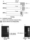
Prostaglandin E2 (PGE2) mediates the stimulatory effect of norepinephrine (NE) on the secretion of luteinizing hormone-releasing hormone (LHRH), the neuropeptide controlling reproductive function. In rodents, this facilitatory effect requires previous exposure to estradiol, suggesting that the steroid affects downstream components in the cascade that leads to PGE2-induced LHRH release. Because astroglia are the predominant cell type contacting LHRH-secreting nerve terminals, we investigated the involvement of hypothalamic astrocytes in the estradiol facilitation of PGE2-induced LHRH release. A subpopulation of LHRH neurons was found to express the mRNA encoding the PGE2 receptor subtype EP1-R, which is coupled to calcium mobilization. The LHRH-producing cell line GT1-1 also contains EP1-R mRNA and, to a lesser extent, the three alternatively spliced forms of EP3-R mRNA (alpha, beta, and gamma) that encode receptors linked to inhibition and stimulation of cAMP formation. Hypothalamic astrocytes treated with estradiol produced a conditioned medium that when applied to GT1-1 cells resulted in a selective upregulation of EP1-R and EP3gamma-R mRNAs. The conditioned medium also enhanced the LHRH response to EP1-R and EP3-R agonists and the cAMP response to EP3-R activation. Thus, one mechanism by which estradiol facilitates the effect of neurotransmitters acting via PGE2 to stimulate LHRH release is by enhancing the glial production of substances that upregulate PGE2 receptors on LHRH neurons. The existence of such a mechanism underscores the emerging importance of glial-neuronal communication in the control of brain neurosecretory activity.
Figures


Similar articles
-
Hypothalamic astrocytes respond to transforming growth factor-alpha with the secretion of neuroactive substances that stimulate the release of luteinizing hormone-releasing hormone.Endocrinology. 1997 Jan;138(1):19-25. doi: 10.1210/endo.138.1.4863. Endocrinology. 1997. PMID: 8977380
-
Prostaglandin E2 releases luteinizing hormone-releasing hormone from the female juvenile hypothalamus through a Ca2+-dependent, calmodulin-independent mechanism.Brain Res. 1988 Feb 16;441(1-2):339-51. doi: 10.1016/0006-8993(88)91412-6. Brain Res. 1988. PMID: 2834003
-
Exploration of prostanoid receptor subtype regulating estradiol and prostaglandin E2 induction of spinophilin in developing preoptic area neurons.Neuroscience. 2007 May 25;146(3):1117-27. doi: 10.1016/j.neuroscience.2007.02.006. Epub 2007 Apr 6. Neuroscience. 2007. PMID: 17408863 Free PMC article.
-
Glia-to-neuron signaling and the neuroendocrine control of female puberty.Recent Prog Horm Res. 2000;55:197-223; discussion 223-4. Recent Prog Horm Res. 2000. PMID: 11036938 Review.
-
LHRF, LHRH, GnRH: what controls the secretion of this hormone?Mol Psychiatry. 1997 Sep;2(5):373-6. doi: 10.1038/sj.mp.4000311. Mol Psychiatry. 1997. PMID: 9322226 Review.
Cited by
-
Sexual differentiation of the rodent brain: dogma and beyond.Front Neurosci. 2012 Feb 21;6:26. doi: 10.3389/fnins.2012.00026. eCollection 2012. Front Neurosci. 2012. PMID: 22363256 Free PMC article.
-
Prostaglandin E2 release from astrocytes triggers gonadotropin-releasing hormone (GnRH) neuron firing via EP2 receptor activation.Proc Natl Acad Sci U S A. 2011 Sep 20;108(38):16104-9. doi: 10.1073/pnas.1107533108. Epub 2011 Sep 6. Proc Natl Acad Sci U S A. 2011. PMID: 21896757 Free PMC article.
-
Deletion of the Ttf1 gene in differentiated neurons disrupts female reproduction without impairing basal ganglia function.J Neurosci. 2006 Dec 20;26(51):13167-79. doi: 10.1523/JNEUROSCI.4238-06.2006. J Neurosci. 2006. PMID: 17182767 Free PMC article.
-
Control of puberty by excitatory amino acid neurotransmitters and its clinical implications.Endocrine. 2005 Dec;28(3):281-6. doi: 10.1385/ENDO:28:3:281. Endocrine. 2005. PMID: 16388117 Review.
-
Unusual suspects: Glial cells in fertility regulation and their suspected role in polycystic ovary syndrome.J Neuroendocrinol. 2022 Jun;34(6):e13136. doi: 10.1111/jne.13136. Epub 2022 Apr 20. J Neuroendocrinol. 2022. PMID: 35445462 Free PMC article. Review.
References
-
- Barraclough CA, Wise PM. The role of catecholamines in the regulation of pituitary-luteinizing hormone and follicle-stimulating hormone secretion. Endocr Rev. 1982;3:91–119. - PubMed
-
- Berg-von der Emde K, Dees WL, Hiney JK, Hill DF, Dissen GA, Costa ME, Moholt-Siebert M, Ojeda SR. Neurotrophins and the neuroendocrine brain: different neurotrophins sustain anatomically and functionally segregated subsets of hypothalamic dopaminergic neurons. J Neurosci. 1995;15:4223–4237. - PMC - PubMed
-
- Brooker G, Harper JF, Terasaki WL, Moylan RD. Radioimmunoassay of cyclic AMP and cyclic GMP. In: Brooker G, Greengard P, editors. Advances in cyclic nucleotide research, Vol 10. Raven; New York: 1979. pp. 1–32. - PubMed
-
- Coleman RA, Kennedy I, Humphrey PPA, Bunce K, Lumley P. Prostanoids and their receptors. In: Hansch C, Sammes PG, Taylor JB, Emmett JC, editors. Comprehensive medicinal chemistry, Vol 3. Pergamon; New York: 1990. pp. 643–714.
-
- Coleman RA, Smith WL, Narumiya S. International union of pharmacology classification of prostanoid receptors: properties, distribution and structure of the receptors and their subtypes. Pharmacol Rev. 1994;46:205–229. - PubMed
Publication types
MeSH terms
Substances
Grants and funding
LinkOut - more resources
Full Text Sources
