Review
. 2024 Jun 7;134(12):1703-1717.
doi: 10.1161/CIRCRESAHA.124.323892. Epub 2024 Jun 6.

Cardiac Fibroblastic Niches in Homeostasis and Inflammation

Affiliations
Review

Cardiac Fibroblastic Niches in Homeostasis and Inflammation

Nadine Cadosch et al. Circ Res. .

Abstract

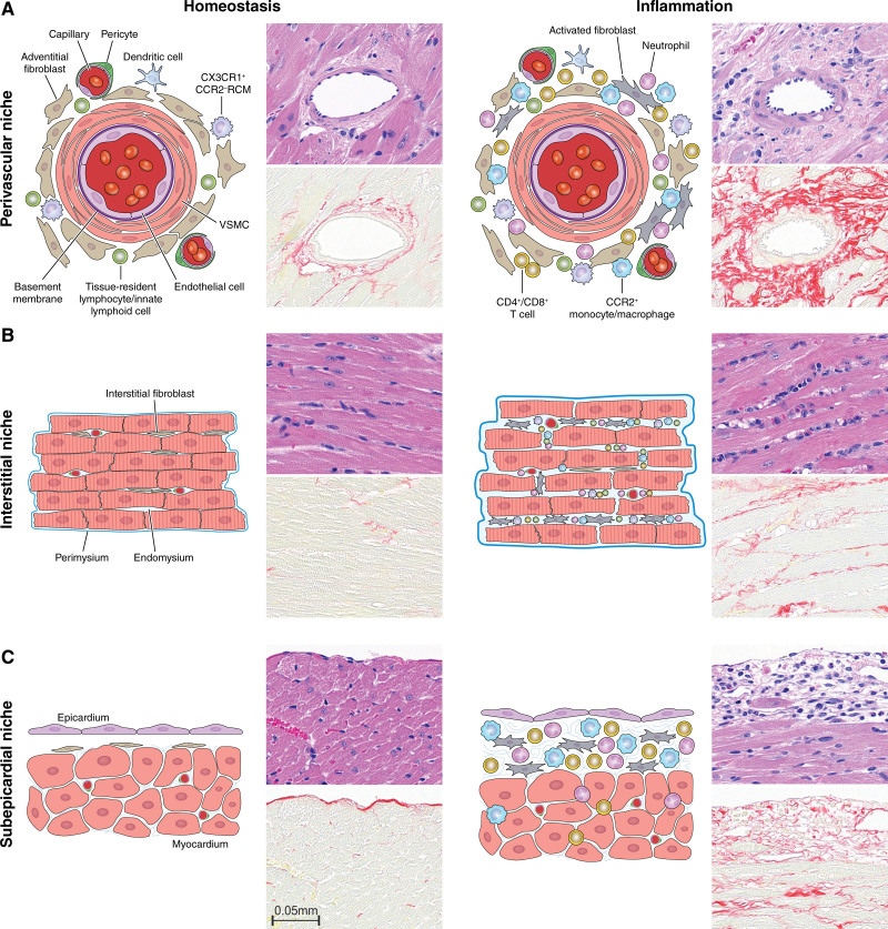
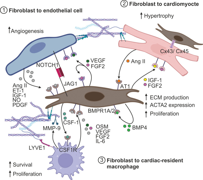
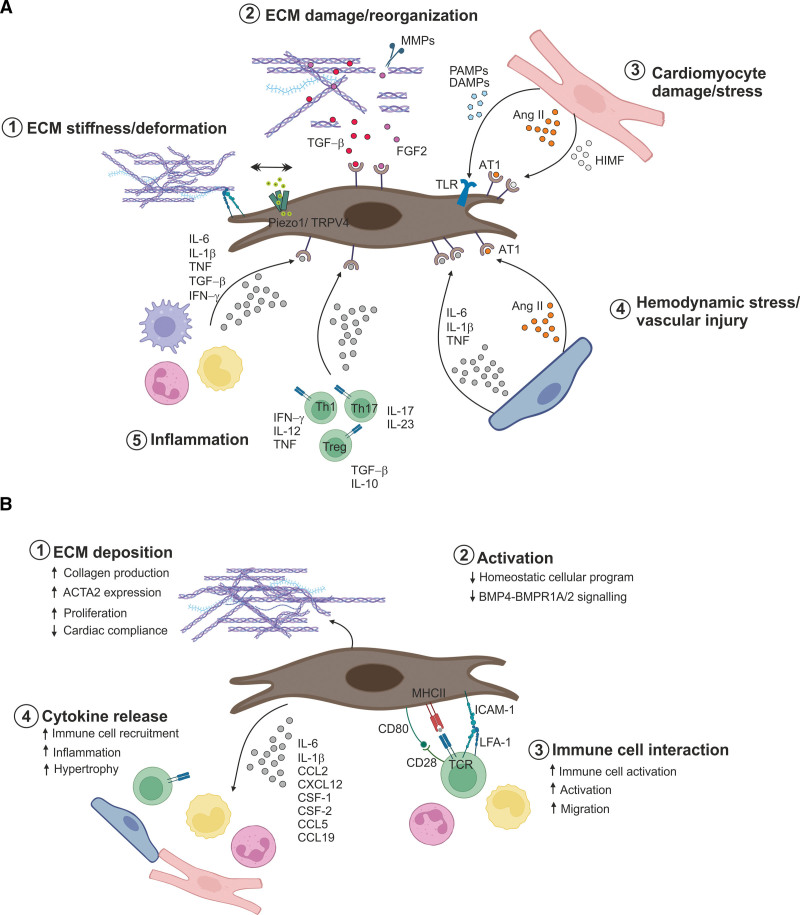
Fibroblasts are essential for building and maintaining the structural integrity of all organs. Moreover, fibroblasts can acquire an inflammatory phenotype to accommodate immune cells in specific niches and to provide migration, differentiation, and growth factors. In the heart, balancing of fibroblast activity is critical for cardiac homeostasis and optimal organ function during inflammation. Fibroblasts sustain cardiac homeostasis by generating local niche environments that support housekeeping functions and by actively engaging in intercellular cross talk. During inflammatory perturbations, cardiac fibroblasts rapidly switch to an inflammatory state and actively communicate with infiltrating immune cells to orchestrate immune cell migration and activity. Here, we summarize the current knowledge on the molecular landscape of cardiac fibroblasts, focusing on their dual role in promoting tissue homeostasis and modulating immune cell-cardiomyocyte interaction. In addition, we discuss potential future avenues for manipulating cardiac fibroblast activity during myocardial inflammation.

Keywords: cardiovascular diseases; endothelial cells; fibroblasts; homeostasis; inflammation; myocarditis.


Conflict of interest statement

Disclosures C. Perez-Shibayama, C. Gil-Cruz, and B. Ludewig are founders and C. Perez-Shibayama, C. Gil-Cruz, and B. Ludewig are shareholders of Stromal Therapeutics AG, Basel, Switzerland. B. Ludewig is a member of the board of Stromal Therapeutics AG, Basel, Switzerland. C. Perez-Shibayama, C. Gil-Cruz, and B. Ludewig are listed as inventors on patent WO 2022/084400 A1. The other author reports no conflicts.

Figures

Figure 1.
Figure 1.
Cardiac fibroblasts underpin different anatomic niches in the heart. Fibroblasts occupy specific anatomic niches within the heart, where their interactions with neighboring cells contribute to the maintenance of physiological homeostatic organ structure and function (left). A, Fibroblasts in the perivascular niche localize in concentric layers around blood vessels, providing critical support for vascular integrity and facilitating immune cell immigration. B, Cardiac fibroblasts in the interstitial space primarily produce and maintain the cardiac ECM (extracellular matrix) components, allowing both cell anchorage and communication with other cardiac cells. C, The subepicardial space is located between the epicardium and the myocardium. Cardiac inflammation alters intercellular communication in the different niche environments (right). Inflammation-induced fibroblast activation triggers increased fibroblast-to-immune-cell communication and ECM deposition (A–C, right). Histological images represent hematoxylin and eosin (top) or picrosirius red stained sections (bottom; scale bars=50 µm) of homeostatic (8-week-old BALB/c mice) or acutely inflamed hearts (8-week-old TCRM mice, a T-cell transgenic mouse model of autoimmune myocarditis). CCR2 indicates C-C chemokine receptor type 2; CX3CR1, C-X3-C motif chemokine receptor 1; RCM, resident cardiac macrophage; and VSMC, vascular smooth muscle cell. Illustration credit: Sceyence Studios.
Figure 2.
Figure 2.
Homeostatic intercellular communication in the heart. Depending on the specific niche environment, cardiac fibroblasts engage in active cross talk with different cell types, including endothelial cells, cardiomyocytes, and tissue-resident immune cells. In the perivascular niche, fibroblasts communicate with endothelial cells via direct cell-to-cell contacts (NOTCH [neurogenic locus notch homolog protein 1]-JAG [jagged]), soluble signaling molecules, such as Ang II (angiotensin II) or ET-1 (endothelin), or indirectly via the ECM (extracellular matrix) (1). Interstitial fibroblasts are intermingled in the myocardium and support homeostatic cardiomyocyte function and survival via the ECM, growth factor secretion (eg, IGF-1 [insulin-like growth factor 1], or FGF2 [fibroblast growth factor-2]) and through direct cell coupling mediated by Cx (connexin) 43 or 45 (2). Fibroblasts support cardiomyocyte fitness by providing a niche for resident cardiac macrophages (resident cardiac macrophages [RCMs]), which in turn regulate ECM stiffness through the secretion of ECM modulators such as MMP (matrix metalloproteinase) (3). ACTA2 indicates alpha smooth muscle actin 2; AT1, type 1 angiotensin receptor; BMP4, bone morphogenic protein-4; BMPR1A/2, bone morphogenic protein receptor 1A and 2; CSF-1, colony-stimulating factor 1; CSF1R, colony-stimulating factor-1 receptor; LYVE1, lymphatic vessel endothelial hyaluronan receptor 1; OSM, oncostatin M; PDGF, platelet-derived growth factors; and VEGF, vascular endothelial growth factor. Created using BioRender.com.
Figure 3.
Figure 3.
Inflammation leads to reprogramming of cardiac fibroblasts. A, Cardiac fibroblasts monitor the environment for signs of inflammation, damage, and cellular stress. Fibroblasts are able to integrate information on ECM (extracellular matrix) physical properties (1), ECM quality (2), cardiomyocyte fitness (3), vascular integrity (4), and inflammation (5) derived from multiple cellular and structural sources. Changes in ECM properties and tissue injury trigger mechanosensitive ion channels (eg, piezo1) and the release of latent growth factors such as TGF-β (transforming growth factor beta). Stressed or dying cardiac cells and infiltrating immune cells further release signaling molecules, including Ang II (angiotensin II), interleukins (IL), and IFN-γ (interferon-γ), which foster local inflammation and trigger fibroblast activation. B, Following stimulation cardiac fibroblasts transition from a homeostatic to a proinflammatory state. This process is characterized by increased ECM deposition (1), sustained activation (2), enhanced communication with immune cells (3), and localized cytokine release (4). ACTA2 indicates alpha smooth muscle actin 2; AT1, type 1 angiotensin receptor; BMP4, bone-morphogenic protein-4; BMPR1A/2, bone morphogenic protein receptor 1A and 2; CCL, C-C motif ligand; CSF-1, colony-stimulating factor 1; CD, cluster of differentiation; CXCL, C-X-C motif ligand; DAMP, danger-associated molecular patterns; FGF2, fibroblast growth factor-2; HIMF, hypoxia-induced mitogenic factor; ICAM-1, intercellular adhesion molecule 1; LFA-1, lymphocyte function-associated antigen 1; MHC II, major histocompatibility complex class II; MMP, matrix metalloproteinase; PAMP, pathogen-associated molecular patterns; TCR, T-cell receptor; Th, T helper cell; TLR, toll-like receptor; TNF, tumor necrosis factor; and Treg, regulatory T cell. Created using BioRender.com.

Similar articles

Cited by

  • Cardiac fibroblasts: answering the call.
    Kleinbongard P, Senyo SE, Lindsey ML, Garvin AM, Simpson JA, de Castro Braz LE. Kleinbongard P, et al. Am J Physiol Heart Circ Physiol. 2024 Sep 1;327(3):H681-H686. doi: 10.1152/ajpheart.00478.2024. Epub 2024 Aug 2. Am J Physiol Heart Circ Physiol. 2024. PMID: 39093000 Free PMC article. Review.

References

    1. Pinto AR, Ilinykh A, Ivey MJ, Kuwabara JT, D’Antoni ML, Debuque R, Chandran A, Wang L, Arora K, Rosenthal NA, et al. . Revisiting cardiac cellular composition. Circ Res. 2016;118:400–409. doi: 10.1161/CIRCRESAHA.115.307778 - PMC - PubMed
    1. Tallquist MD. Cardiac fibroblast diversity. Annu Rev Physiol. 2020;82:63–78. doi: 10.1146/annurev-physiol-021119-034527 - PMC - PubMed
    1. Ngwenyama N, Kaur K, Bugg D, Theall B, Aronovitz M, Berland R, Panagiotidou S, Genco C, Perrin MA, Davis J, et al. . Antigen presentation by cardiac fibroblasts promotes cardiac dysfunction. Nat Cardiovasc Res. 2022;1:761–774. doi: 10.1038/s44161-022-00116-7 - PMC - PubMed
    1. McLellan MA, Skelly DA, Dona MSI, Squiers GT, Farrugia GE, Gaynor TL, Cohen CD, Pandey R, Diep H, Vinh A, et al. . High-resolution transcriptomic profiling of the heart during chronic stress reveals cellular drivers of cardiac fibrosis and hypertrophy. Circulation. 2020;142:1448–1463. doi: 10.1161/CIRCULATIONAHA.119.045115 - PMC - PubMed
    1. Perez-Shibayama C, Gil-Cruz C, Cadosch N, Lütge M, Cheng HW, De Martin A, Frischmann K, Joachimbauer A, Onder L, Papadopoulou I, et al. . Bone morphogenic protein-4 availability in the cardiac microenvironment controls inflammation and fibrosis in autoimmune myocarditis. Nat Cardiovasc Res. 2024;3:301–316. doi: 10.1038/s44161-024-00432-0