

MSCs‑derived exosomes attenuate ischemia-reperfusion brain injury and inhibit microglia apoptosis might via exosomal miR-26a-5p mediated suppression of CDK6
- PMID: 34215174
- PMCID: PMC8254277
- DOI: 10.1186/s10020-021-00324-0
MSCs‑derived exosomes attenuate ischemia-reperfusion brain injury and inhibit microglia apoptosis might via exosomal miR-26a-5p mediated suppression of CDK6
Abstract
Background: This study aimed to explore the role of mesenchymal stromal cells (MSCs)-derived exosomes (MSCs-Exo) in the cerebral ischemia-reperfusion (I/R) injury.
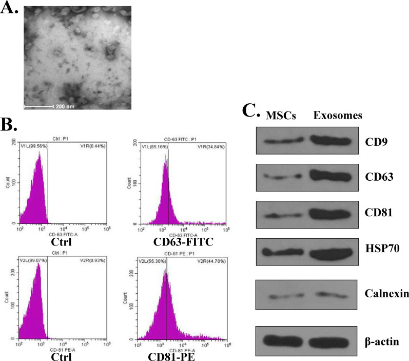
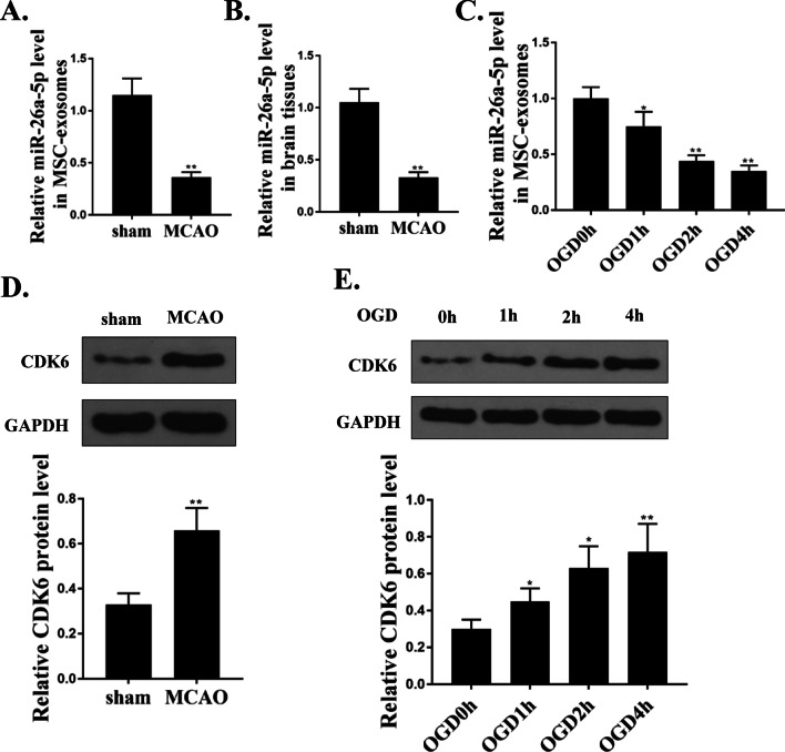
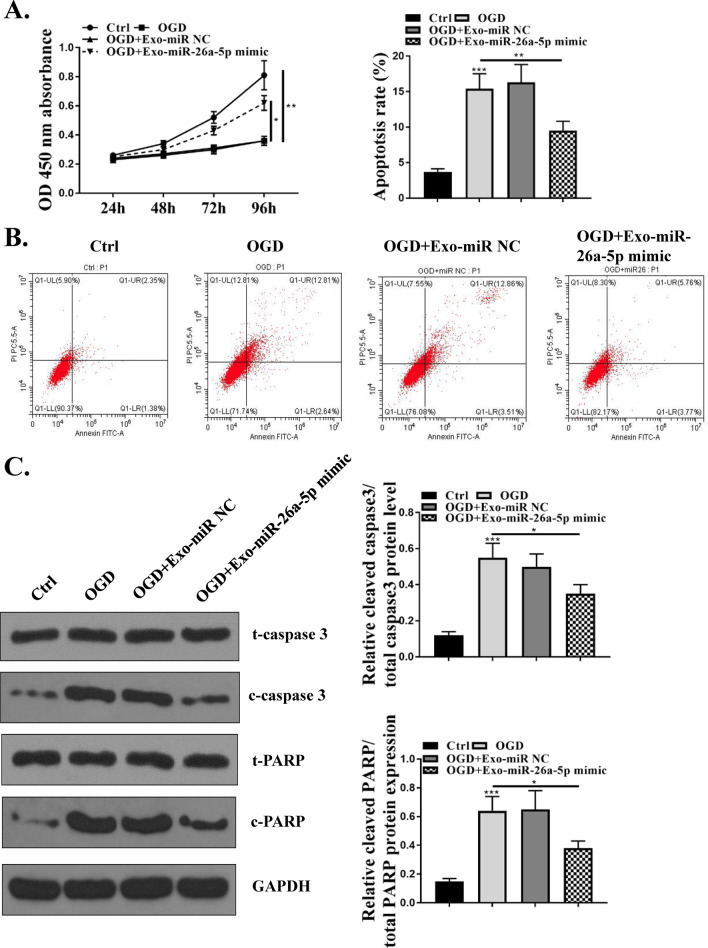
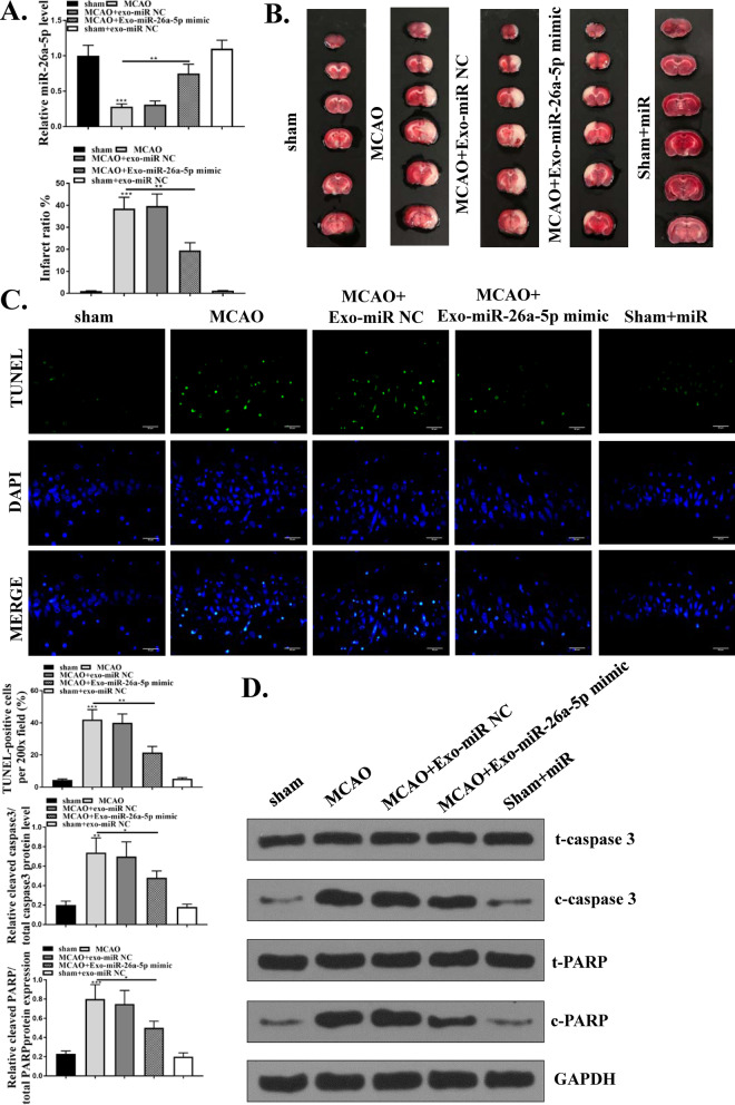
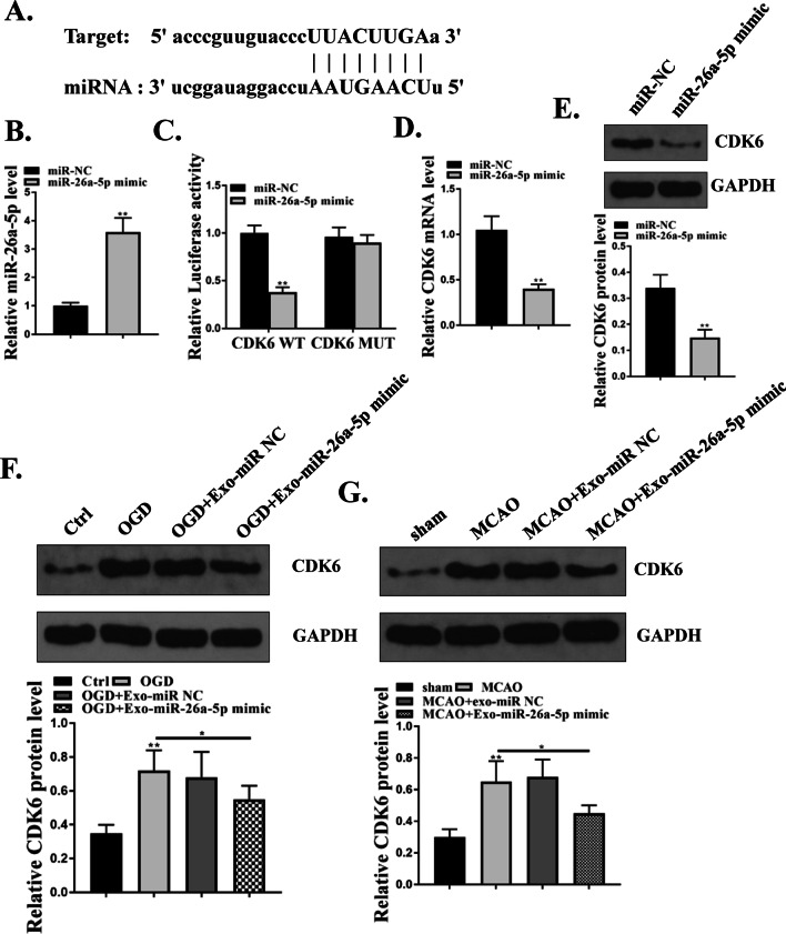
Methods: Exosomes were isolated from MSCs of adult C57BL/6J mice by the gradient centrifugation method. The expression of miR-26a-5p and CDK6 in MSCs-Exo and mice brain tissues were evaluated by qRT-PCR and western blot. miR-26a-5p mimics and miR-NC were transfected into MSCs, and exosomes were isolated from the MSCs stably expressing miR-26a-5p. Then MSCs-Exo-miR-26a-5p mimics or MSCs-Exo-miR-NC was injected into mice through the tail vein, or added into medium to stimulate BV-2 cells. Cell viability was evaluated by CCK-8 assay. Cell apoptosis was detected by flow cytometry. The apoptosis in brain tissues was evaluated by TUNEL staining assay. Bioinformatics analysis and luciferase reporter assay were performed to determine the binding relationship between miR-26a-5p and CDK6.
Results: miR-26a-5p was downregulated and CDK6 was upregulated in MSCs-Exo of MCAO-mice and OGD-induced MSCs. MSCs-Exo-miR-26a-5p mimics significantly reduced cell apoptosis of OGD-injured BV-2 cells. MSCs-Exo-miR-26a-5p mimics significantly reduced the infarct volume of MCAO-induced mice. Luciferase reporter assay revealed that CDK-6 was a target of miR-26a-5p. In addition, MSCs-Exo-miR-26a-5p mimics significantly decreased the expression of CDK6 in both OGD-induced BV-2 cells and the brain tissues of MCAO-treated mice.
Conclusion: Our results indicated that MSCs‑Exo attenuated I/R injury in mice by inhibiting microglia apoptosis might via exosomal miR-26a-5p mediated suppression of CDK6. Our study shed light on the application of MSC-Exo as a potential therapeutic tool for cerebral I/R injury.
Keywords: CDK6; Exosomes; Ischemia–reperfusion injury; Mesenchymal stromal cells; miR-26a-5p.
Conflict of interest statement
The authors declare that they have no competing interests.
Figures
Similar articles
-
MSCs-derived exosomes containing miR-486-5p attenuate cerebral ischemia and reperfusion (I/R) injury.Gene. 2024 May 15;906:148262. doi: 10.1016/j.gene.2024.148262. Epub 2024 Feb 10. Gene. 2024. PMID: 38346456
-
Mesenchymal stromal cells-derived exosomes alleviate ischemia/reperfusion injury in mouse lung by transporting anti-apoptotic miR-21-5p.Eur J Pharmacol. 2019 Jun 5;852:68-76. doi: 10.1016/j.ejphar.2019.01.022. Epub 2019 Jan 23. Eur J Pharmacol. 2019. PMID: 30682335
-
MiR-145 enriched exosomes derived from bone marrow-derived mesenchymal stem cells protects against cerebral ischemia-reperfusion injury through downregulation of FOXO1.Biochem Biophys Res Commun. 2022 Dec 3;632:92-99. doi: 10.1016/j.bbrc.2022.09.089. Epub 2022 Sep 27. Biochem Biophys Res Commun. 2022. PMID: 36206599
-
The therapeutic potential of stem cell-derived exosomes in the ulcerative colitis and colorectal cancer.Stem Cell Res Ther. 2022 Apr 1;13(1):138. doi: 10.1186/s13287-022-02811-5. Stem Cell Res Ther. 2022. PMID: 35365226 Free PMC article. Review.
-
New Therapeutic Strategies for the Inflammatory Rheumatoid Arthritis Disease: Emphasizing Mesenchymal Stem Cells and Associated exo-miRNA or exo-lncRNA.Cell Biochem Biophys. 2024 Sep;82(3):1599-1611. doi: 10.1007/s12013-024-01316-7. Epub 2024 May 31. Cell Biochem Biophys. 2024. PMID: 38822204 Review.
Cited by
-
Immunomodulation: The next target of mesenchymal stem cell-derived exosomes in the context of ischemic stroke.World J Stem Cells. 2023 Mar 26;15(3):52-70. doi: 10.4252/wjsc.v15.i3.52. World J Stem Cells. 2023. PMID: 37007453 Free PMC article. Review.
-
Mesenchymal stem cell-derived exosomes: Shaping the next era of stroke treatment.Neuroprotection. 2023 Dec;1(2):99-116. doi: 10.1002/nep3.30. Epub 2023 Dec 30. Neuroprotection. 2023. PMID: 38283953 Free PMC article.
-
Mesenchymal stem cell-derived exosomes regulate microglia phenotypes: a promising treatment for acute central nervous system injury.Neural Regen Res. 2023 Aug;18(8):1657-1665. doi: 10.4103/1673-5374.363819. Neural Regen Res. 2023. PMID: 36751776 Free PMC article. Review.
-
Exosomal miR-17-92 Cluster from BMSCs Alleviates Apoptosis and Inflammation in Spinal Cord Injury.Biochem Genet. 2024 Jul 3. doi: 10.1007/s10528-024-10876-5. Online ahead of print. Biochem Genet. 2024. PMID: 38961001
-
Exosomes Derived From Mesenchymal Stem Cells: Novel Effects in the Treatment of Ischemic Stroke.Front Neurosci. 2022 May 2;16:899887. doi: 10.3389/fnins.2022.899887. eCollection 2022. Front Neurosci. 2022. PMID: 35585925 Free PMC article. Review.
References
Publication types
MeSH terms
Substances
LinkOut - more resources
Full Text Sources
