. 2021 Apr 27;118(17):e2025998118.
doi: 10.1073/pnas.2025998118.

PIP2 corrects cerebral blood flow deficits in small vessel disease by rescuing capillary Kir2.1 activity

Affiliations

PIP2 corrects cerebral blood flow deficits in small vessel disease by rescuing capillary Kir2.1 activity

Fabrice Dabertrand et al. Proc Natl Acad Sci U S A. .

Abstract

Cerebral small vessel diseases (SVDs) are a central link between stroke and dementia-two comorbidities without specific treatments. Despite the emerging consensus that SVDs are initiated in the endothelium, the early mechanisms remain largely unknown. Deficits in on-demand delivery of blood to active brain regions (functional hyperemia) are early manifestations of the underlying pathogenesis. The capillary endothelial cell strong inward-rectifier K+ channel Kir2.1, which senses neuronal activity and initiates a propagating electrical signal that dilates upstream arterioles, is a cornerstone of functional hyperemia. Here, using a genetic SVD mouse model, we show that impaired functional hyperemia is caused by diminished Kir2.1 channel activity. We link Kir2.1 deactivation to depletion of phosphatidylinositol 4,5-bisphosphate (PIP2), a membrane phospholipid essential for Kir2.1 activity. Systemic injection of soluble PIP2 rapidly restored functional hyperemia in SVD mice, suggesting a possible strategy for rescuing functional hyperemia in brain disorders in which blood flow is disturbed.

Keywords: CADASIL; PIP2; cerebral small vessel diseases; functional hyperemia; potassium channel.


Conflict of interest statement

Competing interest statement: Exogenous PIP2 administration was submitted as patent number 62/823,378 titled “Methods to promote cerebral blood flow in the brain” on 25 March 2019.

Figures

Fig. 1.
Fig. 1.
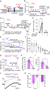
Capillary-to-arteriole electrical signaling during functional hyperemia is abrogated in SVD. (A) Proposed mechanism for pathogenic blunting of EGFR activation by accumulation of TIMP3 in CADASIL at the capillary level. (B) Pipette positions for arteriole stimulation (Left, orange arrow) and capillary stimulation (Right, purple arrow) in CaPA preparations. (C) Representative traces of arteriolar diameter showing effects of pressure ejection of 10 mM K+ onto capillaries (P2, purple triangle) on the diameter of upstream arteriolar segments in control preparations from WT and SVD mice. (D) Summary data from 8 WT and 8 SVD mice (n.s., not significant; ****P < 0.0001, unpaired Student’s t test). (E) Micrograph displaying a micropipette containing 10 mM K+ and TRITC-dextran (red) in close apposition to a capillary (FITC-dextran, green) in SVD mice. RBC flux induced by local ejection of K+ onto the capillary was measured by high-frequency line scanning. (F and G) (Top Panels) Raw recordings showing RBC flux at baseline and after application of 10 mM K+ onto a capillary in WT (F) and SVD (G) mice. RBCs appear as black streaks in plasma (green). x axis, time; y axis, scanned capillary distance, d. (Bottom) Full trace from raw recordings shown above. (H) Summary data showing that K+-evoked hyperemia is crippled in SVD mice (n = 16–17 experiments in seven to eight mice; **P < 0.01, unpaired Student’s t test).
Fig. 2.
Fig. 2.
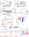
Kir2.1 channel down-regulation underlies deficits in capillary-to-arteriole electrical signaling in SVD. (A) Whisker stimulation experimental scheme. (B) Representative traces showing whisker stimulation-induced changes in CBF in control WT (blue) and SVD (red) mice in the presence (gray traces) and absence of Ba2+ (100 µM). (C) Summary data from five WT and five SVD mice (*P < 0.05, ***P < 0.001, ****P < 0.0001, repeated-measures two-way ANOVA). (D) Representative traces of Kir2.1 currents in cECs from WT (blue) or SVD (red) mice, recorded in the perforated-patch configuration. (E) Summary data showing Kir2.1 currents (n = 5–6 cECs from three mice per group; *P < 0.05, unpaired Student’s t test).
Fig. 3.
Fig. 3.
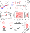
Pathogenic accumulation of TIMP3 reduces Kir2.1 currents and K+-induced dilations in SVD. (A) ADAM17/HB-EGF/EGFR signaling axis. TIMP3 inhibits ADAM17-mediated cleavage of proHB-EGF (black dashed line) and subsequent EGFR activation by HB-EGF (black arrow). (B) Arteriole diameter trace in a CaPA preparation from an SVD mouse showing myogenic tone and arteriolar dilation induced by capillary stimulation with 10 mM K+ in the presence of bath-applied sADAM17. (C) Trace showing block of sADAM17 effects by the EGFR antagonist AG1478. (D) Summary data from two groups of six SVD mice showing arteriolar dilation induced by capillary stimulation with 10 mM K+ (n.s., not significant; ***P < 0.001, paired Student’s t test). (E) Arteriole diameter trace showing effects of bath-applied TIMP3 on capillary-to-arteriole electrical signaling in WT CaPA preparation. (F) Summary data for E from six WT animals. (G) Arteriolar diameter trace in a CaPA preparation showing rescue of capillary-to-arteriole electrical signaling by genetic reduction of TIMP3 expression in SVD mice. (H) Summary data from six TgNotch3R169C;Timp3+/− mice showing inhibitory effect of 100 µM Ba2+ on K+ (10 mM)-induced dilation (****P < 0.0001, paired Student’s t test). Dotted blue line indicates dilation observed in a WT CaPA preparation, as in Fig. 1C. (I) Whisker stimulation-induced CBF increases in five TgNotch3R169C;Timp3+/− mice in the absence and presence of 100 µM Ba2+ (**P < 0.01, paired Student’s t test). Dotted lines indicate whisker stimulation–induced CBF changes in SVD (red) and WT (blue) mice in the absence of Ba2+, as in Fig. 2C. (J) Ba2+-sensitive Kir2.1 current in a TgNotch3R169C;Timp3+/− cEC, recorded in the perforated patch-clamp configuration. (K) Summary data showing currents recorded from TgNotch3R169C;Timp3+/− cECs in the absence and presence of 100 µM Ba2+ (n = 8 cECs from two mice; ****P < 0.0001, paired Student’s t test). (Right) Ba2+-sensitive Kir2.1 current. Dotted lines indicate current density observed in SVD (red) and WT (blue) cECs as in Fig. 2E.
Fig. 4.
Fig. 4.
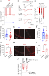
Lower Kir2.1 channel currents in SVD cECs and restoration by HB-EGF. (A and B) Arteriolar diameter traces in SVD CaPA preparations showing effects of bath-applied HB-EGF on myogenic tone and upstream arteriolar dilation in the absence (A) and presence (B) of the EGFR antagonist AG1478. (C) Arteriolar diameter traces in CaPA preparation from an EC-Kir2.1−/− mouse. (D) Summary data showing arteriolar dilation induced by capillary stimulation with 10 mM K+ (n = 5 mice per condition). (E) Ba2+-sensitive Kir2.1 currents in freshly isolated SVD cECs, recorded in the perforated-patch configuration in the absence or presence of bath-applied HB-EGF for 20 min. (F) Summary data showing the effect of HB-EGF on Kir2.1 currents from SVD and WT cECs (n = 4–8 cECs per group from eight mice; n.s., not significant; *P < 0.05, unpaired Student’s t test). (G) Representative traces of whisker stimulation-induced changes in CBF in WT and SVD mice before and after treatment with Ba2+. (H) Summary data showing restoration of Ba2+-sensitive functional hyperemia in SVD mice by HB-EGF (n = 4 mice per group; n.s., not significant; ****P < 0.0001, repeated-measures one-way ANOVA).
Fig. 5.
Fig. 5.
Exogenous PIP2 treatment improves functional hyperemia in SVD mice. (A) Kir2.1 currents recorded from WT and SVD cECs in the conventional (dialyzed cytoplasm) configuration within 3 to 5 min after gaining access (0 mM Mg-ATP in the pipette). (B) Summary data for Kir2.1 currents in WT and SVD cECs as in A (n = 6–11 cECs from three to four mice per group; *P < 0.05, unpaired Student’s t test). (C and D) Representative traces (C) and summary data (D) for Kir2.1 currents from SVD cECs preincubated with or without bath-applied diC16-PIP2 for 20 min (n = 8–13 cECs from three mice; **P < 0.01, unpaired Student’s t test). (E) Arteriolar diameter trace showing restoration of 10 mM K+-induced (capillary applied) dilatory responses by bath-applied diC16-PIP2, and their inhibition by Ba2+, in a CaPA preparation from an SVD mouse. (F) Summary data showing the effect of diC16-PIP2 in nine SVD and five EC-Kir2.1−/− mice. (G) Summary data from six SVD mice showing elimination of K+ (10 mM)-induced dilation by Ba2+ in the presence of diC16-PIP2 (****P < 0.0001, paired Student’s t test). (H) Representative traces showing whisker stimulation-induced changes in CBF in SVD mice before and after systemic injection of diC16-PIP2 and cortical superfusion of Ba2+. (I) Summary data showing increased whisker stimulation-induced functional hyperemia with PIP2 treatment and elimination by Ba2+ (n = 6 animals, *P < 0.05, **P < 0.01, repeated-measures one-way ANOVA).
Fig. 6.
Fig. 6.
Decreased intracellular ATP in capillaries from SVD mice. (A) Summary data for Kir2.1 currents in cECs from SVD mice measured using the perforated-patch configuration. SVD cECs were bath-incubated for ∼15 min with the Gαq/11 inhibitor YM-254890 or the phospholipase C inhibitor U73122 (n = 5–7 cECs from three to four mice per group). Dotted lines indicate current density observed in SVD (red) and WT (blue) cECs, as in Fig. 2E. (B) Kir2.1 currents in SVD cECs dialyzed with 0, 0.1, or 1 mM Mg-ATP in the pipette solution using the conventional configuration. (C) Summary data for B (n = 5–7 cECs from four mice; n.s., not significant, *P < 0.05, one-way ANOVA). (DI) Intracellular ATP levels in WT or SVD cECs, without or with HB-EGF treatment. cEC luminescence (in relative luminescence units [RLU]), reflecting luciferase-induced conversion of ATP to light, is directly proportional to cEC ATP concentration (D and G). cEC fluorescence intensity, measured with the live cell ATP dye ATP-Red1, is proportional to ATP levels (E, F, H, and I). Each data point on scatter plots in F and I represent a capillary segment, and each data point in D and G was obtained using ∼500 cECs manually collected from three WT and three SVD mice (*P < 0.05, unpaired Student’s t test). (J) Kir2.1 currents in cECs from SVD, recorded using the conventional configuration. One group of cECs was dialyzed with 0 mM Mg-ATP, and the other was dialyzed with 0.1 mM Mg-ATP and incubated with HB-EGF for ∼20 min (n = 4–9 cECs from six mice; n.s., not significant, unpaired Student’s t test). Dotted line indicates Kir2.1 current amplitude in SVD cECs incubated with HB-EGF using the perforated configuration (as in Fig. 4F).
Fig. 7.
Fig. 7.
Proposed mechanism by which SVDs silence Kir2.1 channel during functional hyperemia and how treatment with exogenous PIP2 can restore the physiological response. In SVD, a substantial fraction of Kir2.1 channels in cECs are not associated with PIP2, making them not activable by increases in extracellular K+ from neural activity. PIP2 treatment allows Kir2.1 channels subunits to be associated with PIP2 and then be activable by external K+ and hyperpolarization. This produces a regenerative hyperpolarization that spreads to adjacent ECs up to the arteriolar SMCs. Hyperpolarization then closes voltage-dependent Ca2+ channels (VDCCs) to cause arteriolar dilation, promoting an increase in blood flow into the capillaries.

Comment in

Similar articles

Cited by

References

    1. Pantoni L., Cerebral small vessel disease: From pathogenesis and clinical characteristics to therapeutic challenges. Lancet Neurol. 9, 689–701 (2010). - PubMed
    1. Wardlaw J. M., Smith C., Dichgans M., Small vessel disease: Mechanisms and clinical implications. Lancet Neurol. 18, 684–696 (2019). - PubMed
    1. Iadecola C., et al. ., Vascular cognitive impairment and dementia: JACC scientific expert panel. J. Am. Coll. Cardiol. 73, 3326–3344 (2019). - PMC - PubMed
    1. Chabriat H., Joutel A., Dichgans M., Tournier-Lasserve E., Bousser M.-G., Cadasil. Lancet Neurol. 8, 643–653 (2009). - PubMed
    1. Longden T. A., et al. ., Capillary K+-sensing initiates retrograde hyperpolarization to increase local cerebral blood flow. Nat. Neurosci. 20, 717–726 (2017). - PMC - PubMed

Publication types

MeSH terms

Substances

LinkOut - more resources