Myosin Light Chain 9/12 Regulates the Pathogenesis of Inflammatory Bowel Disease
- PMID: 33584659
- PMCID: PMC7878395
- DOI: 10.3389/fimmu.2020.594297
Myosin Light Chain 9/12 Regulates the Pathogenesis of Inflammatory Bowel Disease
Abstract
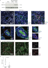
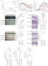
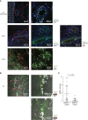
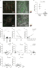
The numbers of patients with inflammatory bowel disease (IBD), such as ulcerative colitis (UC) and Crohn's disease (CD), have been increasing over time, worldwide; however, the pathogenesis of IBD is multifactorial and has not been fully understood. Myosin light chain 9 and 12a and 12b (Myl9/12) are known as ligands of the CD69 molecule. They create "Myl9 nets" that are often detected in inflamed site, which play a crucial role in regulating the recruitment and retention of CD69-expressing effector cells in inflamed tissues. We demonstrated the strong expression of Myl9/12 in the inflamed gut of IBD patients and mice with DSS-induced colitis. The administration of anti-Myl9/12 Ab to mice with DSS-induced colitis ameliorated the inflammation and prolonged their survival. The plasma Myl9 levels in the patients with active UC and CD were significantly higher than those in patients with disease remission, and may depict the disease severity of IBD patients, especially those with UC. Thus, our results indicate that Myl9/12 are involved in the pathogenesis of IBD, and are likely to be a new therapeutic target for patients suffering from IBD.
Keywords: CD69; Crohn’s disease; Myl9; plasma biomarker; ulcerative colitis.
Copyright © 2021 Yokoyama, Kimura, Ito, Hayashizaki, Endo, Wang, Yagi, Nakagawa, Kato, Matsubara and Nakayama.
Conflict of interest statement
The authors declare that the research was conducted in the absence of any commercial or financial relationships that could be construed as a potential conflict of interest.
Figures


Similar articles
-
A new therapeutic target: the CD69-Myl9 system in immune responses.Semin Immunopathol. 2019 May;41(3):349-358. doi: 10.1007/s00281-019-00734-7. Epub 2019 Apr 5. Semin Immunopathol. 2019. PMID: 30953160 Review.
-
Crucial role for CD69 in allergic inflammatory responses: CD69-Myl9 system in the pathogenesis of airway inflammation.Immunol Rev. 2017 Jul;278(1):87-100. doi: 10.1111/imr.12559. Immunol Rev. 2017. PMID: 28658550 Review.
-
Development, validation and implementation of an in vitro model for the study of metabolic and immune function in normal and inflamed human colonic epithelium.Dan Med J. 2015 Jan;62(1):B4973. Dan Med J. 2015. PMID: 25557335 Review.
-
Regulation of CEACAM Family Members by IBD-Associated Triggers in Intestinal Epithelial Cells, Their Correlation to Inflammation and Relevance to IBD Pathogenesis.Front Immunol. 2021 Jul 29;12:655960. doi: 10.3389/fimmu.2021.655960. eCollection 2021. Front Immunol. 2021. PMID: 34394073 Free PMC article.
-
Expression and Clinical Significance of Elafin in Inflammatory Bowel Disease.Inflamm Bowel Dis. 2017 Dec;23(12):2134-2141. doi: 10.1097/MIB.0000000000001252. Inflamm Bowel Dis. 2017. PMID: 29084078
Cited by
-
Increased Myosin light chain 9 expression during Kawasaki disease vasculitis.Front Immunol. 2023 Jan 6;13:1036672. doi: 10.3389/fimmu.2022.1036672. eCollection 2022. Front Immunol. 2023. PMID: 36685558 Free PMC article.
-
MYL9 expressed in cancer-associated fibroblasts regulate the immune microenvironment of colorectal cancer and promotes tumor progression in an autocrine manner.J Exp Clin Cancer Res. 2023 Nov 6;42(1):294. doi: 10.1186/s13046-023-02863-2. J Exp Clin Cancer Res. 2023. PMID: 37926835 Free PMC article.
-
Antioxidant, Immunomodulatory and Potential Anticancer Capacity of Polysaccharides (Glucans) from Euglena gracilis G.A. Klebs.Pharmaceuticals (Basel). 2022 Nov 10;15(11):1379. doi: 10.3390/ph15111379. Pharmaceuticals (Basel). 2022. PMID: 36355551 Free PMC article.
-
Alleviation of LPS-induced Endothelial Injury due to GHRH Antagonist Treatment.Int J Pept Res Ther. 2024;30(6):67. doi: 10.1007/s10989-024-10653-3. Epub 2024 Oct 1. Int J Pept Res Ther. 2024. PMID: 39465062
-
Elevated Myl9 reflects the Myl9-containing microthrombi in SARS-CoV-2-induced lung exudative vasculitis and predicts COVID-19 severity.Proc Natl Acad Sci U S A. 2022 Aug 16;119(33):e2203437119. doi: 10.1073/pnas.2203437119. Epub 2022 Jul 27. Proc Natl Acad Sci U S A. 2022. PMID: 35895716 Free PMC article.
References
Publication types
MeSH terms
Substances
LinkOut - more resources
Full Text Sources
Other Literature Sources

