. 2021 Jan 19:10:e62455.
doi: 10.7554/eLife.62455.

Highly redundant neuropeptide volume co-transmission underlying episodic activation of the GnRH neuron dendron

Affiliations

Highly redundant neuropeptide volume co-transmission underlying episodic activation of the GnRH neuron dendron

Xinhuai Liu et al. Elife. .

Abstract

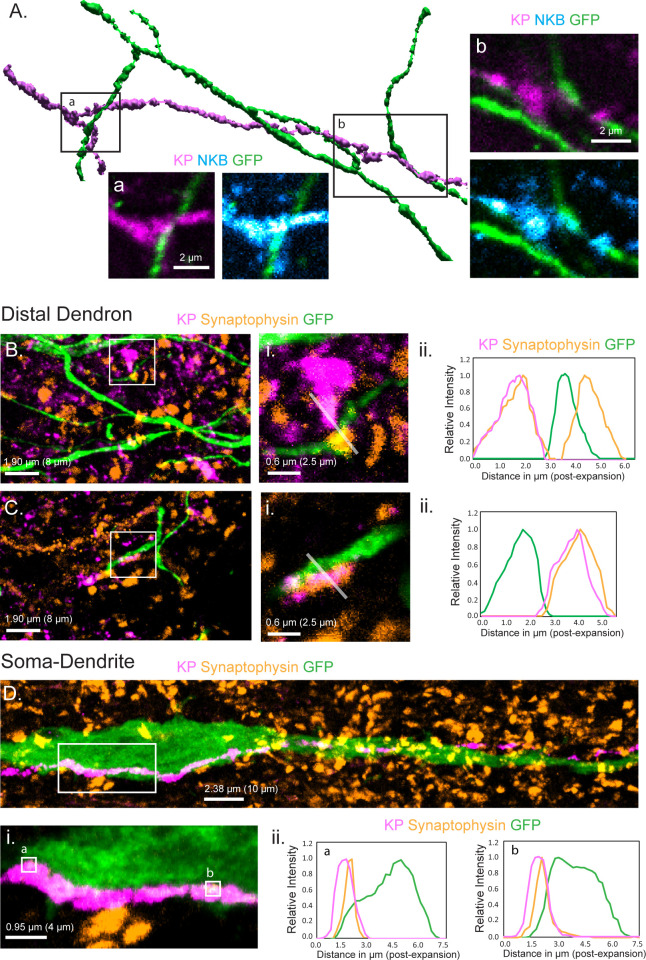
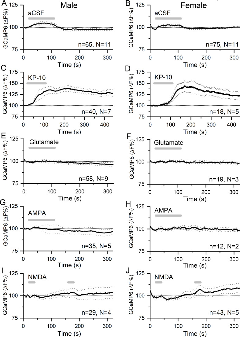
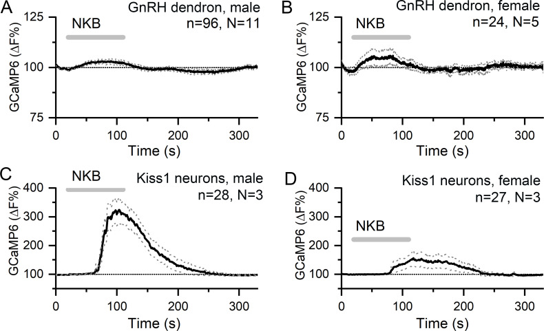
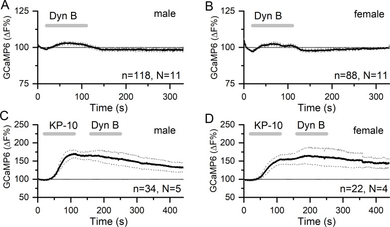
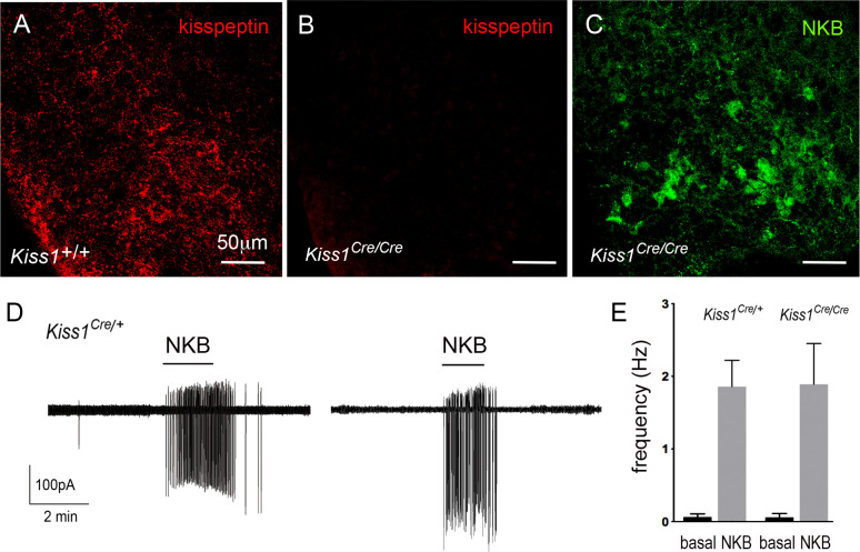
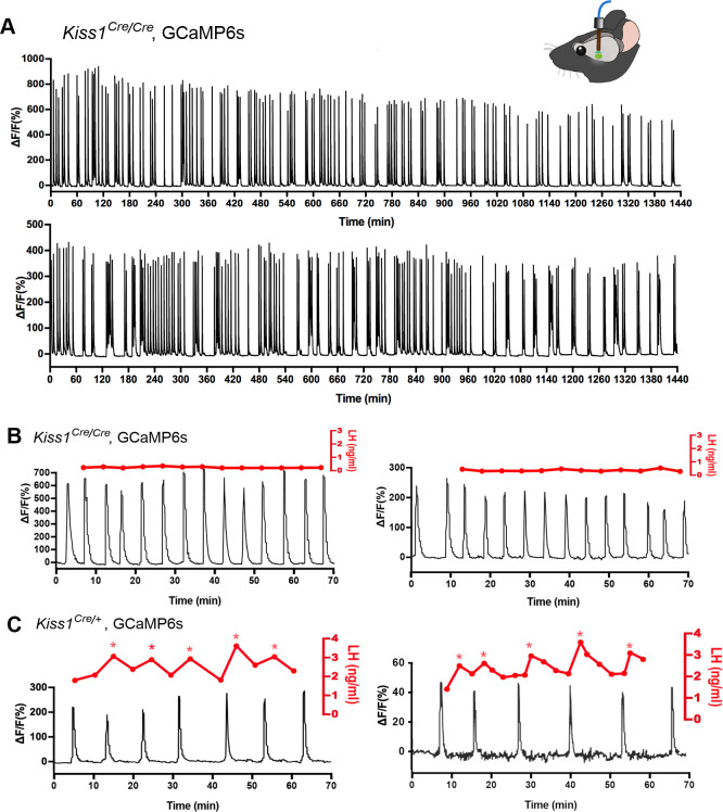
The necessity and functional significance of neurotransmitter co-transmission remains unclear. The glutamatergic 'KNDy' neurons co-express kisspeptin, neurokinin B (NKB), and dynorphin and exhibit a highly stereotyped synchronized behavior that reads out to the gonadotropin-releasing hormone (GnRH) neuron dendrons to drive episodic hormone secretion. Using expansion microscopy, we show that KNDy neurons make abundant close, non-synaptic appositions with the GnRH neuron dendron. Electrophysiology and confocal GCaMP6 imaging demonstrated that, despite all three neuropeptides being released from KNDy terminals, only kisspeptin was able to activate the GnRH neuron dendron. Mice with a selective deletion of kisspeptin from KNDy neurons failed to exhibit pulsatile hormone secretion but maintained synchronized episodic KNDy neuron behavior that is thought to depend on recurrent NKB and dynorphin transmission. This indicates that KNDy neurons drive episodic hormone secretion through highly redundant neuropeptide co-transmission orchestrated by differential post-synaptic neuropeptide receptor expression at the GnRH neuron dendron and KNDy neuron.

Keywords: Dynorphin; GCaMP; GnRH; NKB; kisspeptin; mouse; neuroscience; pulse generator.


Conflict of interest statement

XL, SY, HM, MH, SH, IC, RP, AH No competing interests declared

Figures

Figure 1.
Figure 1.. Relationship of KNDy neuron fibers to GnRH neuron distal dendrons.
(A) 3D reconstruction of regular confocal images showing a KNDy fiber expressing kisspeptin (KP) and neurokinin B (NKB) making close appositions with three GnRH neuron dendrons in the ventrolateral ARN of a GnRH-GFP mouse. (B,C) Expansion microscopy views of GnRH distal dendrons surrounded by kisspeptin fibers and synaptophysin puncta. Insets (i) highlight two examples of synaptophysin-expressing kisspeptin terminals adjacent to GnRH dendrons. Gray lines indicate the line scans used to generate the fluorescence relative intensity profiles shown to the right. (Bii) A GFP-expressing dendron with a chemically unidentified synapse on one side (>0.95 μm overlap between synaptophysin and GFP signals) and a kisspeptin terminal making a close non-synaptic contact on the other (no overlap). (Cii) Another example of kisspeptin terminal (kisspeptin and synaptophysin) making a close non-synaptic (overlap <0.95 μm) contact with a GnRH dendron. (D) Expansion microscopy view of a GnRH neuron cell body and proximal dendrite surrounded by synaptophysin puncta and with a kisspeptin fiber running along its length. Imaging in the z-axis face view shows two locations (ia and b) where kisspeptin/synaptophysin puncta make synapses on the GnRH neuron cell body. (Dii) Fluorescence relative intensity profiles show two synaptophysin-containing kisspeptin boutons exhibiting >1.75 μm overlap with cytoplasmic GFP of the GnRH neuron. Scale bars show pre-expansion units with post-expansion values in brackets.
Figure 2.
Figure 2.. Horizontal brain slice preparation for examining GnRH neuron distal dendrons.
(A) View looking down on a thick horizontal brain slice prepared from a Gnrh-GFP mouse showing the laterally positioned GnRH neuron cell bodies in the anterior hypothalamic area (AHA) and the concentrated GnRH neuron projections in the median eminence (ME). 3V, third ventricle. (B) Higher-power view of the same orientation of the ME region in a living brain slice prepared from a GCaMP6s AAV-injected Gnrh1-Cre mouse showing the recording location (dotted square) and position of the puff pipette.
Figure 3.
Figure 3.. Kisspeptin but not glutamate regulates [Ca2+] in GnRH neuron distal dendrons.
(A,B) Effects of 90 s puffs of aCSF on GCaMP6 fluorescence in GnRH neuron distal dendrons in male and female Gnrh1-Cre::GCaMP6s mice. (C,D) Puffs of kisspeptin-10 (100 nM) generate large, sustained increases in [Ca2+] in both sexes. Note the altered x- and y-axes. (E–J) Long (90 s) or short (20 s) puffs of glutamate (600 μM), AMPA (80 μM), and NMDA (200 μM) have no significant effects on [Ca2+] in dendrons. Dotted lines indicate 95% confidence intervals. Numbers of dendrons (n) and mice (N) are given for each treatment and each sex.
Figure 4.
Figure 4.. NKB increases [Ca2+] in KNDy neurons but not in GnRH neuron distal dendrons.
(A,B) Ninety second puffs of 100 nM NKB have no significant effect on GCaMP6 fluorescence in GnRH neuron distal dendrons in male and female Gnrh1-Cre::GCaMP6s mice. (C,D) Ninety second puffs of 100 nM NKB evoke large increases in [Ca2+] in KNDy neurons of male and female Kiss1Cre/+;GCaMP6f mice. Note the altered y-axis. Dotted lines indicate 95% confidence intervals. Numbers of dendrons (n) and mice (N) are given for each treatment and each sex.
Figure 5.
Figure 5.. Dynorphin has no effect on [Ca2+] in GnRH neuron distal dendrons.
(A,B) Ninety second puffs of 200 nM dynorphin have no significant effect on GCaMP6 fluorescence in GnRH neuron distal dendrons in male and female Gnrh1-Cre::GCaMP6s mice. (C,D) Similarly, dynorphin has no effect on kisspeptin-10-evoked increases in GCaMP6 fluorescence in either sex. Dotted lines indicate 95% confidence intervals. Numbers of dendrons (n) and mice (N) are given for each treatment and each sex.
Figure 6.
Figure 6.. NKB and dynorphin are released from KNDy neuron terminals in the vicinity of the GnRH neuron dendrons.
(A) Optogenetic blue light (473 nm) activation of a transduced KNDy neuron at 20 Hz and 10 Hz. Rate meter trace below shows sustained 20 Hz and 10 Hz firing during the majority of the 10 s stimuli. Expanded inset shows individual action currents. (B) Cell-attached recording (above) and rate meter histogram (below) of an unidentified ventrolateral ARN neuron exhibiting a slow excitatory response to 10 Hz blue light (blue bar) that is reversibly suppressed by addition of the NK3R antagonist SB222200 (20 μM) shown as the dark bar. (C) Cell-attached recording of another unidentified ventrolateral ARN neuron exhibiting an inhibitory response to 10 Hz blue light (blue bar) that is reversibly suppressed by addition of the kappa-opioid antagonist nor-binaltorphimine (NBI) (12.5 μM) shown as the dark bar. The inhibitory response to optogenetic activation returns after ~20 min.
Figure 7.
Figure 7.. Characterization of Kiss1-null mice.
(A-C) Immunofluorescence for kisspeptin (A,B) and NKB (C) in the ARN of wild-type (A) and Kiss1-null (B,C) female mice. (D) Cell-attached recordings showing the effects of 100 nM NKB on firing of KNDy neurons in acute brain slices prepared from female heterozygous Kiss1Cre/+ and homozygous (null) Kiss1Cre/Cre;Rosa26-tdT mice. (E) Mean ± SEM changes in KNDy neuron firing evoked by 100 nM NKB in heterozygous Kiss1Cre/+and homozygous (null) Kiss1Cre/Cre;Rosa26-tdT mice.
Figure 8.
Figure 8.. Mice with deleted Kiss1 exhibit KNDY neuron synchronization events but fail to generate pulsatile LH secretion.
(A) Representative examples of 24 hr in vivo GCaMP6 fiber photometry recordings of KNDy neuron synchronization events from two female Kiss1Cre/Cre; Rosa26-tdT::GCaMP6s mice. (B) Representative examples of combined 5 min tail-tip bleeding for LH levels (red) and GCaMP6 fiber photometry (black) recordings from two female Kiss1Cre/Cre; Rosa26-tdT::GCaMP6s mice. (C) Representative GCaMP6 photometry, 3–5 min tail-tip bleeding LH levels from two ovariectomized heterozygous female Kiss1Cre/+;Rosa26-tdT::GCaMP6s mice.
Figure 9.
Figure 9.. Schematic diagram depicting the proposed patterns of co-transmission that occur at the KNDy neuron recurrent collaterals (upper inset) and at their non-synaptic projections to the GnRH neuron dendrons where only kisspeptin is active (lower inset).
NKR, neurokinin receptors; GluR, glutamate receptors.

Similar articles

Cited by

References

    1. Clarkson J, Han SY, Piet R, McLennan T, Kane GM, Ng J, Porteous RW, Kim JS, Colledge WH, Iremonger KJ, Herbison AE. Definition of the hypothalamic GnRH pulse generator in mice. PNAS. 2017;114:E10216–E10223. doi: 10.1073/pnas.1713897114. - DOI - PMC - PubMed
    1. Constantin S, Reynolds D, Oh A, Pizano K, Wray S. Nitric oxide resets kisspeptin-excited GnRH neurons via PIP2 replenishment. PNAS. 2021;118:e2012339118. doi: 10.1073/pnas.2012339118. - DOI - PMC - PubMed
    1. de Croft S, Piet R, Mayer C, Mai O, Boehm U, Herbison AE. Spontaneous kisspeptin neuron firing in the adult mouse reveals marked sex and brain region differences but no support for a direct role in negative feedback. Endocrinology. 2012;153:5384–5393. doi: 10.1210/en.2012-1616. - DOI - PubMed
    1. de Croft S, Boehm U, Herbison AE. Neurokinin B activates arcuate kisspeptin neurons through multiple tachykinin receptors in the male mouse. Endocrinology. 2013;154:2750–2760. doi: 10.1210/en.2013-1231. - DOI - PubMed
    1. Eccles J. From electrical to chemical transmission in the central nervous system. Notes and Records of the Royal Society of London. 1976;30:219–230. doi: 10.1098/rsnr.1976.0015. - DOI - PubMed

Publication types