. 2020 Dec 1;13(660):eaaz4051.
doi: 10.1126/scisignal.aaz4051.

Unlike LGR4, LGR5 potentiates Wnt-β-catenin signaling without sequestering E3 ligases

Affiliations

Unlike LGR4, LGR5 potentiates Wnt-β-catenin signaling without sequestering E3 ligases

Soohyun Park et al. Sci Signal. .

Abstract

LGR4 and LGR5 encode two homologous receptors with critical, yet distinct, roles in organ development and adult stem cell survival. Both receptors are coexpressed in intestinal crypt stem cells, bind to R-spondins (RSPOs) with high affinity, and potentiate Wnt-β-catenin signaling, presumably by the same mechanism: forming RSPO-bridged complexes with the E3 ligases RNF43 and ZNRF3 to inhibit ubiquitylation of Wnt receptors. However, direct evidence for RSPO-bound, full-length LGR5 interacting with these E3 ligases in whole cells has not been reported, and only LGR4 is essential for the self-renewal of intestinal stem cells. Here, we examined the mechanisms of action of LGR4 and LGR5 in parallel using coimmunoprecipitation, proximity ligation, competition binding, and time-resolved FRET assays in whole cells. Full-length LGR4 formed a tight complex with ZNRF3 and RNF43 even without RSPO, whereas LGR5 did not interact with either E3 ligase with or without RSPO. Domain-swapping experiments with LGR4 and LGR5 revealed that the seven-transmembrane domain of LGR4 conferred interaction with the E3 ligases. Native LGR4 and LGR5 existed as dimers on the cell surface, and LGR5 interacted with both FZD and LRP6 of the Wnt signalosome to enhance LRP6 phosphorylation and potentiate Wnt-β-catenin signaling. These findings provide a molecular basis for the weaker activity of LGR5 in the potentiation of Wnt signaling that may underlie the distinct roles of LGR4 and LGR5 in organ development, as well as the self-renewal and fitness of adult stem cells.


Conflict of interest statement

Competing interests: Q.J.L. and K.S.C., and the University of Texas have patents issued related to LGR4 and LGR5 signaling and patent applications files on anti-LGR4 antibodies.

Figures

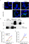
Fig. 1.
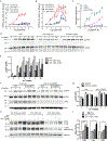
Fig. 1.. LGR5 potentiates Wnt–β-catenin signaling without increasing Wnt receptor level.
(A and B) TOPFlash activity of Super TOPFlash LGR4 knockout cells stably transfected with empty vector (STF-4KO-vect) or Myc-LGR5 construct (STF-4KO-LGR5) after stimulation with RSPO1 or RSPO4 (A) or with RSPO2 or RSPO3 (B) at the indicated concentrations in Wnt3a-conditioned media (CM). TOPFlash activity was normalized to baseline before RSPO addition in Wnt3a CM. (C) Normalized TOPFlash activity of Super TOPFlash LGR4 knockout (STF-LGR4KO) cells transiently transfected with empty vector, Myc-LGR5FL, or Myc-LGR5ECDTM after stimulation with the indicated concentrations of RSPO1 in Wnt3a CM. The TOPFlash activities are representatives of 6–10 independent experiments, and error bars show SEM of 2–3 technical repeats. (D and E) Western blot (D) and quantification (E) of phosphorylated LRP6 (pLRP6) and total LRP6 (tLRP6) in parental STF (endogenous LGR4 expression), STF-4KO-vect, and STF-4KO-LGR5 cell lines after treatment with Wnt3A CM + RSPO1. n = 3 independent experiments. Data represent mean and standard deviation (SD) of the quantified pLRP6 normalized to tLRP6. Two-way Anova Bonferroni posttest was performed, and * represents P < 0.05 against STF cells. (F and G) Western blot (F) and quantification (G) of pLRP6 and tLRP6 in CHP-212 and CHP-212 LGR5KO cells after treatment with Wnt3a CM only or Wnt3a CM + RSPO1. Actin is a loading control. Data represent mean and standard deviation (SD) of the quantified pLRP6 normalized to tLRP6. n = 3 independent experiments. Two-way Anova Bonferroni posttest was performed against non-treated cells, and no significance was found. (H and I) Western blot (H) and quantification (I) of RNF43 and FZD5 in STF-4KO-LGR5 cells co-expressing HA-RNF43 and FLAG-FZD5 (LGR5 overexpression) and STF-4KO-vect cells coexpressing HA-tagged LGR4,RNF43 and FZD5 (LGR4 overexpression) HA-RNF43 and FLAG-FZD5 after Wnt3a CM, Wnt3a CM + RSPO1, or Wnt3a CM + R2Fu-F109A treatment. STF-4KO-vect cells (no LGR4 or LGR5) co-expressing HA-RNF43 and FLAG-FZD5 are a negative control. GAPDH is a loading control. n = 3–4 independent experiments. Data represent mean and SD of FZD/GAPDH normalized to the untreated condition of each cell line. One-way Anova Dunnett’s multiple comparison test against the untreated cells was performed, and * represents P < 0.05.
Fig. 2.
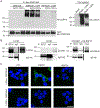
Fig. 2.. LGR5 does not interact with ZNRF3 or RNF43.
(A) Co-immunoprecipitation of LGR4 and LGR5 with experiments with ZNRF3ΔR. Myc immunoprecipitates from HEK293T cells expressing Myc-ZNRF3ΔR only or co-transfected with HA-tagged full-length LGR4 or LGR5 in the presence or absence of RSPO2 and RSPO4 as indicated were blotted for HA and Myc. Total lysates were also probed with the HA antibody. Blot is representative of 5 independent experiments. (B) Co-immunoprecipitation of LGR4 and LGR5 with RNF43. SC37 immunoprecipitates (endogenous RNF43) from LS180 cells were blotted for LGR4 and LGR5, and RNF43 immunoprecipitates from HEK293T cells transiently transfected with HA-tagged RNF43 were blotted for HA. IgG was used as a negative control. Blots are representative of 4–6 independent experiments. (C) Confocal microscopy images of PLA (green) between LGR4 or LGR5 using the antibodies 8D2 and 8F2, respectively and Myc-ZNRF3ΔR in the absence or presence of RSPO1 in HEK293T cells transiently co-expressing Myc-ZNRF3ΔR and either LGR4 or LGR5. CTL is the negative control without primary antibody in the absence of RSPO1. Nuclei were stained with TO-PRO-3(blue). Images are representative of 2 independent experiments. Scale bar, 25 μm.
Fig. 3.
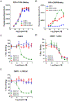
Fig. 3.. LGR5 does not enable binding of RSPO1 or RSPO4 to ZNRF3.
(A and B) Saturation binding analyses of R2Fu-F109A (A) and R4Fu-Q65R (B) to HEK293T cells transfected with ZNRF3ΔR, ZNRF3ΔR + LGR4FL, or ZNRF3ΔR + LGR5ΔC. (C to E) Competition binding curves for R2Fu-F109A with increasing concentrations of recombinant RSPO1, RSP2, or RSPO4 to HEK293T cells transfected with ZNRF3ΔR alone (C), ZNRF3ΔR + LGR4FL (D), and ZNRF3ΔR + LGR5ΔC (E). All error bars are SEM from 2–3 technical replicates, and the curves are representative of 3–5 independent experiments.
Fig. 4.
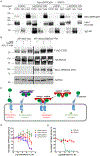
Fig. 4.. The 7TM domain of LGR4 confers interaction with ZNRF3, and the LGR4-ECD does not antagonize RSPO activity.
(A) Co-immunoprecipitation of ZNRF3ΔR with LGR4 and LGR5 full-length and ECD constructs. Western blotting for Myc and HA in LGR4 and LGR5 immunoprecipitates (8D2 or 8F2 antibody, respectively) from HEK293T cells co-transfected with Myc-ZNRF3ΔR and HA-tagged LGR4-FL, LGR4ECD-5TM, LGR5FL, or LGR5ECD-4TM as indicated in the presence of 1 μg/mL RSPO1. IgG was used as a negative control. Blot is representative of 3 independent experiments. (B) Western blotting for FLAG, HA, and Myc in STF-LGR4KO cells transiently transfected with vector or Myc-tagged LGR5ECD-4TM, HA-tagged RNF43, and FLAG-tagged FZD5. Cells were treated with Wnt3a CM, Wnt3a CM+RSPO1, or Wnt3a CM+R2Fu-F109A overnight. GAPDH is a loading control. Blot is representative of 2 independent experiments. (C) A schematic model illustrating the distinct interactions among RSPO-LGRECDs and RNF43/ZNRF3. RSPO and LGR5ECD form a 2:2 dimer that can no longer bind to RNF43/ZNRF3 due to steric hindrance of the LGR5ECD in trans. RSPO and LGR4ECD form a 2:2 dimer that remains capable of binding to RNF43/ZNRF3. RSPO and ZNRF3ECD forms a 1:1 dimer that cannot bind to native ZNRF3 or RNF43. (D and E) TOPFlash results for the effect of LGR4ECD-Fc and LGR5ECD-Fc (D) and ZNRF3ECD-Fc (E) on the activity of 0.01 μg/mL RSPO2 or 0.2 μg/mL RSPO4 in 293-STF cells. The TOPFlash activities are representatives of 5 independent experiments with the error bars representing SEM of 2–3 technical repeats.
Fig. 5.
Fig. 5.. Both LGR4 and LGR5 exist primarily as dimers with or without ligand binding.
(A) Western blot for endogenous LGR4 in extracts from OV90 cells with and without the addition of β-mercaptoethanol (BME) to the samples. (B) Western blot for endogenous LGR5 in extracts from LoVo cells with and without BS3 cross-linker pretreatment and with or without or BME added to the samples. (C) Western blot for recombinant LGR5 in HEK293-LGR5 cells with or without BS3 pre-treatment. Blots are representative of 3 independent experiments. (D) Schematic illustration of fluorescence resonance energy transfer (FRET) between two RSPO ligands bound to LGR4 or LGR5. A 1:1 mixture of Europium (Eu)- and Ulight-labelled His tag–specific antibodies were added to the cells. FRET would occur if two ligands were within 10 nm of one another. (E) Baseline-corrected time-resolved FRET (TR-FRET) signal of R1Fu-His bound to HEK293-LGR4 or HEK293-LGR5ΔC cells with increasing concentrations of the ligand. (F) Baseline-corrected TR-FRET signal of LGR4 and LGR5ΔC bound R1Fu-His molecules in the presence of increasing concentrations of unlabeled RSPO3. TR-FRET curves are representatives of 3 independent experiments, and error bars are SEM of 2 technical repeats.
Fig. 6.
Fig. 6.. LGR5 interacts with both FZD and LRP6 of the Wnt signalosome and promotes signaling IQGAP1-dependently.
(A) Confocal microscopy images of PLA between LGR5 and endogenous FZD and between LGR5 and HA-LRP6 in the absence or presence of RSPO1 during primary antibody incubation in STF-4KO-LGR5 cells transiently transfected with HA-LRP6. CTL is negative control without primary antibody. Nuclei were stained with TO-PRO-3 (blue). Images are representative of 2 independent experiments. Scale bar, 25 μm. (B) Coimmunoprecipitation of LGR5 with FZD. Western blot for LGR5 and FLAG in FLAG immunoprecipitates from HEK293T cells overexpressing FLAG-FZD5, FLAG-FZD5 + LGR5, or LGR5. Blot is representative of 2 independent experiments. (C and D) Normalized TOPFlash activity of STF-4KO-LGR5 cells transfected with vector or IQGAP1ΔIQ (dominant negative IQGAP1) stimulated with RSPO1 (C) or R2Fu-F109A (D). Curves are representatives of 3 independent experiments and error bars represent SEM of 2 technical repeats.
Fig. 7.
Fig. 7.. A schematic model illustrating the mechanistic differences between LGR4 and LGR5 in mediating RSPO-induced potentiation of Wnt–β-catenin signaling.
(A). LGR4 binds to the E3 ligases RNF43 and ZNRF3 (R/F-E3) as a 2:2 dimer without inhibiting ligase activity. Upon RSPO binding and trimer formation, E3 ligase activity is lost. LGR4 dimers that are not engaged with E3 ligases can also enhance LRP6 phosphorylation through direct interaction with the Wnt signalosome and IQGAP1 (not shown). (B) LGR5 exists as a homodimer that is not bound to an E3 ligase but instead interacts with Wnt receptors (not shown). Through an unknown mechanism, RSPO binding to LGR5 enhances LRP6 phosphorylation in an IQGAP1-dependent manner (not shown). LGR4-RSPO was modeled after PDB structures 4KT1 and 4KNG, and LGR5-RSPO was modeled after 4UFR, and the 7TM was modeled after 4OR2.

Similar articles

Cited by

References

    1. Barker N, van Es JH, Kuipers J, Kujala P, van den Born M, Cozijnsen M, Haegebarth A, Korving J, Begthel H, Peters PJ, Clevers H, Identification of stem cells in small intestine and colon by marker gene Lgr5. Nature 449, 1003–1007 (2007); published online EpubOct 25 (10.1038/nature06196). - DOI - PubMed
    1. Koo BK, Clevers H, Stem cells marked by the R-spondin receptor LGR5. Gastroenterology 147, 289–302 (2014); published online EpubAug (10.1053/j.gastro.2014.05.007). - DOI - PubMed
    1. McDonald T, Wang R, Bailey W, Xie G, Chen F, Caskey CT, Liu Q, Identification and cloning of an orphan G protein-coupled receptor of the glycoprotein hormone receptor subfamily. Biochem Biophys Res Commun 247, 266–270 (1998); published online EpubJun 18 (S0006–291X(98)98774–5 [pii]10.1006/bbrc.1998.8774). - DOI - PubMed
    1. Hsu SY, Liang SG, Hsueh AJ, Characterization of two LGR genes homologous to gonadotropin and thyrotropin receptors with extracellular leucine-rich repeats and a G protein-coupled, seven-transmembrane region. Mol Endocrinol 12, 1830–1845 (1998); published online EpubDec (10.1210/mend.12.12.0211) - DOI - PubMed
    1. Barker N, Clevers H, Leucine-rich repeat-containing G-protein-coupled receptors as markers of adult stem cells. Gastroenterology 138, 1681–1696 (2010); published online EpubMay (S0016–5085(10)00336–7 [pii] 10.1053/j.gastro.2010.03.002). - DOI - PubMed

Publication types

MeSH terms

LinkOut - more resources