Recent advances in managing and understanding Stevens-Johnson syndrome and toxic epidermal necrolysis
- PMID: 32595945
- PMCID: PMC7308994
- DOI: 10.12688/f1000research.24748.1
Recent advances in managing and understanding Stevens-Johnson syndrome and toxic epidermal necrolysis
Abstract
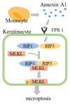
Stevens-Johnson syndrome (SJS) and toxic epidermal necrolysis (TEN) are life-threatening diseases characterized by detachment of the epidermis and mucous membrane. SJS/TEN are considered to be on the same spectrum of diseases with different severities. They are classified by the percentage of skin detachment area. SJS/TEN can also cause several complications in the liver, kidneys, and respiratory tract. The pathogenesis of SJS/TEN is still unclear. Although it is difficult to diagnose early stage SJS/TEN, biomarkers for diagnosis or severity prediction have not been well established. Furthermore, optimal therapeutic options for SJS/TEN are still controversial. Several drugs, such as carbamazepine and allopurinol, are reported to have a strong relationship with a specific human leukocyte antigen (HLA) type. This relationship differs between different ethnicities. Recently, the usefulness of HLA screening before administering specific drugs to decrease the incidence of SJS/TEN has been investigated. Skin detachment in SJS/TEN skin lesions is caused by extensive epidermal cell death, which has been considered to be apoptosis via the Fas-FasL pathway or perforin/granzyme pathway. We reported that necroptosis, i.e. programmed necrosis, also contributes to epidermal cell death. Annexin A1, released from monocytes, and its interaction with the formyl peptide receptor 1 induce necroptosis. Several diagnostic or prognostic biomarkers for SJS/TEN have been reported, such as CCL-27, IL-15, galectin-7, and RIP3. Supportive care is recommended for the treatment of SJS/TEN. However, optimal therapeutic options such as systemic corticosteroids, intravenous immunoglobulin, cyclosporine, and TNF-α antagonists are still controversial. Recently, the beneficial effects of cyclosporine and TNF-α antagonists have been explored. In this review, we discuss recent advances in the pathophysiology and management of SJS/TEN.
Keywords: Stevens-Johnson syndrome; drug reaction; erythema multiforme; necroptosis; toxic epidermal necrolysis.
Copyright: © 2020 Hasegawa A and Abe R.
Conflict of interest statement
No competing interests were disclosed.No competing interests were disclosed.No competing interests were disclosed.
Figures


Similar articles
-
Stevens-Johnson syndrome and toxic epidermal necrolysis: Updates in pathophysiology and management.Chin Med J (Engl). 2024 Oct 5;137(19):2294-2307. doi: 10.1097/CM9.0000000000003250. Epub 2024 Sep 5. Chin Med J (Engl). 2024. PMID: 39238098 Free PMC article. Review.
-
Current Perspectives on Stevens-Johnson Syndrome and Toxic Epidermal Necrolysis.Clin Rev Allergy Immunol. 2018 Feb;54(1):147-176. doi: 10.1007/s12016-017-8654-z. Clin Rev Allergy Immunol. 2018. PMID: 29188475 Review.
-
Stevens-johnson Syndrome and Toxic Epidermal Necrolysis: An Overview of Diagnosis, Therapy Options and Prognosis of Patients.Recent Adv Inflamm Allergy Drug Discov. 2023;17(2):110-120. doi: 10.2174/2772270817666230821102441. Recent Adv Inflamm Allergy Drug Discov. 2023. PMID: 37605396
-
Toxic epidermal necrolysis and Stevens-Johnson syndrome.Orphanet J Rare Dis. 2010 Dec 16;5:39. doi: 10.1186/1750-1172-5-39. Orphanet J Rare Dis. 2010. PMID: 21162721 Free PMC article. Review.
-
Stevens-Johnson syndrome and toxic epidermal necrolysis.Chem Immunol Allergy. 2012;97:149-66. doi: 10.1159/000335627. Epub 2012 May 3. Chem Immunol Allergy. 2012. PMID: 22613860
Cited by
-
Coronavirus-disease-2019-associated Stevens-Johnsons syndrome in a 15-year-old boy: a case report and review of the literature.J Med Case Rep. 2024 Oct 11;18(1):493. doi: 10.1186/s13256-024-04842-3. J Med Case Rep. 2024. PMID: 39390502 Free PMC article. Review.
-
Development of Esophageal Epidermoid Metaplasia in a Pediatric Patient After Stevens-Johnson Syndrome.ACG Case Rep J. 2024 Sep 13;11(9):e01508. doi: 10.14309/crj.0000000000001508. eCollection 2024 Sep. ACG Case Rep J. 2024. PMID: 39280887 Free PMC article.
-
Anti-seizure medication prescription preferences: a Moroccan multicenter study.Front Neurol. 2024 Aug 23;15:1435075. doi: 10.3389/fneur.2024.1435075. eCollection 2024. Front Neurol. 2024. PMID: 39246605 Free PMC article.
-
Stevens-Johnson syndrome and toxic epidermal necrolysis: Updates in pathophysiology and management.Chin Med J (Engl). 2024 Oct 5;137(19):2294-2307. doi: 10.1097/CM9.0000000000003250. Epub 2024 Sep 5. Chin Med J (Engl). 2024. PMID: 39238098 Free PMC article. Review.
-
Allopurinol: Clinical Considerations in the Development and Treatment of Stevens-Johnson Syndrome, Toxic Epidermal Necrolysis, and Other Associated Drug Reactions.Cureus. 2024 Jul 16;16(7):e64654. doi: 10.7759/cureus.64654. eCollection 2024 Jul. Cureus. 2024. PMID: 39149682 Free PMC article. Review.
References
-
- Griffiths C, Barker J, Bleiker T, et al. : Rook’s Textbook of Dermatology. 9th ed. Wiley.2016. 10.1002/9781118441213 - DOI
Publication types
MeSH terms
Grants and funding
LinkOut - more resources
Full Text Sources
Research Materials
Miscellaneous

