. 2020 Feb 6;4(4):606-626.
doi: 10.1002/hep4.1483. eCollection 2020 Apr.

Primary Alcohol-Activated Human and Mouse Hepatic Stellate Cells Share Similarities in Gene-Expression Profiles

Affiliations

Primary Alcohol-Activated Human and Mouse Hepatic Stellate Cells Share Similarities in Gene-Expression Profiles

Xiao Liu et al. Hepatol Commun. .

Abstract

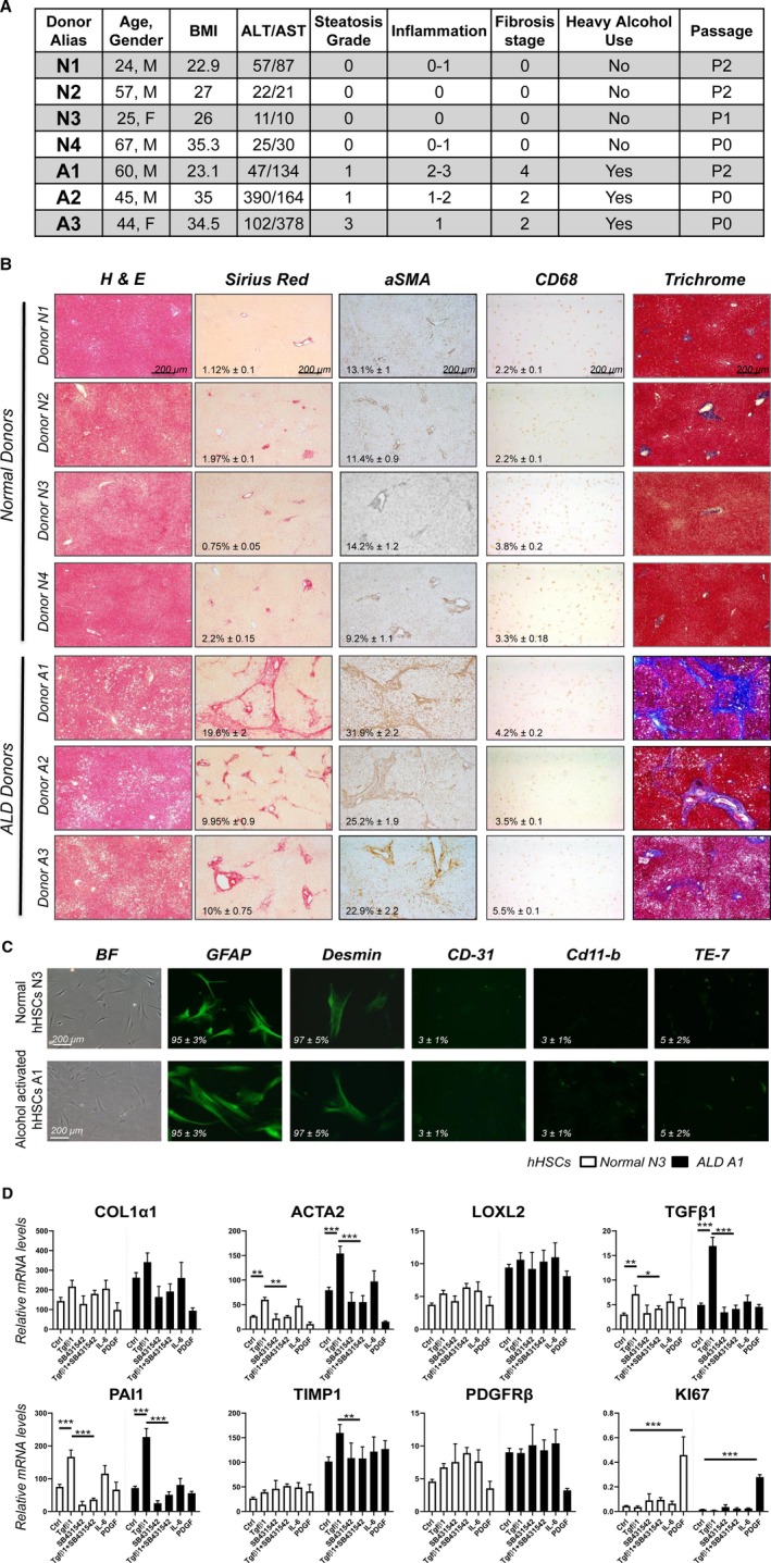
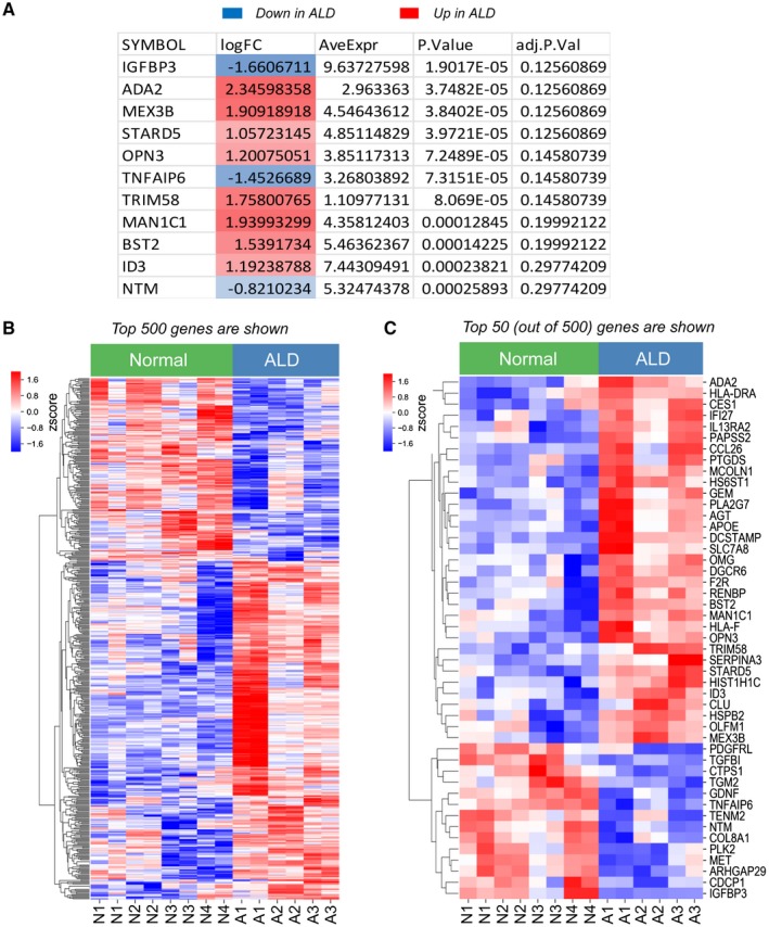
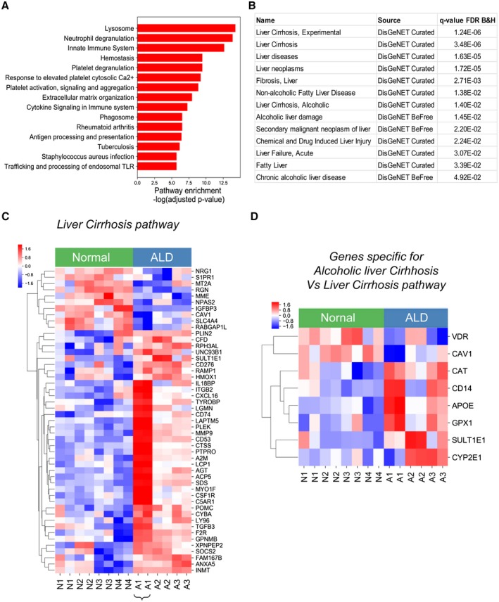
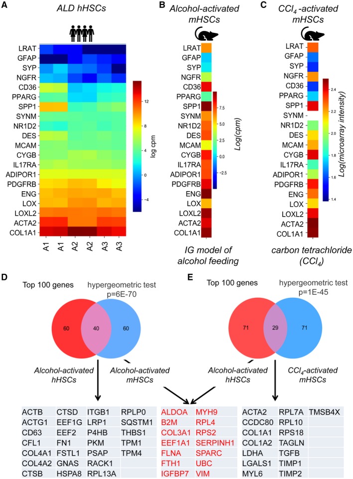
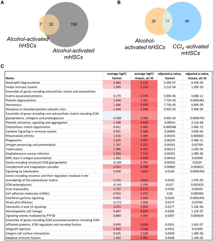
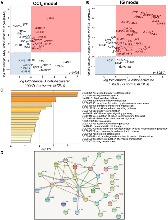
Alcoholic liver disease (ALD) is a leading cause of cirrhosis in the United States, which is characterized by extensive deposition of extracellular matrix proteins and formation of a fibrous scar. Hepatic stellate cells (HSCs) are the major source of collagen type 1 producing myofibroblasts in ALD fibrosis. However, the mechanism of alcohol-induced activation of human and mouse HSCs is not fully understood. We compared the gene-expression profiles of primary cultured human HSCs (hHSCs) isolated from patients with ALD (n = 3) or without underlying liver disease (n = 4) using RNA-sequencing analysis. Furthermore, the gene-expression profile of ALD hHSCs was compared with that of alcohol-activated mHSCs (isolated from intragastric alcohol-fed mice) or CCl4-activated mouse HSCs (mHSCs). Comparative transcriptome analysis revealed that ALD hHSCs, in addition to alcohol-activated and CCl4-activated mHSCs, share the expression of common HSC activation (Col1a1 [collagen type I alpha 1 chain], Acta1 [actin alpha 1, skeletal muscle], PAI1 [plasminogen activator inhibitor-1], TIMP1 [tissue inhibitor of metalloproteinase 1], and LOXL2 [lysyl oxidase homolog 2]), indicating that a common mechanism underlies the activation of human and mouse HSCs. Furthermore, alcohol-activated mHSCs most closely recapitulate the gene-expression profile of ALD hHSCs. We identified the genes that are similarly and uniquely up-regulated in primary cultured alcohol-activated hHSCs and freshly isolated mHSCs, which include CSF1R (macrophage colony-stimulating factor 1 receptor), PLEK (pleckstrin), LAPTM5 (lysosmal-associated transmembrane protein 5), CD74 (class I transactivator, the invariant chain), CD53, MMP9 (matrix metallopeptidase 9), CD14, CTSS (cathepsin S), TYROBP (TYRO protein tyrosine kinase-binding protein), and ITGB2 (integrin beta-2), and other genes (compared with CCl4-activated mHSCs). Conclusion: We identified genes in alcohol-activated mHSCs from intragastric alcohol-fed mice that are largely consistent with the gene-expression profile of primary cultured hHSCs from patients with ALD. These genes are unique to alcohol-induced HSC activation in two species, and therefore may become targets or readout for antifibrotic therapy in experimental models of ALD.


Figures

Figure 1
Figure 1
Primary cultured alcohol‐activated hHSCs exhibit a more activated phenotype than normal hHSCs. (A) Donor information: donors without underlying liver disease (N1‐N4). Donors with ALD (A1‐A3) were diagnosed based on liver function, histology, and history of alcohol abuse. (B) Human livers (healthy [n = 4] or ALD [n = 3]) were stained for H&E, sirius red, α‐SMA, CD68, and trichrome. Positive area was calculated as a percentage; representative micrographs are shown (×4 objective). (C) Primary human HSCs were immunostained for GFAP, DES, CD31, CD11b, and elastin (TE‐7 antibody). (D) Alcohol‐activated hHSCs (A1) and normal hHSCs (N3) were stimulated ± TGFβ1 (5 ng/mL), SB431542 (10 µM), IL‐6 (10 ng/mL), and PDGFβ (10 ng/mL) for 24 hours, and analyzed by quantitative real‐time PCR (*P < 0.05, **P < 0.01, ***P < 0.001), Student t test, and one‐way analysis of variance. Abbreviations: ALT, alanine aminotransferase; AST, aspartate aminotransferase; BF, Bright‐Field; Ctrl, control.
Figure 2
Figure 2
Eleven most dysregulated genes that best discriminate ALD hHSCs from normal hHSCs. (A) Eleven genes that are significantly differentially expressed (red, up‐regulated; blue, down‐regulated) between ALD and normal hHSCs. (B,C) Heatmap showing the relative gene expression between the top 500 (B) or top 50 (C) most differentially expressed genes in normal and ALD hHSCs. The genes were grouped by similarity to each other using hierarchical clustering. The Benjamini‐Hochberg method for multiple test corrections was applied to differentiate the expression results. Eleven genes were changed significantly with the Benjamini‐Hochberg method (adjusted P < 0.3). Abbreviations: adj.P.Val, multiple‐test‐corrected P value using the method of Benjamini‐Hochberg; AveExpr, average expression across all samples; logFC, log fold change in expression between ALD and normal hHSCs; P.Value, P value for the comparison. Abbreviations: ADA2, adenosine deaminase 2; AGT, angiotensinogen; APOE, apolipoprotein E; ARHGAP29, rho GTPase activating protein 29; BST2, bone marrow stromal cell antigen 2; CCL26, C‐C motif chemokine ligand 26; CDCP1, CUB domain containing protein 1; CES1, carboxylesterase 1; CLU, clusterin; COL8A1, collagen type VIII alpha 1 chain; CTPS1, CTP synthase 1; DCSTAMP, dendrocyte expressed seven transmembrane protein; DGCR6, DiGeorge syndrome critical region gene 6; F2R, coagulation factor II thrombin receptor; GDNF, glial cell derived neurotrophic factor; GEM, GTP binding protein overexpressed in skeletal muscle; HIST1H1C, H1.2 linker histone, cluster member; HLA‐DRA, major histocompatibility complex, class II, DR alpha; HLA‐F, major histocompatibility complex, class I, F; HS6ST1, heparan sulfate 6‐O‐sulfotransferase 1; HSPB2, heat shock protein family B (small) member 2; ID3, inhibitor of DNA binding 3; IFI27, interferon alpha inducible protein 27; IGFBP3, insulin like growth factor binding protein 3; IL13RA2, interleukin 13 receptor subunit alpha 2; MAN1C1, mannosidase alpha class 1C member 1; MCOLN1, mucolipin 1; MET, MET proto‐oncogene, receptor tyrosine kinase; MEX3B, mex‐3 RNA binding family member B; NTM, neurotrimin; OLFM1, olfactomedin 1; OMG, oligodendrocyte myelin glycoprotein; OPN3, opsin 3; PAPSS2, 3,‐phosphoadenosine 5,‐phosphosulfate synthase 2; PDGFRL, platelet derived growth factor receptor like; PLA2G7, phospholipase A2 group VII; PLK2, polo like kinase 2; PTGDS, prostaglandin D2 synthase; RENBP, renin binding protein; SERPINA3, serpin family a member 3; SLC7A8, solute carrier family 7 member 8; STARD5, StAR related lipid transfer domain containing 5; TENM2, teneurin transmembrane protein 2; TGFBI, transforming growth factor beta induced; TGM2, transglutaminase 2; TNFAIP6, tumor necrosis factor‐alpha‐induced protein; TRIM58, Tripartite motif containing 58.
Figure 3
Figure 3
Fibrogenic and immune pathways are dysregulated in alcohol‐activated hHSCs (compared with normal hHSCs). (A) Functional enrichment analysis of the top 500 most differentially expressed genes between ALD and normal hHSCs identified 39 dysregulated pathways. The top 15 up‐regulated pathways (out of 39) are shown. The significance of the pathway enrichment is shown on the x axis (negative log Benjamini‐Hochberg–adjusted P value). (B) The top 500 differentially expressed genes demonstrate a significant association with curated liver disease gene sets (DisGeNet, http://www.disgenet.org/dbinfo). The q‐value false discovery rate is the Benjamini‐Hochberg–adjusted P value for the gene‐set enrichment. (C,D) Heatmap showing the relative expression between ALD and healthy HSCs for genes in the liver cirrhosis experimental DisGeNet gene set (C) or differentially expressed genes in the alcoholic liver cirrhosis DisGeNet gene set (compared with the liver cirrhosis experimental DisGeNet gene set). (D). The genes were grouped by similarity to each other using hierarchical clustering. Pathway significance and disease gene‐set significance was assessed using hypergeometric tests to compare the significance of the observed overlap in genes. P values are corrected for multiple testing with the Benjamini‐Hochberg method (adjusted P < 0.05). Abbreviations: B&H, Benjamini‐Hochberg; FDR, false discovery rate. Abbreviations: A2M, alpha‐2‐macroglobulin; ACP5, acid phosphatase 5; AGT, angiotensinogen; ANXA5, annexin A5; APOE, apolipoprotein E; C5AR1, complement C5a receptor 1; CAT, catalase; CAV1, caveolin 1; CD14, CD14 molecule; CD276, CD276 molecule; CD53, CD53 molecule; CD74, CD74 molecule; CFD, complement factor D; CSF1R, colony stimulating factor 1 receptor; CTSS, cathepsin S; CXCL16, C‐X‐C motif chemokine ligand 16; CYBA, cytochrome b‐245 alpha chain; CYP2E1, cytochrome P450 family 2 subfamily E member 1; F2R, coagulation factor II thrombin receptor; FAM167B, family with sequence similarity 167 member B; GPNMB, glycoprotein nmb; GPX1, glutathione peroxidase 1; HMOX1, heme oxygenase 1; IGFBP3, insulin like growth factor binding protein 3; IL18BP, interleukin 18 binding protein; INMT, indolethylamine N‐methyltransferase; ITGB2, integrin subunit beta 2; LAPTM5, lysosomal protein transmembrane 5; LCP1, lymphocyte cytosolic protein 1; LGMN, legumain; LY96, lymphocyte antigen 96; MME, membrane metalloendopeptidase; MMP9, matrix metallopeptidase 9; MT2A, metallothionein 2A; MYO1F, myosin IF; NPAS2, neuronal PAS domain protein 2; NRG1, neuregulin 1; PLEK, pleckstrin; PLIN2, perilipin 2; POMC, proopiomelanocortin; PTPRO, protein tyrosine phosphatase receptor type O; RABGAP1L, RAB GTPase activating protein 1 like; RAMP1, receptor activity modifying protein 1; RGN, regucalcin; RPH3AL, rabphilin 3A like; S1PR1, sphingosine‐1‐phosphate receptor 1; SDS, serine dehydratase; SLC4A4, solute carrier family 4 member 4; SOCS2, suppressor of cytokine signaling 2; SULT1E1, sulfotransferase family 1E member 1; TGFB3, transforming growth factor beta 3; TYROBP, TYRO protein tyrosine kinase binding protein; UNC93B1, unc‐93 homolog B1; VDR, vitamin D receptor; XPNPEP2, X‐prolyl minopeptidase 2.
Figure 4
Figure 4
Expression of activation markers is conserved among ALD hHSCs, alcohol‐activated, and CCl4‐activated mHSCs. (A‐C) Heatmap displaying the expression of HSC‐specific markers in ALD hHSC (A), alcohol‐activated (B), and CCl4‐activated mHSCs (C). (D,E) Venn diagrams demonstrate the overlapping genes (from the top 100 most highly expressed genes) between alcohol‐activated hHSCs and mHSCs (D) (P = 6−70) or alcohol‐activated hHSCs and CCl4‐activated mHSCs (E) (P = 1−45). Genes expressed in both pools of overlapping genes are shown in red; unique genes are shown in black. Overlap significance was assessed with the hypergeometric test, to compare the observed overlap with what is expected, given the randomly selected gene sets. Abbreviations: ACTA2, actin alpha 2, smooth muscle; ACTB, actin beta; ACTG1, actin gamma 1; ADIPOR1, adiponectin receptor 1; ALDOA, aldolase, fructose‐bisphosphate A; B2M, beta‐2‐microglobulin; CCDC80, coiled‐coil domain containing 80; CD36, CD36 molecule; CD63, CD63 molecule; CFL1, cofilin 1; COL1A1, collagen type I alpha 1 chain; COL1A2, collagen type I alpha 2 chain; COL3A1, collagen type III alpha 1 chain; COL4A1, collagen type IV alpha 1 chain; COL4A2, collagen type IV alpha 2 chain; CTSB, cathepsin B; CTSD, cathepsin D; CYGB, cytoglobin; DES, desmin; EF1A1, eukaryotic translation elongation factor 1 alpha 1; EEF1G, eukaryotic translation elongation factor 1 gamma; EEF2, eukaryotic translation elongation factor 2; ENG, endoglin; FLNA, filamin A; FN1, fibronectin 1; FSTL1, follistatin like 1; FTH1, ferritin heavy chain 1; GFAP, glial fibrillary acidic protein; GNAS, GNAS complex locus; HSPA8, heat shock protein family A (Hsp70) member 8; IGFBP7, insulin like growth factor binding protein 7; IL17RA, interleukin 17 receptor A; ITGB1, integrin subunit beta 1; LDHA, lactate dehydrogenase A; LGALS1, galectin 1; LOX, lysyl oxidase; LOXL2, lysyl oxidase like 2; LRAT, lecithin retinol acyltransferase; LRP1, LDL receptor related protein 1; MCAM, melanoma cell adhesion molecule; MYH9, myosin heavy chain 9; MYL6, myosin light chain 6; NGFR, nerve growth factor receptor; NR1D2, nuclear receptor subfamily 1 group D member 2; P4HB, prolyl 4‐hydroxylase subunit beta; PDGFRB, platelet derived growth factor receptor beta; PKM, pyruvate kinase M1/2; PPARG, peroxisome proliferator activated receptor gamma; PSAP, prosaposin; RACK1, receptor for activated C kinase 1; RPL7A, ribosomal protein L7a; RPL10, ribosomal protein L10; RPL13A, ribosomal protein L13a; RPL4, ribosomal protein L4; RPLP0, ribosomal protein lateral stalk subunit P0; RPS18, ribosomal protein S18; RPS2, ribosomal protein S2; SERPINH1, serpin family H member 1; SPARC, secreted protein acidic and cysteine rich; SPP1, secreted phosphoprotein 1; SQSTM1, sequestosome 1; SYNM, synemin; SYP, synaptophysin; TAGLN, transgelin; TGFB, TGFB2 overlapping transcript 1; TIMP1, TIMP metallopeptidase inhibitor 1; TIMP2, TIMP metallopeptidase inhibitor 2; TMSB4X, thymosin beta 4 X‐linked; TPM1, tropomyosin 1; TPM4, tropomyosin 4; UBC, ubiquitin C; VIM, vimentin.
Figure 5
Figure 5
Pathways are shared between alcohol‐activated hHSCs and mHSCs. (A,B) Pathways commonly dysregulated in alcohol‐activated hHSCs and mHSCs (A) in alcohol‐activated hHSCs and CCl4‐activated mHSCs (B), shown as Venn diagrams. (Pathways that were significantly enriched in the top 500 most differentially expressed genes between alcohol‐activated and control HSCs were identified using the hypergeometric test–adjusted P < 0.05). (C) Thirty‐two pathways shared between alcohol‐activated hHSCs and mHSCs. The top 500 differentially expressed genes were used for pathway analysis. Abbreviations: Average logFC human, average log fold change between ALD and normal hHSCs for genes in the pathway; average logFC mouse, alc IG, average log fold change between alcohol‐activated and quiescent mHSCs (from IG alcohol‐fed mice) for genes in the pathway; adjusted P value, human, significance of the pathway enrichment in human, adjusted for multiple tests using the method of Benjamini‐Hochberg; adjusted P value, mouse, alc IG, significance of the pathway enrichment in mouse intragastric model, adjusted for multiple tests using the method of Benjamini‐Hochberg.
Figure 6
Figure 6
Expression of 35 genes distinguishes alcohol‐activated hHSCs and mHSCs from CCl4‐activated mHSCs. (A) Scatterplot shows 33 overlapping genes from the top 500 genes in ALD (vs. normal) hHSCs and CCl4‐activated (vs. quiescent) mHSCs (hypergeometric test, P = 0.002). Fourteen genes were dysregulated in the same direction. Genes located in the pink square are up‐regulated, and those in the blue square are down‐regulated, in both hHSCs and mHSCs. (B) Scatterplot shows 44 overlapping genes from the top 500 genes ALD (vs. normal) hHSCs and alcohol‐activated (vs. quiescent) mHSCs (P = 3.9−7). Thirty‐five genes dysregulated in the same direction are shown in the pink (up‐regulated) and blue (down‐regulated) squares in both alcohol‐activated hHSCs and mHSCs. (C) GO term/pathway analysis of 35 genes overlapping between alcohol‐activated hHSCs/mHSCs. (D) Thirty‐five genes were grouped in a cross‐species protein–protein interaction network (https://https//string-db.org). Significantly more connections were found among the genes than expected by chance (P < 1−16, permutation test). Abbreviations: ABI3BP, ABI family member 3 binding protein; ADARB1, adenosine deaminase RNA specific B1; AOX1, aldehyde oxidase 1; BOC, BOC cell adhesion associated; BST2, bone marrow stromal cell antigen 2; CARD9, caspase recruitment domain family member 9; CAV1, caveolin 1; CD14, CD14 molecule; CD44, CD44 molecule; CD52, CD52 molecule; CD53, CD53 molecule; CD74, CD74 molecule; CD84, CD84 molecule; CD9, CD9 molecule; COL8A1, collagen type VIII alpha 1 chain; CPXM1, carboxypeptidase X, M14 family member 1; CSF1R, colony stimulating factor 1 receptor; CTHRC1, collagen triple helix repeat containing 1; CTSS, cathepsin S; CYBA, cytochrome b‐245 alpha chain; DCLK1, doublecortin like kinase 1; DMPK, DM1 protein kinase; EMILIN2, elastin microfibril interfacer 2; FGF2, fibroblast growth factor 2; FMOD, fibromodulin; FSTL3, follistatin like 3; GEM, GTP binding protein overexpressed in skeletal muscle; GLRX, glutaredoxin; GPC1, glypican 1; HAS2, hyaluronan synthase 2; HIST1H1C, H1.2 linker histone, cluster member; HS6ST1, heparan sulfate 6‐O‐sulfotransferase 1; HSPA1A, heat shock protein family A (Hsp70) member 1A; ID1, inhibitor of DNA binding 1; IGFBP3, insulin like growth factor binding protein 3; IL11, interleukin 11; IL1R2, interleukin 1 receptor type 2; INHBA, inhibin subunit beta A; IRF7, interferon regulatory factor 7; ITGB2, integrin subunit beta 2; KLF10, kruppel like factor 10; LAPTM5, lysosomal protein transmembrane 5; LCP1, lymphocyte cytosolic protein 1; MAFB, MAF bZIP transcription factor B; MMP9, matrix metallopeptidase 9; MSR1, macrophage scavenger receptor 1; NCAM1, neural cell adhesion molecule 1; PGM5, phosphoglucomutase 5; PLA2G7, phospholipase A2 group VII; PLCB2, phospholipase C beta 2; PLCB4, phospholipase C beta 4; PLEK, pleckstrin; PLXNC1, plexin C1; RASGRP3, RAS guanyl releasing protein 3; RGS14, regulator of G protein signaling 14; RIMS3, regulating synaptic membrane exocytosis 3; RSPO3, R‐spondin 3; SEPT5, Septin 5; SERINC2, serine incorporator 2; SOX17, SRY‐box transcription factor 17; SPP1, secreted phosphoprotein 1; TGFB3, transforming growth factor beta 3; TIMP3, TIMP metallopeptidase inhibitor 3; TMEM140, transmembrane protein 140; TNC, tenascin C; TREM2, triggering receptor expressed on myeloid cells 2; TYROBP, TYRO protein tyrosine kinase binding protein; VCAN, versican; VDR, vitamin D receptor; VEGFC, vascular endothelial growth factor C; WT1, WT1 transcription factor.
Figure 7
Figure 7
Fourteen genes were identified as the signature genes of alcohol‐activated hHSCs/mHSCs. (A) Fourteen genes with 2 or fewer connections within the network were identified as signature genes of alcohol‐activated hHSCs/mHSCs. The P value and Benjamini‐Hochberg–adjusted P value of alcohol‐activated (vs. normal) hHSCs are shown. (B) Functional enrichment analysis identified pathways associated with these 14 genes (six pathways are shown) and were grouped in a cross‐species protein–protein interaction network (https://https//string-db.org). The pathways shown are significantly enriched for these 14 genes with Benjamini‐Hochberg–adjusted P < 0.05. (C) Expression of selected genes was analyzed by quantitative real‐time PCR in normal hHSCs (n = 3, N5, N6, N7) and ALD hHSCs (n = 3, A3, A4, A5) isolated from a different set of donors (*P < 0.05, **P < 0.01, ***P < 0.001; Student t test and one‐way analysis of variance). (D) Donor information: N5‐N7, donors without underlying liver disease; A4‐A6, donors with ALD who were diagnosed based on liver function, histology, and history of alcohol abuse. Abbreviations: ALT, alanine aminotransferase; AST, aspartate aminotransferase. Abbreviations: CD14, CD14 molecule; CD53, CD53 molecule; CD74, CD74 molecule; CSF1R, Colony stimulating factor 1 receptor; CTSS, Cathepsin S; CYBA, Cytochrome b‐245 alpha chain; IRF7, Interferon regulatory factor 7; ITGB2, Integrin subunit beta 2; LAPTM5, Lysosomal protein transmembrane 5; MMP9, Matrix metallopeptidase 9; PLEK, Pleckstrin; SPP1, Secreted phosphoprotein 1; TREM2, Teneurin transmembrane protein 2; TYROBP, TYRO protein tyrosine kinase binding protein.

Similar articles

Cited by

References

    1. Gao B, Bataller R. Alcoholic liver disease: pathogenesis and new therapeutic targets. Gastroenterology 2011;141:1572‐1585. - PMC - PubMed
    1. Brandl K, Hartmann P, Jih LJ, Pizzo DP, Argemi J, Ventura‐Cots M, et al. Dysregulation of serum bile acids and FGF19 in alcoholic hepatitis. J Hepatol 2018;69:396‐405. - PMC - PubMed
    1. Shang L, Hosseini M, Liu X, Kisseleva T, Brenner DA. Human hepatic stellate cell isolation and characterization. J Gastroenterol 2018;53:6‐17. - PubMed
    1. Xu J, Liu X, Koyama Y, Wang P, Lan T, Kim IG, et al. The types of hepatic myofibroblasts contributing to liver fibrosis of different etiologies. Front Pharmacol 2014;5:1‐12. - PMC - PubMed
    1. Ueno A, Lazaro R, Wang PY, Higashiyama R, Machida K, Tsukamoto H. Mouse intragastric infusion (iG) model. Nat Protoc 2012;7:771‐781. - PMC - PubMed