

Melatonin reverses nasopharyngeal carcinoma cisplatin chemoresistance by inhibiting the Wnt/β-catenin signaling pathway
- PMID: 32203052
- PMCID: PMC7138577
- DOI: 10.18632/aging.102968
Melatonin reverses nasopharyngeal carcinoma cisplatin chemoresistance by inhibiting the Wnt/β-catenin signaling pathway
Abstract
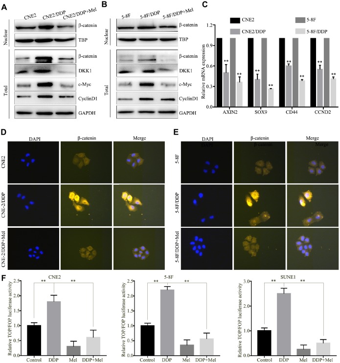
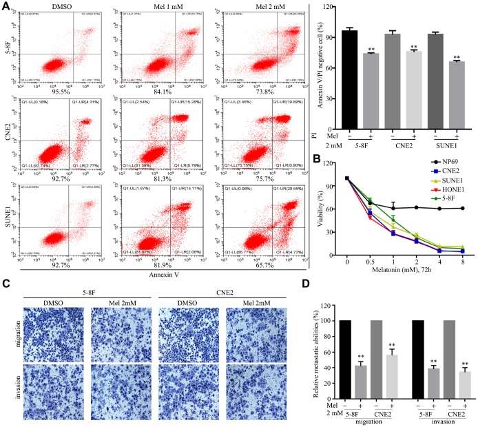
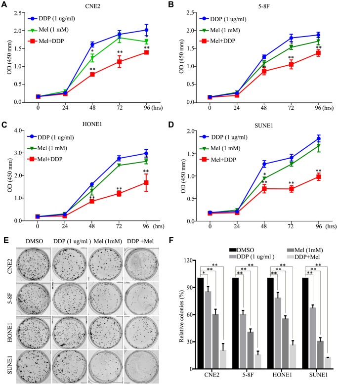
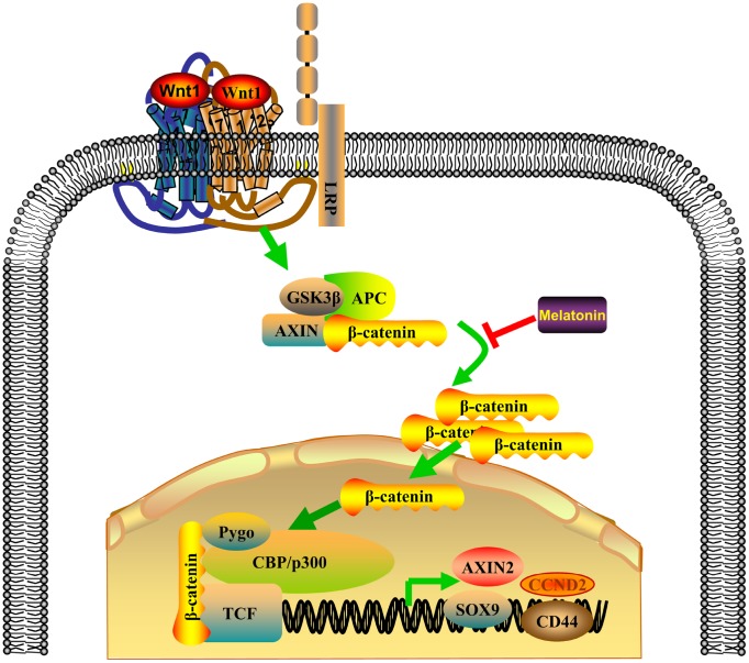
Cisplatin (DDP)-based concurrent chemo-radiotherapy is a standard approach to treat locoregionally advanced nasopharyngeal carcinoma (NPC). However, many patients eventually develop recurrence and/or distant metastasis due to chemoresistance. In this study, we aimed to elucidate the effects of melatonin on DDP chemoresistance in NPC cell lines in vitro and vivo, and we explored potential chemoresistance mechanisms. We found that DDP chemoresistance in NPC cells is mediated through the Wnt/β-catenin signaling pathway. Melatonin not only reversed DDP chemoresistance, but also enhanced DDP antitumor activity by suppressing the nuclear translocation of β-catenin, and reducing expression of Wnt/β-catenin response genes in NPC cells. In vivo, combined treatment with DDP and melatonin reduced tumor burden to a greater extent than single drug-treatments in an orthotopic xenograft mouse model. Our findings provide novel evidence that melatonin inhibits the Wnt/β-catenin pathway in NPC, and suggest that melatonin could be applied in combination with DDP to treat NPC.
Keywords: Wnt/β-catenin; chemoresistance; cisplatin; melatonin; nasopharyngeal carcinoma.
Conflict of interest statement
Figures


Similar articles
-
Reversal of cisplatin sensitization and abrogation of cisplatin-enriched cancer stem cells in 5-8F nasopharyngeal carcinoma cell line through a suppression of Wnt/β-catenin-signaling pathway.Mol Cell Biochem. 2021 Apr;476(4):1663-1672. doi: 10.1007/s11010-020-04045-6. Epub 2021 Jan 9. Mol Cell Biochem. 2021. PMID: 33423190
-
Cinobufotalin powerfully reversed EBV-miR-BART22-induced cisplatin resistance via stimulating MAP2K4 to antagonize non-muscle myosin heavy chain IIA/glycogen synthase 3β/β-catenin signaling pathway.EBioMedicine. 2019 Oct;48:386-404. doi: 10.1016/j.ebiom.2019.08.040. Epub 2019 Oct 5. EBioMedicine. 2019. PMID: 31594754 Free PMC article.
-
Casein kinase 1α-dependent inhibition of Wnt/β-catenin selectively targets nasopharyngeal carcinoma and increases chemosensitivity.Anticancer Drugs. 2019 Aug;30(7):e0747. doi: 10.1097/CAD.0000000000000747. Anticancer Drugs. 2019. PMID: 31305293
-
Melatonin as a Promising Agent for Cancer Treatment: Insights into its Effects on the Wnt/beta-catenin Signaling Pathway.Curr Med Chem. 2024;31(11):1315-1331. doi: 10.2174/0929867330666230409141957. Curr Med Chem. 2024. PMID: 37031385 Review.
-
Chemotherapy and chemo-resistance in nasopharyngeal carcinoma.Eur J Med Chem. 2020 Dec 1;207:112758. doi: 10.1016/j.ejmech.2020.112758. Epub 2020 Aug 20. Eur J Med Chem. 2020. PMID: 32858472 Review.
Cited by
-
HOXA11-AS induces cisplatin resistance by modulating the microRNA-98/PBX3 axis in nasopharyngeal carcinoma.Oncol Lett. 2021 Jun;21(6):493. doi: 10.3892/ol.2021.12754. Epub 2021 Apr 26. Oncol Lett. 2021. PMID: 33968209 Free PMC article.
-
Alkylating Agent-Induced Toxicity and Melatonin-Based Therapies.Front Pharmacol. 2022 Mar 23;13:873197. doi: 10.3389/fphar.2022.873197. eCollection 2022. Front Pharmacol. 2022. PMID: 35401215 Free PMC article. No abstract available.
-
Increased MCL-1 synthesis promotes irradiation-induced nasopharyngeal carcinoma radioresistance via regulation of the ROS/AKT loop.Cell Death Dis. 2022 Feb 8;13(2):131. doi: 10.1038/s41419-022-04551-z. Cell Death Dis. 2022. PMID: 35136016 Free PMC article.
-
Investigations on the Role of the MicroRNA-338-5p/Wnt Family Member 2B (WNT2B) Axis in Regulating the Pathogenesis of Nasopharyngeal Carcinoma (NPC).Front Oncol. 2021 Jun 29;11:684462. doi: 10.3389/fonc.2021.684462. eCollection 2021. Front Oncol. 2021. PMID: 34268117 Free PMC article.
-
High VCAM-1 Predicts Poor Prognosis and is Associated with Chemotherapy Resistance in Nasopharyngeal Carcinoma.Onco Targets Ther. 2021 Mar 2;14:1633-1641. doi: 10.2147/OTT.S292259. eCollection 2021. Onco Targets Ther. 2021. PMID: 33688210 Free PMC article.
References
-
- Lai SZ, Li WF, Chen L, Luo W, Chen YY, Liu LZ, Sun Y, Lin AH, Liu MZ, Ma J. How does intensity-modulated radiotherapy versus conventional two-dimensional radiotherapy influence the treatment results in nasopharyngeal carcinoma patients? Int J Radiat Oncol Biol Phys. 2011; 80:661–68. 10.1016/j.ijrobp.2010.03.024 - DOI - PubMed
-
- Chen SH, Kuo CC, Li CF, Cheung CH, Tsou TC, Chiang HC, Yang YN, Chang SL, Lin LC, Pan HY, Chang KY, Chang JY. O(6) -methylguanine DNA methyltransferase repairs platinum-DNA adducts following cisplatin treatment and predicts prognoses of nasopharyngeal carcinoma. Int J Cancer. 2015; 137:1291–305. 10.1002/ijc.29486 - DOI - PubMed
Publication types
MeSH terms
Substances
LinkOut - more resources
Full Text Sources
Miscellaneous
