. 2019 Nov 14;4(22):e128799.
doi: 10.1172/jci.insight.128799.

Investigation of a dilated cardiomyopathy-associated variant in BAG3 using genome-edited iPSC-derived cardiomyocytes

Affiliations

Investigation of a dilated cardiomyopathy-associated variant in BAG3 using genome-edited iPSC-derived cardiomyocytes

Chris McDermott-Roe et al. JCI Insight. .

Abstract

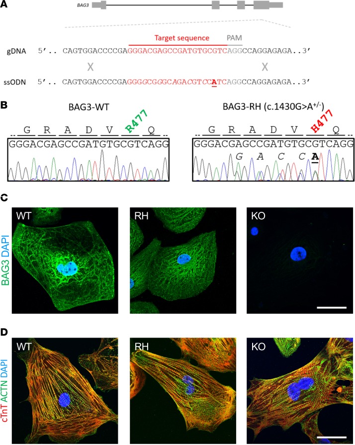
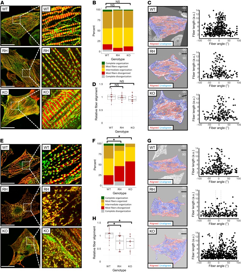
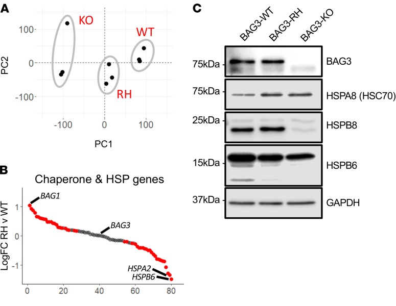
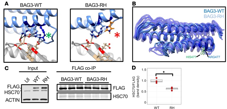
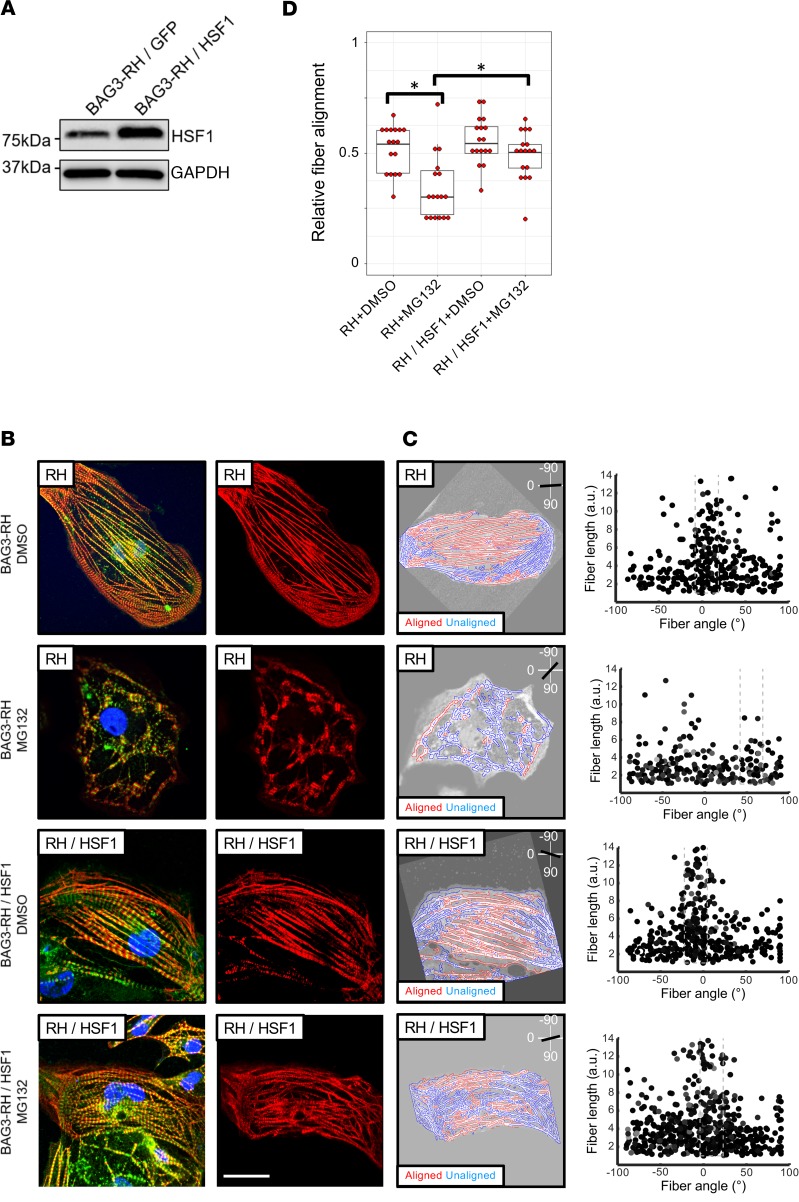
Mutations in B cell lymphoma 2-associated athanogene 3 (BAG3) are recurrently associated with dilated cardiomyopathy (DCM) and muscular dystrophy. Using isogenic genome-edited human induced pluripotent stem cell-derived cardiomyocytes (iPSC-CMs), we examined how a DCM-causing BAG3 mutation (R477H), as well as complete loss of BAG3 (KO), impacts myofibrillar organization and chaperone networks. Although unchanged at baseline, fiber length and alignment declined markedly in R477H and KO iPSC-CMs following proteasome inhibition. RNA sequencing revealed extensive baseline changes in chaperone- and stress response protein-encoding genes, and protein levels of key BAG3 binding partners were perturbed. Molecular dynamics simulations of the BAG3-HSC70 complex predicted a partial disengagement by the R477H mutation. In line with this, BAG3-R477H bound less HSC70 than BAG3-WT in coimmunoprecipitation assays. Finally, myofibrillar disarray triggered by proteasome inhibition in R477H cells was mitigated by overexpression of the stress response protein heat shock factor 1 (HSF1). These studies reveal the importance of BAG3 in coordinating protein quality control subsystem usage within the cardiomyocyte and suggest that augmenting HSF1 activity might be beneficial as a means to mitigate proteostatic stress in the context of BAG3-associated DCM.

Keywords: Cardiology; Genetics; Heart failure; iPS cells.


Conflict of interest statement

Conflict of interest: The authors have declared that no conflict of interest exists.

Figures

Figure 1
Figure 1. Production and preliminary analysis of BAG3-R477H and BAG3-KO induced pluripotent stem cell (iPSC)-derived cardiomyocytes.
(A) Schematic representation of the BAG3 gene, WT genomic DNA (gDNA) sequence, and the central part of the single-stranded oligonucleotide (ssODN) sequence used to introduce the c.1430G>A (R477H) mutation, which causes DCM in humans (3), into iPSCs. (B) Sanger sequence traces and corresponding amino acid sequences of an unedited iPSC line (BAG3-WT, left) and an iPSC line heterozygous for the c.1430G>A (BAG3-RH) mutation (right). In A and B, underlined/bolded and italicized nucleotides denote the variant of interest and synonymous Cas9-blocking mutations, respectively. (C) BAG3 localization in BAG3-WT (WT), BAG3-R477H (RH), and BAG3-KO (KO) iPSC–derived cardiomyocytes. Green, BAG3; blue, DAPI. Scale bar: 20 μm. (D) Visualization of myofibrillar organization in BAG3-WT (WT), BAG3-R477H (RH), and BAG3-KO (KO) iPSC–derived cardiomyocytes. Red, cardiac troponin T; green, α-actinin; blue, DAPI. Scale bar: 20 μm. Data are representative of 3 independent experiments.
Figure 2
Figure 2. Proteasome inhibition causes myofibrillar disarray in BAG3-R477H and BAG3-KO induced pluripotent stem cell–derived cardiomyocytes (iPSC-CMs).
(A) Visualization of myofibrillar organization in unedited (WT), BAG3-R477H knock-in (RH), and BAG3-KO (KO) iPSC-CMs, maintained under standard culture conditions. Red, cardiac troponin T; green, α-actinin. (B) Quantification of myofibrillar disorganization/organization in BAG3-WT, BAG3-RH, and BAG3-KO iPSC-CMs based on manual classification (n > 150 cells per genotype). (C) Quantification of myofibrillar disorganization/organization in BAG3-WT, BAG3-RH, and BAG3-KO iPSC-CMs from corresponding cells in A. Left panels show individual myofiber boundaries (identified from cardiac troponin channel) defined using an edge detection algorithm in MATLAB (see Methods) and are displayed as red or blue if aligned or unaligned, respectively, with the predicted long axis of the parent cell, indicated in the top right corner. Right panels display each myofiber from the corresponding parent cell (left panels) as a single point and are plotted according to their length (y axis) and angle relative to the predicted long axis (x axis) of the parent cell. The dashed box represents ±15°of the predicted long axis of the cell. (D) Relative myofiber alignment based on edge detection measurements in BAG3-WT, BAG3-RH, and BAG3-KO iPSC-CMs. E–H are as described in AD, respectively, but with the addition of MG132 (25 μM, 15 hours). In A and E, scale bar: 20 μm. In D and H, each data point represents average myofiber alignment from a single cell. For each genotype, 15 randomly selected cells were analyzed. Boxplots show median and interquartile range (IQR), whiskers extend 1.5 times the IQR, and individual data points are displayed as red points. *P < 0.05 (unpaired 2-tailed t test) following Bonferroni correction for multiple comparisons. Images in A and E display representative cells from 1 experiment, and data are representative of 3 independent experiments.
Figure 3
Figure 3. Dysregulated expression and stability of chaperones and heat shock genes/proteins in BAG3-R477H and BAG3-KO induced pluripotent stem cell–derived cardiomyocytes (iPSC-CMs).
(A) Principal components analysis of RNA-Seq gene expression profiles from unedited (WT), BAG3-R477H (RH), and BAG3-KO (KO) iPSC-CM. PC1 and PC2 denote principal components 1 and 2, respectively. (B) Relative transcript abundance of all chaperone and heat shock protein (HSP) genes in BAG3-RH vs. BAG3-WT iPSC-CMs (expressed as log2 fold change). Red and gray points denote transcripts at P < 0.01 and FDR < 0.1 calculated by quasi-likelihood F-test (QLF test) in edgeR (28) and have been adjusted for multiple testing. (C) Western blot for BAG3, key binding partners, and GAPDH (loading control) in BAG3-WT (WT), BAG3-RH (RH), and BAG3-KO (KO) iPSC-CMs.
Figure 4
Figure 4. Molecular dynamics simulations and coimmunoprecipitation assays implicate the BAG3-R477H mutation as weakening the BAG3-HSC70 interaction.
(A) Ribbon representations of theoretically derived models (see Methods) of WT and R477H BAG3-HSC70 complexes generated using I-TASSER (30). The BAG domain 3-helix bundle is shown in blue, and the HSC70 ATPase domain is shown in gray. Molecular dynamics simulations predict that ARG477 (green asterisk) hydrogen bonds to GLU283 in HSC70 but HIS477 (red asterisk) does not. (B) Superimposed first normal mode motions (see Methods) of BAG3-WT (dark blue, opaque) and BAG3-R477H (light blue, transparent) BAG domain trajectories over a 20-ns molecular dynamics simulation. Ball-and-stick representation displays position of ARG477 (blue) and HIS477 (green). Note increased motion of the RH protein predictive of weaker interaction with HSC70. (C) Coimmunoprecipitation analysis of BAG3-HSC70 binding. Left panel shows Western blot detection of stably expressed FLAG-tagged BAG3-WT and BAG3-RH in HL-1 cardiomyocytes. Right panel shows coimmunoprecipitation (co-IP) analysis of how R477H substitution affects BAG3-HSC70 interaction. Each lane corresponds to a distinct sample. (D) Quantification of band densities in C. Individual data points are displayed as red points, and boxplot shows median and interquartile range; whiskers extend 1.5 times the IQR. *P < 0.05 (unpaired 2-tailed t test).
Figure 5
Figure 5. Overexpression of HSF1 mitigates proteasome inhibition–induced myofibrillar disarray in induced pluripotent stem cell–derived cardiomyocytes (iPCS-CMs) with the BAG3-R477H (RH) mutation.
(A) Western blot for HSF1 and GAPDH (loading control) in BAG3-RH iPCS-CMs transduced with HSF1 and GFP lentivirus. (B) Visualization of myofibrillar organization in untransduced and HSF1-transduced BAG3-RH iPSC-CMs following treatment with MG132 (25 μM, 15 hours) or DMSO. Red, cardiac troponin T; green, α-actinin; blue, DAPI. Images display representative cells. Right panels show cardiac troponin channel only for clarity. Scale bar: 20 μm. (C) Quantification of myofibrillar disorganization/disorganization in cells in B. Left panels show individual myofiber boundaries (identified from cardiac troponin channel) defined using an edge detection algorithm in MATLAB (see Methods) and are displayed as red or blue if aligned or unaligned, respectively, with the predicted long axis of the parent cell, indicated in the top right corner. Right panels display each myofiber from the corresponding parent cell (left panels) as a single point and are plotted according to their length (y axis) and angle relative to the predicted long axis (x axis) of the parent cell. The dashed box represents ±15° of the predicted long axis of the cell. (D) Relative myofiber alignment based on edge detection measurements in untransduced BAG3-RH iPSC-CMs and BAG3-RH iPSC-CMs overexpressing HSF1 following addition of MG132 (25 μM, 15 hours) or DMSO. Each data point represents average myofiber alignment from a single cell. For each condition, 17 randomly selected cells per analyzed. Boxplots show median and interquartile range (IQR); whiskers extend 1.5 times the IQR. *P < 0.05 (unpaired 2-tailed t test) following Bonferroni correction for multiple comparisons. Data is representative of 3 independent experiments.

Similar articles

Cited by

References

    1. McNally EM, Golbus JR, Puckelwartz MJ. Genetic mutations and mechanisms in dilated cardiomyopathy. J Clin Invest. 2013;123(1):19–26. doi: 10.1172/JCI62862. - DOI - PMC - PubMed
    1. Villard E, et al. A genome-wide association study identifies two loci associated with heart failure due to dilated cardiomyopathy. Eur Heart J. 2011;32(9):1065–1076. doi: 10.1093/eurheartj/ehr105. - DOI - PMC - PubMed
    1. Norton N, et al. Genome-wide studies of copy number variation and exome sequencing identify rare variants in BAG3 as a cause of dilated cardiomyopathy. Am J Hum Genet. 2011;88(3):273–282. doi: 10.1016/j.ajhg.2011.01.016. - DOI - PMC - PubMed
    1. Takayama S, Xie Z, Reed JC. An evolutionarily conserved family of Hsp70/Hsc70 molecular chaperone regulators. J Biol Chem. 1999;274(2):781–786. doi: 10.1074/jbc.274.2.781. - DOI - PubMed
    1. Rosati A, Graziano V, De Laurenzi V, Pascale M, Turco MC. BAG3: a multifaceted protein that regulates major cell pathways. Cell Death Dis. 2011;2:e141. doi: 10.1038/cddis.2011.24. - DOI - PMC - PubMed

Publication types

MeSH terms

Substances