Anti-Migration and Anti-Invasion Effects of Curcumin via Suppression of Fascin Expression in Glioblastoma Cells
- PMID: 31062527
- PMCID: PMC6504753
- DOI: 10.14791/btrt.2019.7.e28
Anti-Migration and Anti-Invasion Effects of Curcumin via Suppression of Fascin Expression in Glioblastoma Cells
Abstract
Background: The natural compound curcumin was known to inhibit migration and invasion of glioblastoma (GBM) cells. Fascin, a kind of actin-binding proteins, is correlated with migration and invasion of GBM cells. The purpose of this study was to investigate anti-migration and anti-invasion effects of curcumin via suppression of fascin expression in GBM cells.
Methods: U87 cell line was used as an experimental model of GBM. Fascin was quantified by Western blot analysis. And, the signal transducer and activator of transcription 3 (STAT3), known to play an important role in migration and invasion of tumor cells, were analyzed by sandwich-ELISA. Migration and invasion capacities were assessed by attachment, migration and invasion assays. Cellular morphology was demonstrated by immunofluorescence.
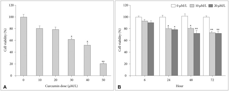
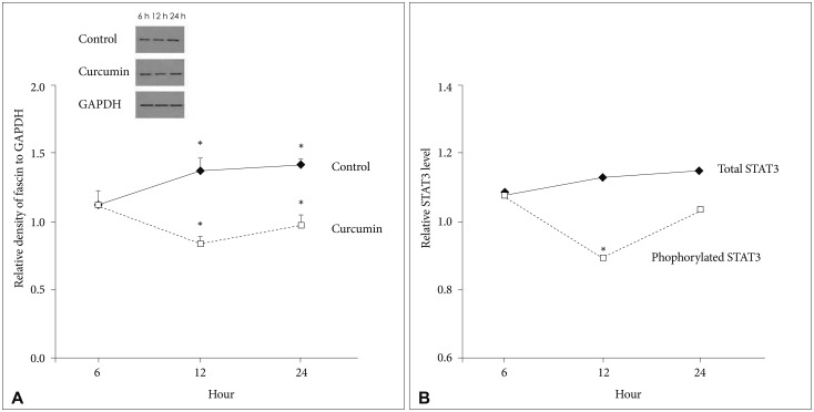
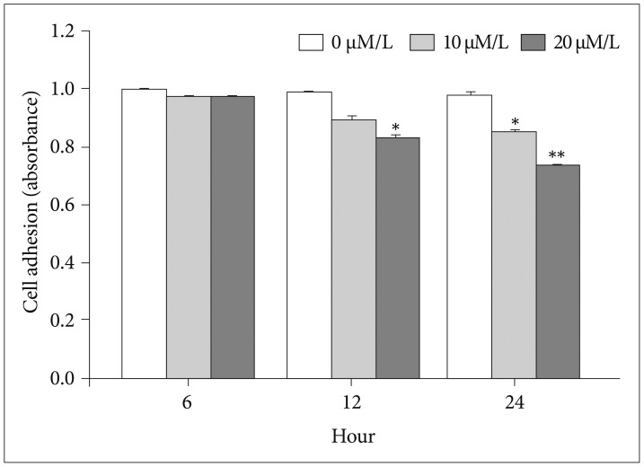
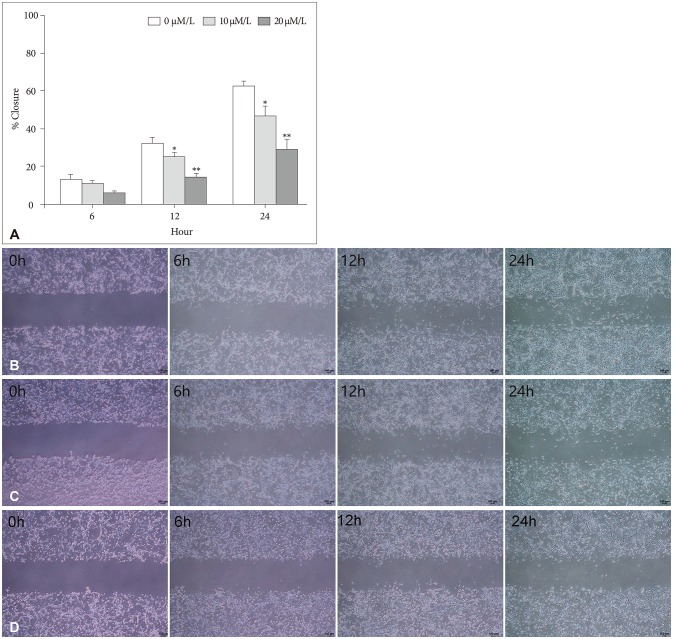
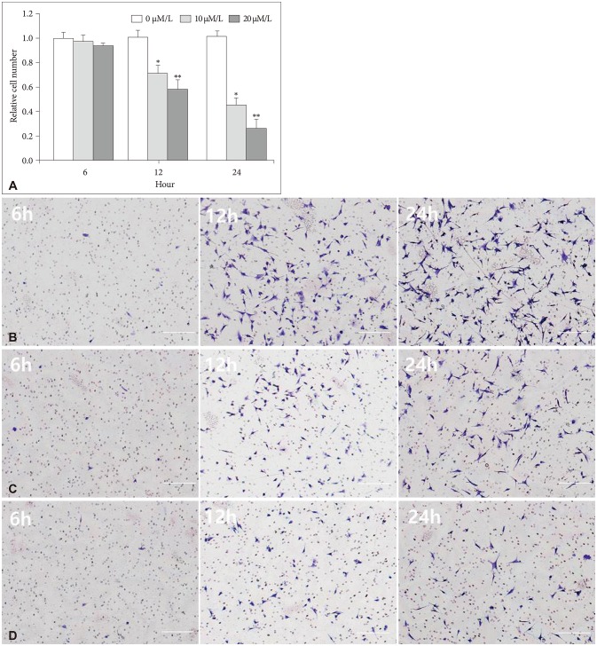
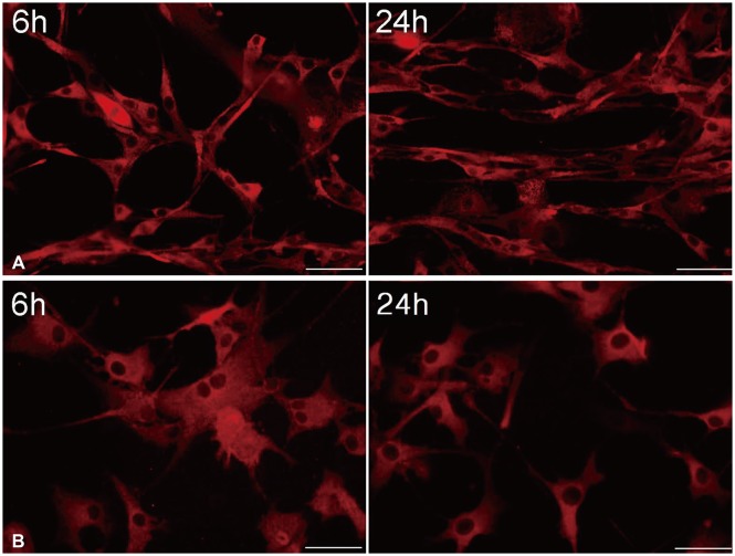
Results: At various concentrations of curcumin and exposure times, fascin expression decreased. After temporarily exposure to 10 μM/L curcumin during 6 hours as less invasive concentration and time, fascin expression temporarily decreased at 12 hours (18.4%, p=0.024), and since then recovered. And, the change of phosphrylated STAT3 level also reflected the temporarily decreased pattern of fascin expression at 12 hours (19.7%, p=0.010). Attachment, migration, and invasion capacities consistently decreased at 6, 12, and 24 hours. And, immunofluorescence showed the change of shape and the reduction of filopodia formation in cells.
Conclusion: Curcumin is likely to suppress the fascin expression in GBM cells, and this might be a possible mechanism for anti-migration and anti-invasion effects of Curcumin via inhibition of STAT3 phosphorylation.
Keywords: Curcumin; Fascin; Glioblastoma; STAT3 transcription factor.
Copyright © 2019 The Korean Brain Tumor Society, The Korean Society for Neuro-Oncology, and The Korean Society for Pediatric Neuro-Oncology.
Conflict of interest statement
The authors have no potential conflicts of interest.
Figures
Similar articles
-
The inhibitory effect of curcumin via fascin suppression through JAK/STAT3 pathway on metastasis and recurrence of ovary cancer cells.BMC Womens Health. 2020 Nov 19;20(1):256. doi: 10.1186/s12905-020-01122-2. BMC Womens Health. 2020. PMID: 33213437 Free PMC article.
-
Signal transducer and activator of transcription 3 signaling upregulates fascin via nuclear factor-κB in gastric cancer: Implications in cell invasion and migration.Oncol Lett. 2014 Mar;7(3):902-908. doi: 10.3892/ol.2014.1804. Epub 2014 Jan 15. Oncol Lett. 2014. PMID: 24527098 Free PMC article.
-
Verbascoside Inhibits Glioblastoma Cell Proliferation, Migration and Invasion While Promoting Apoptosis Through Upregulation of Protein Tyrosine Phosphatase SHP-1 and Inhibition of STAT3 Phosphorylation.Cell Physiol Biochem. 2018;47(5):1871-1882. doi: 10.1159/000491067. Epub 2018 Jun 29. Cell Physiol Biochem. 2018. PMID: 29961065
-
miR-519a enhances chemosensitivity and promotes autophagy in glioblastoma by targeting STAT3/Bcl2 signaling pathway.J Hematol Oncol. 2018 May 29;11(1):70. doi: 10.1186/s13045-018-0618-0. J Hematol Oncol. 2018. PMID: 29843746 Free PMC article.
-
Phytosomal curcumin causes natural killer cell-dependent repolarization of glioblastoma (GBM) tumor-associated microglia/macrophages and elimination of GBM and GBM stem cells.J Exp Clin Cancer Res. 2018 Jul 25;37(1):168. doi: 10.1186/s13046-018-0792-5. J Exp Clin Cancer Res. 2018. PMID: 30041669 Free PMC article.
Cited by
-
The inhibitory effect of curcumin via fascin suppression through JAK/STAT3 pathway on metastasis and recurrence of ovary cancer cells.BMC Womens Health. 2020 Nov 19;20(1):256. doi: 10.1186/s12905-020-01122-2. BMC Womens Health. 2020. PMID: 33213437 Free PMC article.
-
Visible light potentiates rapid cell destruction and death by curcumin in vitro.Photochem Photobiol Sci. 2024 Oct;23(10):1893-1914. doi: 10.1007/s43630-024-00639-x. Epub 2024 Sep 27. Photochem Photobiol Sci. 2024. PMID: 39333349
-
Curcumin piperidone derivatives induce anti-proliferative and anti-migratory effects in LN-18 human glioblastoma cells.Sci Rep. 2022 Jul 30;12(1):13131. doi: 10.1038/s41598-022-16274-4. Sci Rep. 2022. PMID: 35907913 Free PMC article.
-
Novel Therapeutic Delivery of Nanocurcumin in Central Nervous System Related Disorders.Nanomaterials (Basel). 2020 Dec 22;11(1):2. doi: 10.3390/nano11010002. Nanomaterials (Basel). 2020. PMID: 33374979 Free PMC article. Review.
-
Natural Compounds as Promising Adjuvant Agents in The Treatment of Gliomas.Int J Mol Sci. 2022 Mar 20;23(6):3360. doi: 10.3390/ijms23063360. Int J Mol Sci. 2022. PMID: 35328780 Free PMC article. Review.
References
LinkOut - more resources
Full Text Sources
Miscellaneous