Review
. 2018 Aug 28:6:97.
doi: 10.3389/fcell.2018.00097. eCollection 2018.

CD44/CD44v6 a Reliable Companion in Cancer-Initiating Cell Maintenance and Tumor Progression

Affiliations
Review

CD44/CD44v6 a Reliable Companion in Cancer-Initiating Cell Maintenance and Tumor Progression

Zhe Wang et al. Front Cell Dev Biol. .

Abstract

Metastasis is the leading cause of cancer death, tumor progression proceeding through emigration from the primary tumor, gaining access to the circulation, leaving the circulation, settling in distant organs and growing in the foreign environment. The capacity of a tumor to metastasize relies on a small subpopulation of cells in the primary tumor, so called cancer-initiating cells (CIC). CIC are characterized by sets of markers, mostly membrane anchored adhesion molecules, CD44v6 being the most frequently recovered marker. Knockdown and knockout models accompanied by loss of tumor progression despite unaltered primary tumor growth unraveled that these markers are indispensable for CIC. The unexpected contribution of marker molecules to CIC-related activities prompted research on underlying molecular mechanisms. This review outlines the contribution of CD44, particularly CD44v6 to CIC activities. A first focus is given to the impact of CD44/CD44v6 to inherent CIC features, including the crosstalk with the niche, apoptosis-resistance, and epithelial mesenchymal transition. Following the steps of the metastatic cascade, we report on supporting activities of CD44/CD44v6 in migration and invasion. These CD44/CD44v6 activities rely on the association with membrane-integrated and cytosolic signaling molecules and proteases and transcriptional regulation. They are not restricted to, but most pronounced in CIC and are tightly regulated by feedback loops. Finally, we discuss on the engagement of CD44/CD44v6 in exosome biogenesis, loading and delivery. exosomes being the main acteurs in the long-distance crosstalk of CIC with the host. In brief, by supporting the communication with the niche and promoting apoptosis resistance CD44/CD44v6 plays an important role in CIC maintenance. The multifaceted interplay between CD44/CD44v6, signal transducing molecules and proteases facilitates the metastasizing tumor cell journey through the body. By its engagement in exosome biogenesis CD44/CD44v6 contributes to disseminated tumor cell settlement and growth in distant organs. Thus, CD44/CD44v6 likely is the most central CIC biomarker.

Keywords: CD44; EMT; apoptosis resistance; cancer initiating cells; metastasis; migration; tumor exosomes.


Figures

Figure 1
Figure 1
CD44 molecules, prominent ligands and associated molecules.(A) Genomic organization and protein structure of CD44s and CD44v, glycosylation sites, the location in the cell membrane and some frequently observed CD44v exon product combinations are shown. (B) Most prominent matrix protein and cellular ligands of the globular N-terminal domains and the binding sites of the C-terminal domain to the cytoskeletal linker proteins ERM and ankyrin are indicated. Only CD44v3, CD44v6, and CD44v10 have binding sites for cytokines and chemokines.
Figure 2
Figure 2
Selective contributions of CD44v in modulating a niche for SC and CIC: CD44v6 contributes to matrix assembly and modulation by transcription of HAS and several proteases. CD44v3, CD44v6, and CD44v10 are engaged in cytokine/chemokine harboring, which become deposited in the matrix. Major components of CD44v-promoted changes in matrix composition and the contribution of CD44v-promoted deposits to matrix modulation are shown.
Figure 3
Figure 3
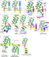
CD44/CD44v6 and CIC apoptosis resistance: (A–H) CD44 becomes engaged in apoptosis resistance by several mechanisms. (A) CD44v6 interferes with receptor-mediated apoptosis induction by preventing FAS trimerization. Antibody blocking of CD44v6 rescues FAS receptor initiated apoptosis; (B) Upon HA-crosslinking, CD44-associated src becomes activated, the CD44 complex which additionally contains HSP70 and CDC37 phosphorylates ERBB2/ERBB3, which binds neuregulin and activates the anti-apoptotic PI3K/Akt signaling pathways. In addition, Cox2 transcription becomes promoted. The latter stimulates PEC2 and blocks caspase 3 activation, which prohibits the execution phase of apoptosis. (C) Activation of c-Met via CD44v6-bound HGF promotes activation of the PI3K/Akt and the PKC-Ras-ERK pathway, activated ERK1/2 contributing to Akt phosphorylation, supporting anti-apoptotic Bcl2 and BclXl liberation. (D) Under stress, Merlin binds to the CD44-ICD leading to activation of the Hippo pathways that promotes caspase 3 cleavage, which initiates the execution phase of apoptosis. In the presence of CD44v3, merlin does not associated with the CD44-ICD; activation of the Hippo pathway is attenuated accompanied by increased apoptosis resistance. (E) CD44v3 contributes to apoptosis protection by binding proHBEGF that becomes cleaved via recruited MMP7. After loading on ERBB4, anti-apoptotic pathways become activated. (F) CD44v6-associated MMP14 accounts for proMMP9 cleavage. Activated MMP9 interferes with TGFβ activation, prohibiting TGFβ-promoted apoptosis induction. (G) Both CD44 and HA contribute to drug resistance. MDR genes are associated with CD44 and CD44 regulates expression of drug transporters by HA-activated CD44 binding to Gab1, which promotes PI3K activation. Activated PI3K stimulates HA production as well as MDR transporter expression. Alternatively, HA binding to CD44 upregulates p300 expression and its acetyltransferase activity, which sustains acetylating β-catenin and NFκB-p65. β-catenin and NFκB are cotranscription factors for MDR1. HA crosslinking stabilizes the CD44-MDR1 complex. Instead, upon Hyal activation and HA degradation the complex becomes internalized, all components including S100A and Annexin II being recovered in the cytoplasm. (H) Via PKC activation CD44v3 promotes Nanog phosphorylation, which induced miR-21 production accounting for MDR1 release from repression.
Figure 4
Figure 4
The multiple pathways of CD44/CD44v6-promoted CIC motility: CD44/CD44v6 drive CIC motility by activation of signaling cascades as well as protease activation. (A) Motility can become promoted by the association with RTK. CD44v-EGFR binding induces EGFR activation. The activated EGFR in cooperation with CD44-associated ERM promotes initiates activation of the Stat3 pathway and EMT gene transcription factors. Through the association of CD44v4-v10 with MET, oncogenes become activated, which is accompanied by transformation of epithelial cells in the colon. Also via the CD44v6 association with MET, ERM promotes MET and downstream MAPK and β-catenin activation, which contributes to activation and nuclear localization of several EMT transcription factors. (B) CD44 also associates with GPCR, a prominent example being the CD44-CXCR4 axis. Depending on HA crosslinking, SDF1-bound, CD44v-associated CXCR4 promotes activation of the MAPK pathway, which supports tumor cell and EC motility. (C) CD44v contributes to EMT. As demonstrated for activation of the Wnt signaling cascade, the supportive CD44 activity can rely on CD44-associated molecules. CD44-associated LRP6 binds to Frizzled, which strengthens Wnt signaling. WNT signaling upregulates SNAI1 to repress epithelial genes, such as E-cadherin. (D) Through the association of CD44 with integrins, CD44 gains access to focal adhesion kinase (FAK), and integrins gain access to Src kinases and ERM proteins, so that the integrin-paxillin association becomes weakened and the GEM-integrated CD44-ezrin-integrin-FAK complex moves toward the leading edge of the cell, promoting cell migration. (E) The connection between CD44 and cytoskeletal linker protein provides another pathway promoting motility, where RHOA plays a major role. The RHOA-specific GEF p115RHOGEF, which interacts with CD44, activates the serine/threonine Rho kinase (ROCK), a downstream target of RHOA. ROCK phosphorylates CD44, leading to enhanced ankyrin binding and guiding CD44 to the leading edge of migrating cells. (F) The crosstalk with proteases adds to the engagement of CD44 in motility. Three examples are shown. CD44-associated PLCγ triggers via IP3 Ca++ influx. Ca++ promotes proADAM10 dissociation from calmodulin; membrane bound ADAM10 contribution to cleavage of the CD44 extracellular regions. ADAM17 colocalizes with CD44 at Rac-regulated membrane ruffling areas and becomes activated by PKC and Rac. Finally, activated ADAM17 also promotes CD44 extracellular region cleavage. After ectodomain cleavage, CD44 becomes accessible to the presenilin/γ-secretase complex, which triggers intramembrane CD44 cleavage, setting free the CD44-ICD. The CD44-ICD binding to a DNA consensus sequence in the promoter regions of CD44 and MMP-9 gene potentiates CD44 and MMP9 transcription.
Figure 5
Figure 5
The contribution of CD44 to the biogenesis of metastasis-promoting exosomes. (A) Overview of Exo biogenesis, where CD44v6 contributes to loading of ILV derived from GEM-internalized membrane domains (red star). (B) Molecules engaged in RNA processing that coimmunoprecipitate with CD44v6, but not CD44s in CIC and CIC-TEX. With very few exceptions RNA transport and processing engaged proteins are recovered in TEX, but not cells, which supports the suggestion of an active engagement of CD44v6 in mRNA recruitment.

Similar articles

Cited by

References

    1. Abd Elmageed Z. Y., Yang Y., Thomas R., Ranjan M., Mondal D., Moroz K., et al. . (2014). Neoplastic reprogramming of patient-derived adipose stem cells by prostate cancer cell-associated exosomes. Stem Cells 32, 983–997. 10.1002/stem.1619 - DOI - PMC - PubMed
    1. Abels E. R., Breakefield X. O. (2016). Introduction to extracellular vesicles: biogenesis, rna cargo selection, content, release, and uptake. Cell. Mol. Neurobiol. 36, 301–312. 10.1007/s10571-016-0366-z - DOI - PMC - PubMed
    1. Adamia S., Maxwell C. A., Pilarski L. M. (2005). Hyaluronan and hyaluronan synthases: potential therapeutic targets in cancer. Curr. Drug Targets Cardiovasc. Haematol. Disord. 5, 3–14. 10.2174/1568006053005056 - DOI - PubMed
    1. Adorno-Cruz V., Kibria G., Liu X., Doherty M., Junk D. J., Guan D., et al. . (2015). Cancer stem cells: targeting the roots of cancer, seeds of metastasis, and sources of therapy resistance. Cancer Res. 75, 924–929. 10.1158/0008-5472.CAN-14-3225 - DOI - PMC - PubMed
    1. Alamoudi A. A., Alnoury A., Gad H. (2017). miRNA in tumour metabolism and why could it be the preferred pathway for energy reprograming. Brief Funct Genomics. 39 (supl 1), S45–S49. 10.1093/bfgp/elx023 - DOI - PubMed

LinkOut - more resources