. 2018 Jul 10;8(1):10379.
doi: 10.1038/s41598-018-28666-6.

AU040320 deficiency leads to disruption of acrosome biogenesis and infertility in homozygous mutant mice

Affiliations

AU040320 deficiency leads to disruption of acrosome biogenesis and infertility in homozygous mutant mice

Luiz G Guidi et al. Sci Rep. .

Abstract

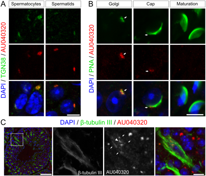
Study of knockout (KO) mice has helped understand the link between many genes/proteins and human diseases. Identification of infertile KO mice provides valuable tools to characterize the molecular mechanisms underlying gamete formation. The KIAA0319L gene has been described to have a putative association with dyslexia; surprisingly, we observed that homozygous KO males for AU040320, KIAA0319L ortholog, are infertile and present a globozoospermia-like phenotype. Mutant spermatozoa are mostly immotile and display a malformed roundish head with no acrosome. In round spermatids, proacrosomal vesicles accumulate close to the acroplaxome but fail to coalesce into a single acrosomal vesicle. In wild-type mice AU040320 localises to the trans-Golgi-Network of germ cells but cannot be detected in mature acrosomes. Our results suggest AU040320 may be necessary for the normal formation of proacrosomal vesicles or the recruitment of cargo proteins required for downstream events leading to acrosomal fusion. Mutations in KIAA0319L could lead to human infertility; we screened for KIAA0319L mutations in a selected cohort of globozoospermia patients in which no genetic abnormalities have been previously identified, but detected no pathogenic changes in this particular cohort.


Conflict of interest statement

The authors declare no competing interests.

Figures

Figure 1
Figure 1
Absence of AU040320 leads to male infertility. (A) Average number of pups born from matings with males wild-type (n = 10), heterozygous (+/−; n = 12) and homozygous (−/−; n = 12) for the AU040320 KO allele. (B) Analysis by Western blotting demonstrating expression of AU040320 in testis of wild-type (+/+) and its absence in AU040320 KO mice (−/−). The protein is also detected in wild-type lysates of the epididymis but not in isolated spermatozoa. Actin was used as loading control. The cropped blots are displayed; full-length blots and Ponceau staining of membranes are shown in Supplementary Fig. S5A. 20 µg loaded per lane. (C) X-gal staining of testis sections of AU040320 −/− mice carrying the lacZ reporter gene. Representative image shows the ß-galactosidase activity (blue dots) resulting from the reporter gene which can be seen in the majority of cells of seminiferous tubules (inset, C’). As expected, wild-type control samples do not display any signal in seminiferous tubules, although some likely unspecific signal is detected in the interstitial region corresponding to the Leydig cells (C”). Nuclei are counterstained with Nuclear FastRed. Tubules are outlined in KO samples (C, C’). (D) Images of testes of wild-type and AU040320 −/− mice show no major morphological abnormalities or differences in size. (E) Average weight of individual testis is comparable between wild-type and mutant mice (n = 8). (F) Quantification of sperm number (x106 cells/ml) revealed a reduction in the number of spermatozoa released from the cauda epididymis of AU040320 KOs (*denotes p < 0.05; n = 3 per genotype). (G–J) Bouins-fixed, H&E-stained histological sections of wild-type and AU040320 KO mice from testis (G,H) and epididymis (I,J), with selected individual tubules shown at larger magnification (G’-J’), including seminiferous tubules containing spermatids in late stages (13–16) of development (G’, H’). Scale bars: 25 μm for all panels.
Figure 2
Figure 2
AU040320-deficient spermatozoa exhibit globozoospermia-like features. (A) SEM images of wild-type and AU040320-deficient spermatozoa demonstrate the rounded nuclear shape and lack of acrosome in all spermatozoa analysed (100% spermatozoa of each animal; n = 5). (B) Immunostaining of dissociated epididymal spermatozoa shows that cells from AU040320-deficient mice have rounded nuclei (DAPI, blue) instead of characteristic sickle shape, lack an acrosome (PNA, green) and tails (acetylated tubulin, red) coiled around the sperm head. (C) Mitochondrial midpiece (MitoTracker, red) in mutant spermatozoa is disarranged instead of forming the normal elongated thin structure at the base of the cell nucleus and extending with the flagellum (acetylated tubulin, green). (D) Ultrastructure of spermatozoa obtained with TEM reveals the absence of the acrosome in rounded-head mutant spermatozoa in all cases, with disarranged mitochondria and coiled tail in some instances (right panel, arrowheads). Additional examples of images from these analyses are shown in Supplementary Fig. S2. Ac, acrosome; m, mitochondria; n, nucleus. (E) Normal ciliary axoneme is detected in spermatozoa from AU040320 KO mice, with the microtubular organisation containing the 9 outer doublets and the central pair of microtubules. Scale bars: (A,D) 2 μm; (B,C) 5 μm; (E) 100 nm.
Figure 3
Figure 3
Acrosome formation is impaired in AU040320-deficient mice. Adult testis sections from wild-type (wt, left panels) and KO (null, right panels) samples stained with PNA (green) to label developing acrosome and DAPI for cell nuclei (blue) to identify stage-dependent cell types; development stage of shown tubules appears in the main panels and that of analysed spermatids in inset panels (Golgi/Cap/Acrosome phases) is shown on the left. PNA staining in wild-type spermatids in the Golgi phase accumulates homogeneously as a single structure (A), whilst spermatids from AU040320-deficient mice have a diffuse pattern (B). In the cap phase, wild-type spermatids display the characteristic thin layer of acrosomal caps (C) while mutant spermatids have a punctate PNA staining that does not form a single acrosomal vesicle (D). In acrosome phase, PNA-labelled acrosomes elongate along with cell nuclei in wild-type testis samples (E) but this pattern is not observed in mutant samples (F), where PNA staining appears disarranged and absent in many cases. Similar pattern is also observed in maturation phase; asterisks in inset panels A’ and B’ indicate examples of spermatids in maturation phase present in these tubules. For each condition, n = 5. Scale bars: main panels 25 μm, insets 5 μm.
Figure 4
Figure 4
Proacrosomal vesicle fusion is impaired in AU040320-null mice. TEM images of developing spermatids in seminiferous tubules of wild-type and mutant mice. Wild-type sperm exhibit the characteristic developmental path of the acrosome, with proacrosomal vesicles fusing together to form a large acrosomal granule (A) which becomes flattened along the acroplaxome and the nuclear face in the cap phase (C), extends further over the elongating nucleus in the acrosome phase (E), and matures into a structure covering nearly all the now heavily condensed nucleus in the maturation phase (G). A couple of spermatocytes in cap phase appearing alongside those in maturation phase are shown in panel G. In mutant spermatids, vesicles containing proacrosomal material (B’, white arrows) are observed approaching the nucleus and forming multiple points of indentations on it (B’, black arrows) but fail to coalesce into a single acrosomal granule. As they develop, mutant round spermatids continue to show no acrosomal structure despite the continuous arrival of proacrosomal vesicles (D’, arrows); the curvatures observed at the edge of the acroplaxome in these cells (D, arrowheads) may indicate the presence of the manchette. A similar pattern appears in mutant spermatids in the acrosome phase (F) which display no developing acrosome, despite the more elongated cell morphology and the apparent presence of the acroplaxome revealed by the flattened nuclear surface (F’, arrows), while larger proacrosomic vesicles can be observed aligning to the acroplaxome. At maturation phase, mutant spermatids show a condensed globular, not elongated, nucleus but no acrosome (H), although an apparently normal developing tail (arrow) can be detected. While mutant spermatids do not develop an acrosome, they appear on occasions, at the cap/acrosome phases, to contain a pseudoacrosome-like structure (I,J) consisting of saccules organised in a layered fashion (black arrows) in close proximity to the acroplaxome (white arrows). n, nucleus; g, Golgi; ac, acrosome; apx, acroplaxome; ag, acrosomal granule; v, proacrosomal vesicles; M, midpiece of developing sperm tail. For each condition, n = 5. Scale bars: (A–H) 2 μm, insets 1 μm; (I,J) 1 μm, insets 200 nm.
Figure 5
Figure 5
AU040320 localises to the TGN in developing spermatids. (A) Double immunohistochemistry for detection of endogenous AU040320 (custom antibody #78, red) and trans-Golgi marker TGN38 (green) shows co-localisation in spermatocytes and round spermatids. (B) Comparison of AU040320 localisation (#78, red) with the PNA pattern (green) over the course of acrosome development reveals association of the AU040320 protein with early acrosomal structures in Golgi phase spermatids but this seems to be reduced in Cap phase spermatids (white arrows); no clear signal for AU040320 can be detected at later stages (acrosome-maturation phases). (C) AU040320 protein (#78, red) exhibits a pattern with some overlap (arrows) with the structure of Sertoli cells (β-tubulin III, green), including a stronger juxta-nuclear signal compatible with TGN localisation (arrowhead), suggesting AU040320 is also present in these cells. For each condition, n = 3. Scale bars: 5 μm in all panels with the exception of in C (25 μm).

Similar articles

Cited by

References

    1. Clermont Y. Kinetics of spermatogenesis in mammals: seminiferous epithelium cycle and spermatogonial renewal. Physiol Rev. 1972;52:198–236. doi: 10.1152/physrev.1972.52.1.198. - DOI - PubMed
    1. Hess, R. A. & Renato de Franca, L. Spermatogenesis and cycle of the seminiferous epithelium. Adv Exp Med Biol.636, 1–15, 10.1007/978-0-387-09597-4_1 (2008). - PubMed
    1. Xiao X, Mruk DD, Wong CK, Cheng CY. Germ cell transport across the seminiferous epithelium during spermatogenesis. Physiology (Bethesda). 2014;29:286–298. - PMC - PubMed
    1. Griswold MDS. The Commitment to Meiosis. Physiol Rev. 2016;96:1–17. doi: 10.1152/physrev.00013.2015. - DOI - PMC - PubMed
    1. Abou-Haila A, Tulsiani DR. Mammalian sperm acrosome: formation, contents, and function. Arch Biochem Biophys. 2000;379:173–182. doi: 10.1006/abbi.2000.1880. - DOI - PubMed

Publication types