

Subcellular localization of MC4R with ADCY3 at neuronal primary cilia underlies a common pathway for genetic predisposition to obesity
- PMID: 29311635
- PMCID: PMC5805646
- DOI: 10.1038/s41588-017-0020-9
Subcellular localization of MC4R with ADCY3 at neuronal primary cilia underlies a common pathway for genetic predisposition to obesity
Abstract
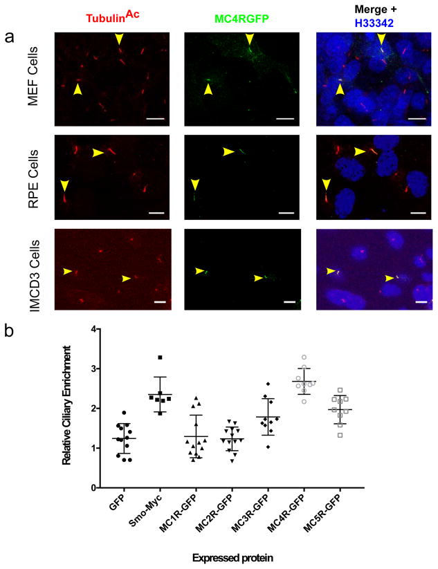
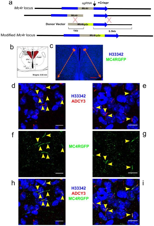
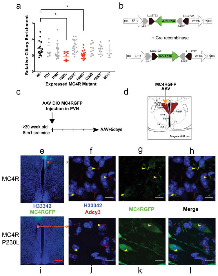
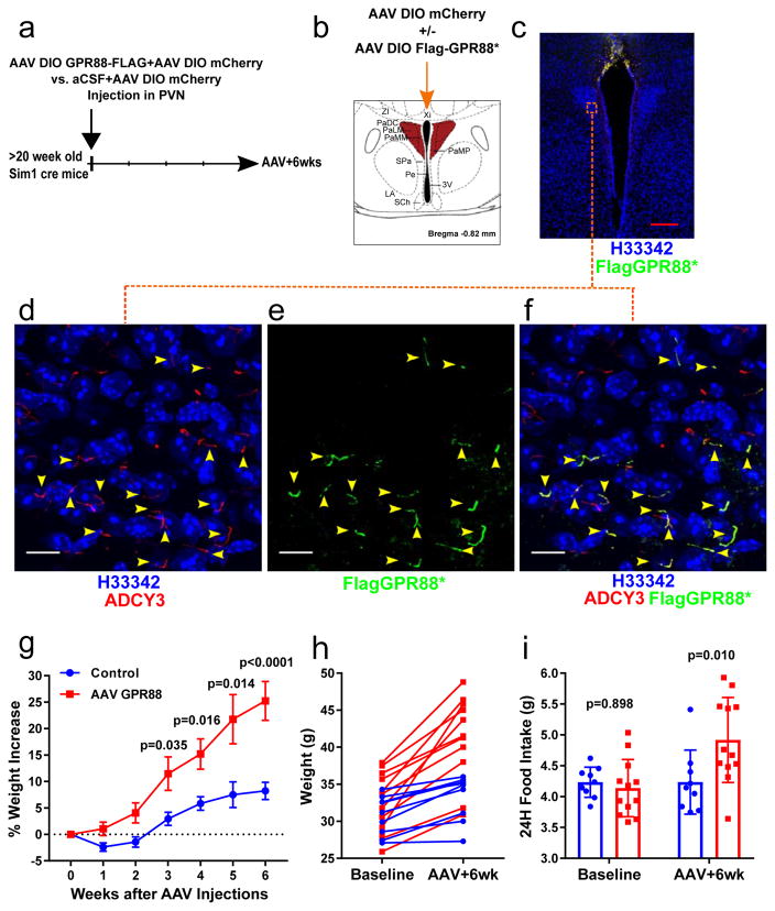
Most monogenic cases of obesity in humans have been linked to mutations in genes encoding members of the leptin-melanocortin pathway. Specifically, mutations in MC4R, the melanocortin-4 receptor gene, account for 3-5% of all severe obesity cases in humans1-3. Recently, ADCY3 (adenylyl cyclase 3) gene mutations have been implicated in obesity4,5. ADCY3 localizes to the primary cilia of neurons 6 , organelles that function as hubs for select signaling pathways. Mutations that disrupt the functions of primary cilia cause ciliopathies, rare recessive pleiotropic diseases in which obesity is a cardinal manifestation 7 . We demonstrate that MC4R colocalizes with ADCY3 at the primary cilia of a subset of hypothalamic neurons, that obesity-associated MC4R mutations impair ciliary localization and that inhibition of adenylyl cyclase signaling at the primary cilia of these neurons increases body weight. These data suggest that impaired signaling from the primary cilia of MC4R neurons is a common pathway underlying genetic causes of obesity in humans.
Conflict of interest statement
The authors declare no competing financial interests.
Figures
Comment in
-
New ADCY3 Variants Dance in Obesity Etiology.Trends Endocrinol Metab. 2018 Jun;29(6):361-363. doi: 10.1016/j.tem.2018.02.004. Epub 2018 Feb 14. Trends Endocrinol Metab. 2018. PMID: 29454745
-
Neuronal Cilia: Another Player in the Melanocortin System.Trends Mol Med. 2018 Apr;24(4):333-334. doi: 10.1016/j.molmed.2018.02.004. Epub 2018 Feb 28. Trends Mol Med. 2018. PMID: 29501261
Similar articles
-
Mechanisms of Weight Control by Primary Cilia.Mol Cells. 2022 Apr 30;45(4):169-176. doi: 10.14348/molcells.2022.2046. Mol Cells. 2022. PMID: 35387896 Free PMC article. Review.
-
Melanocortin 4 receptor signals at the neuronal primary cilium to control food intake and body weight.J Clin Invest. 2021 May 3;131(9):e142064. doi: 10.1172/JCI142064. J Clin Invest. 2021. PMID: 33938449 Free PMC article.
-
Signal pathway analysis of selected obesity-associated melanocortin-4 receptor class V mutants.Biochim Biophys Acta Mol Basis Dis. 2020 Aug 1;1866(8):165835. doi: 10.1016/j.bbadis.2020.165835. Epub 2020 May 8. Biochim Biophys Acta Mol Basis Dis. 2020. PMID: 32423884
-
Age-related ciliopathy: Obesogenic shortening of melanocortin-4 receptor-bearing neuronal primary cilia.Cell Metab. 2024 May 7;36(5):1044-1058.e10. doi: 10.1016/j.cmet.2024.02.010. Epub 2024 Mar 6. Cell Metab. 2024. PMID: 38452767
-
Melanocortin pathways: suppressed and stimulated melanocortin-4 receptor (MC4R).Physiol Res. 2020 Sep 30;69(Suppl 2):S245-S254. doi: 10.33549/physiolres.934512. Physiol Res. 2020. PMID: 33094623 Free PMC article. Review.
Cited by
-
Emerging mechanistic understanding of cilia function in cellular signalling.Nat Rev Mol Cell Biol. 2024 Jul;25(7):555-573. doi: 10.1038/s41580-023-00698-5. Epub 2024 Feb 16. Nat Rev Mol Cell Biol. 2024. PMID: 38366037 Free PMC article. Review.
-
Mechanisms of Weight Control by Primary Cilia.Mol Cells. 2022 Apr 30;45(4):169-176. doi: 10.14348/molcells.2022.2046. Mol Cells. 2022. PMID: 35387896 Free PMC article. Review.
-
The subcellular organisation of Saccharomyces cerevisiae.Curr Opin Chem Biol. 2019 Feb;48:86-95. doi: 10.1016/j.cbpa.2018.10.026. Epub 2018 Nov 29. Curr Opin Chem Biol. 2019. PMID: 30503867 Free PMC article. Review.
-
Role of lipids in the control of autophagy and primary cilium signaling in neurons.Neural Regen Res. 2024 Feb;19(2):264-271. doi: 10.4103/1673-5374.377414. Neural Regen Res. 2024. PMID: 37488876 Free PMC article. Review.
-
Hypothalamic primary cilium: A hub for metabolic homeostasis.Exp Mol Med. 2021 Jul;53(7):1109-1115. doi: 10.1038/s12276-021-00644-5. Epub 2021 Jul 1. Exp Mol Med. 2021. PMID: 34211092 Free PMC article. Review.
References
-
- Lubrano-Berthelier C. Melanocortin 4 Receptor Mutations in a Large Cohort of Severely Obese Adults: Prevalence, Functional Classification, Genotype-Phenotype Relationship, and Lack of Association with Binge Eating. Journal of Clinical Endocrinology & Metabolism. 2006;91:1811–1818. - PubMed
-
- Vaisse C, Clement K, Guy-Grand B, Froguel P. A frameshift mutation in human MC4R is associated with a dominant form of obesity. Nat Genet. 1998;20:113–4. - PubMed
Publication types
MeSH terms
Substances
Grants and funding
LinkOut - more resources
Full Text Sources
Other Literature Sources
Medical
Molecular Biology Databases
