. 2018 Feb;7(2):380-396.
doi: 10.1002/cam4.1276. Epub 2017 Dec 26.

Induction of autophagy and autophagy-dependent apoptosis in diffuse large B-cell lymphoma by a new antimalarial artemisinin derivative, SM1044

Affiliations

Induction of autophagy and autophagy-dependent apoptosis in diffuse large B-cell lymphoma by a new antimalarial artemisinin derivative, SM1044

Chunyan Cheng et al. Cancer Med. 2018 Feb.

Abstract

Diffuse large B-cell lymphoma (DLBCL) is the most common form of non-Hodgkin's lymphoma. R-CHOP is currently the standard therapy for DLBCL, but the prognosis of refractory or recurrent patients remains poor. In this study, we synthesized a new water-soluble antimalarial drug artemisinin derivative, SM1044. The treatment of DLBCL cell lines with SM1044 induces autophagy-dependent apoptosis, which is directed by an accelerated degradation of the antiapoptosis protein Survivin, via its acetylation-dependent interaction with the autophagy-related protein LC3-II. Additionally, SM1044 also stimulates the de novo synthesis of ceramide, which in turn activates the CaMKK2-AMPK-ULK1 axis, leading to the initiation of autophagy. Our findings not only elucidate the mechanism of autophagy-dependent apoptosis in DLBCL cells, but also suggest that SM1044 is a promising therapeutic molecule for the treatment of DLBCL, along with R-CHOP regimen.

Keywords: DLBCL; Apoptosis; Survivin; artemisinin derivative; autophagy.


Figures

Figure 1
Figure 1
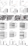
SM1044 induces caspase‐dependent apoptosis in DLBCL cell lines. (A) Structural formula of SM1044. (B) SUDHL‐4 cells were treated with the indicated concentrations of SM1044, DHA, ART, ARS, and ARM for 24 h, respectively. Cell viability was measured by CCK‐8 and the percent inhibitions were calculated (mean ± SEM, n = 3). (C) SUDHL‐4, SUDHL‐10, and OCILY3 cells were treated with the indicated concentrations of SM1044 for 24 h. The cell viability was measured by CCK‐8 and the percent inhibitions were calculated (mean ± SEM, n = 3). (D) SUDHL‐4, SUDHL‐10, and OCILY3 cells were treated with the indicated concentrations of SM1044 for 24 h. Cell apoptosis was detected by flow cytometry using the FITC‐Annexin V/PI apoptosis detection kit (mean ± SEM, n = 3). SUDHL‐4: 0 μmol/L versus 0.05 μmol/L, P = 0.042; 0.1 μmol/L, P < 0.001; 0.5 μmol/L, P < 0.001; 1 μmol/L, P = 0.0061. SUDHL‐10: 0 μmol/L versus 0.1 μmol/L, P = 0.023; 0.5 μmol/L, P < 0.001; 1 μmol/L, P < 0.001; 5 μmol/L, P < 0.001. OCILY3: 0 μmol/L versus 0.05 μmol/L, P = 0.034; 0.1 μmol/L, P < 0.001; 0.5 μmol/L, P < 0.001; 1 μmol/L, P < 0.001. (E) SUDHL‐4 cells were treated with SM1044 for 24 h. Representative electron microscopy photomicrographs are shown. The red arrows point to the apoptosis bodies. (F) The expressions of caspase‐8, ‐9, ‐3, and PARP were detected by western blot in SUDHL‐4, SUDHL‐10, and OCILY3 cells treated with the indicated concentrations of SM1044 for the indicated time courses. (G) Apoptosis was measured in SUDHL‐4, SUDHL‐10, and OCILY3 cells pretreated with caspase inhibitor Z‐VADFMK for 1 h, and then treated with SM1044 for another 24 h (mean ± SEM, n = 3). SUDHL‐4: SM1044 versus control, P < 0.001; SM1044 versus Z‐VADFMK plus SM1044, P < 0.001. SUDHL‐10: SM1044 versus control, P < 0.001; SM1044 versus Z‐VADFMK plus SM1044, P < 0.001. OCILY3: SM1044 versus control, P < 0.001; SM1044 versus Z‐VADFMK plus SM1044, P < 0.001. *P < 0.05, **P < 0.01, ***P < 0.001.
Figure 2
Figure 2
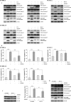
SM1044‐induced autophagy. (A) SUDHL‐4 cells were treated with SM1044 for 24 h. Representative electron microscopy photomicrographs are shown. The green triangles indicate autophagosomes and the yellow triangles indicate autophagolysosomes. (B) GFPLC3 plasmids were transfected into SUDHL‐4 cells. Forty‐eight hours later, cells were pretreated by CQ for 1 h, followed by a SM1044 treatment for another 24 h. LC3 puncta are shown by fluorescence microscope. (C) SUDHL‐4, SUDHL‐10, and OCILY3 cells were treated with the indicated concentrations of SM1044 for the indicated time courses. The expression of LC3 was detected by western blot. (D) SUDHL‐4, SUDHL‐10, and OCILY3 cells were pretreated with CQ or Baf A1 for 1 h, followed by a SM1044 treatment for another 24 h. The expression of LC3 was detected by western blot.
Figure 3
Figure 3
SM1044‐induced apoptosis is dependent on the generation of autophagy. (A) SUDHL‐4, SUDHL‐10, and OCILY3 cells were pretreated with CQ or Baf A1 for 1 h, followed by SM1044 treatment for another 24 h. The respective expressions of caspase‐8, ‐9, ‐3, and PARP were detected by western blot. (B) Apoptosis was measured by flow cytometry (mean ± SEM, n = 3). SUDHL‐4: SM1044 versus control, P < 0.001; SM1044 versus CQ plus SM1044, P < 0.001; SM1044 versus Baf A1, P < 0.001. SUDHL‐10: SM1044 versus control, P < 0.001; SM1044 versus CQ plus SM1044, P = 0.0077; SM1044 versus Baf A1, P < 0.001. OCILY3: SM1044 versus control, P < 0.001; SM1044 versus Baf A1, P = 0.041. (C) SUDHL‐4 cells were stably transfected with scramble or LC3 shRNA, then treated with SM1044 for 24 h. Apoptosis was measured by flow cytometry (mean ± SEM, n = 3). Scramble + SM1044 versus sh‐LC3 + SM1044, P = 0.0012. (D) The respective expressions of caspase‐8, ‐9, ‐3, and PARP were detected by western blot in the stably transfected cells after SM1044 treatment for 24 h. *P < 0.05, **P < 0.01, ***P < 0.001.
Figure 4
Figure 4
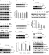
SM044 promotes the acetylation of Survivin, enhances the interaction between Survivin and LC3‐II, leading to the degradation of Survivin and apoptosis. (A) SUDHL‐4 cells were pretreated with CQ for 1 h, followed by SM1044 treatment for another 24 h. The respective expressions of antiapoptosis proteins were detected by western blot. (B) SUDHL‐4 cells were pretreated with Baf A1 for 1 h, followed by SM1044 treatment for another 24 h. The expression of Survivin was detected by western blot (upper panel). Cells with a stable knockdown of LC3 or control cells (scramble shRNA) were treated with SM1044 for 24 h. The expression of Survivin was detected by western blot (lower panel). (C) SUDHL‐4 cells were treated with SM1044 for the indicated time courses. The protein and mRNA levels of Survivin were, respectively, detected by western blot (upper panel) and real‐time PCR (lower panel, mean ± SEM, n = 3). (D) SUDHL‐4 cells were pretreated with MG132 or CQ for 1 h, followed by a SM1044 treatment for another 24 h. The expression of Survivin was detected by western blot. (E) SUDHL‐4 cells were pretreated with CQ for 1 h, followed by SM1044 treatment for another 12 h. Survivin was immunoprecipitated with its antibody. Acetylation of Survivin was detected by western blot. (F) SUDHL‐4 cells were pretreated with the p300 inhibitor C646 for 1 h, followed by SM1044 treatment for another 24 h. Acetylation of Survivin was detected by western blot after Survivin was immunoprecipitated with its antibody (upper panel). The protein and mRNA levels of Survivin were detected by western blot (middle panel) and Real‐time PCR (lower panel, mean ± SEM, n = 3), respectively. For real‐time PCR, C646: 0 versus 5 μmol/L, P = 0.0086; 10 μmol/L, P < 0.001; 20 μmol/L, P < 0.001. The ratio of Survivin (with/without SM1044) at the protein level in different concentration groups of C646 was calculated using ImageJ software. (G) SUDHL‐4 cells were treated with SM1044 for 12 h. Cell lysates were immunoprecipitated using anti‐Survivin antibody (IP: Survivin) or normal rabbit IgG. The respective levels of Survivin and LC3 were detected by western blot. (H) SUDHL‐4 cells were pretreated with C646 for 1 h, followed by SM1044 treatment for another 12 h. Cell lysates were immunoprecipitated using the anti‐Survivin antibody. The levels of Survivin and LC3 were detected by western blot. (I) SUDHL‐4 cells were stably transfected with empty vector, Survivin, or xLIR motif mutated Survivin (PLVX, PLVX‐S, PLVX‐S‐M), respectively. The stable transfected cells were treated with SM1044 for 24 h. Apoptosis was measured by flow cytometry (mean ± SEM, n = 3). PLVX + SM1044 versus PLVX‐S‐M + SM1044, P < 0.001. (J) The stable transfected cells were treated with SM1044 (SM) for 24 h. The levels of Survivin, caspase‐3 and PARP were detected by western blot. **P < 0.01, ***P < 0.001 compared with the control group without C646 treatment. NS, no significance.
Figure 5
Figure 5
SM1044 induces CaMKK2–AMPKULK1 pathway‐mediated autophagy through promoting de novo synthesis of ceramide. (A) SUDHL‐4 cells were treated with SM1044 for the indicated time courses. The active ULK1, AMPK and mTOR were detected by western blot. (B) SUDHL‐4 cells were pretreated with AMPK inhibitor compound C for 1 h, followed by SM1044 treatment for another 24 h. The levels of LC3, Survivin, and p‐AMPK were detected by western blot. (C) SUDHL‐4 cells were pretreated with CaMKK2 inhibitor STO‐609 for 1 h, followed by SM1044 treatment for another 24 h. The levels of p‐AMPK, Survivin, caspase‐8, ‐9, ‐3, and PARP were detected by western blot. (D) SUDHL‐4 cells were treated with SM1044 for 3 h and the level of ceramide was detected by HPLCMS/MS. C24: Control versus SM1044, P = 0.011. C26: Control versus SM1044, P = 0.023. (E) SUDHL‐4 cells were pretreated with ceramide inhibitor S1P for 1 h, followed by SM1044 treatment for another 24 h. The levels of p‐AMPK and Survivin were detected by western blot. (F) SUDHL‐4 cells were pretreated with ceramide de novo synthesis inhibitor l‐cycloserine for 1 h, followed by SM1044 treatment for another 24 h. Cell viability was detected by CCK‐8 (upper panel, mean ± SEM, n = 3) and apoptosis was measured by flow cytometry (lower panel, mean ± SEM, n = 3). Cell viability: SM1044 versus control, P < 0.001, SM1044 versus l‐cycloserine plus SM1044, P < 0.001. Percent apoptosis: SM1044 versus control, P < 0.001, SM1044 versus l‐cycloserine plus SM1044, P < 0.001. (G) SUDHL‐4 cells were pretreated with l‐cycloserine for 1 h, followed by SM1044 treatment for another 24 h. The levels of p‐AMPK, LC3, Survivin, caspase‐8, ‐9, ‐3, and PARP were detected by western blot. *P < 0.05, ***P < 0.001.
Figure 6
Figure 6
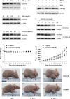
Degradation of Survivin by ARTs and in vivo effect of SM1044. (A) SUDHL‐4 cells were treated with ART and several ART derivatives (as indicated). The expression of Survivin was detected by western blot. (B) SUDHL‐4 cells were pretreated with CQ or Baf A1 for 1 h, followed by DHA treatment for another 24 h. The expression of Survivin was detected by western blot. (C) SUDHL‐10 and OCILY3 cells were treated with SM1044 for the indicated time courses. The expression of Survivin was detected by western blot. (D) Nude mice with grafted tumors were treated with SM1044 (n = 12, 5 mg/kg per day) or vehicle control (n = 11, control) every day. Body weights and tumor volumes were measured every other day and plotted on the curve diagram. On day 0, the mean body weights of normal saline‐treated and SM1044‐treated groups were (19.4 ± 0.5) g and (19.3 ± 0.6) g (P = 0.89), respectively, and the mean tumor volumes were (73.6 ± 13.6) mm3 and (77.6 ± 14.2) mm3 (P = 0.84), respectively. However, on day 24, the mean body weights of normal saline‐treated and SM1044‐treated groups were (19.3 ± 0.5) g and (18.9 ± 0.6) g (P = 0.60), respectively, while the mean tumor volumes were (1315.2 ± 200.4) mm3 and (734.3 ± 177.4) mm3 (P = 0.04), respectively. *< 0.05. (E) Representative images of control and SM1044‐treated tumors.

Similar articles

Cited by

References

    1. Chaganti, S. , Illidge T., Barrington S., McKay P., Linton K., Cwynarski K., et al. 2016. Guidelines for the management of diffuse large B‐cell lymphoma. Br. J. Haematol. 174:43–56. - PubMed
    1. Friedberg, J. W. 2011. Relapsed/refractory diffuse large B‐cell lymphoma. Hematol. Am. Soc. Hematol. Edu. Program 2011:498–505. - PubMed
    1. Perry, A. M. , Mitrovic Z., and Chan W. C.. 2012. Biological prognostic markers in diffuse large B‐cell lymphoma. Cancer Control 19:214–226. - PubMed
    1. Liu, Z. , Xu‐Monette Z. Y., Cao X., Manyam G. C., Wang X., Tzankov A., et al. 2015. Prognostic and biological significance of Survivin expression in patients with diffuse large B‐cell lymphoma treated with rituximab‐CHOP therapy. Modern Pathol. 28: 1297–1314. - PubMed
    1. Kawasaki, H. , Altieri D. C., Lu C. D., Toyoda M., Tenjo T., and Tanigawa N.. 1998. Inhibition of apoptosis by Survivin predicts shorter survival rates in colorectal cancer. Can. Res. 58:5071–5074. - PubMed

Publication types

LinkOut - more resources