. 2018 Jan 2;215(1):91-113.
doi: 10.1084/jem.20171163. Epub 2017 Nov 20.

Maternal IgG immune complexes induce food allergen-specific tolerance in offspring

Affiliations

Maternal IgG immune complexes induce food allergen-specific tolerance in offspring

Asa Ohsaki et al. J Exp Med. .

Abstract

The role of maternal immune responses in tolerance induction is poorly understood. To study whether maternal allergen sensitization affects offspring susceptibility to food allergy, we epicutaneously sensitized female mice with ovalbumin (OVA) followed by epicutaneous sensitization and oral challenge of their offspring with OVA. Maternal OVA sensitization prevented food anaphylaxis, OVA-specific IgE production, and intestinal mast cell expansion in offspring. This protection was mediated by neonatal crystallizable fragment receptor (FcRn)-dependent transfer of maternal IgG and OVA immune complexes (IgG-IC) via breast milk and induction of allergen-specific regulatory T (T reg) cells in offspring. Breastfeeding by OVA-sensitized mothers or maternal supplementation with IgG-IC was sufficient to induce neonatal tolerance. FcRn-dependent antigen presentation by CD11c+ dendritic cells (DCs) in offspring was required for oral tolerance. Human breast milk containing OVA-IgG-IC induced tolerance in humanized FcRn mice. Collectively, we demonstrate that interactions of maternal IgG-IC and offspring FcRn are critical for induction of T reg cell responses and control of food-specific tolerance in neonates.


Figures

Figure 1.
Figure 1.
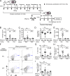
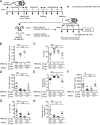
Maternal allergen sensitization protects offspring against food allergy. (A) Experimental protocol. (B) Serum OVA-IgE levels. (C) Serum IL-4 levels. (D) Core body temperature change 30 min after challenge. (E) Serum mMCP1 levels after challenge. (F) Representative flow cytometric analysis of jejunal mast cells (c-kit+IgE+lineageCD45+) from two independent experiments. Numbers indicate percentages. (G–I) Mast cell frequencies (G), numbers (H), and Il13 mRNA (I) in the jejunum. Il13 mRNA levels are expressed as fold induction relative to jejunum of saline (SAL)-exposed offspring of saline-exposed mothers. Groups of animals were compared using nonparametric one-way ANOVA. Data are mean ± SEM of two independent experiments (B–E and G–I). *, P < 0.05; **, P < 0.01; ***, P < 0.001; ****, P < 0.0001; ns, not significant.
Figure 2.
Figure 2.
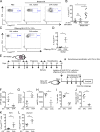
Allergen-specific T reg cells expand in offspring of allergen-sensitized mothers. (A–C) Flow cytometric analysis (A), frequencies (B), and numbers (C) of CD4+Foxp3+ cells in offspring MLN cells. (D) Analysis of OVA-specific Foxp3+ cells expanded from offspring MLN cells. (E and F) Flow cytometric analysis (E) and frequencies (F) of proliferation among CD4+Foxp3+ MLN cells labeled with CellTrace Violet cultured in vitro for 5 d. n = 4 (D and F). (G and H) Flow cytometric analysis (G) and percent suppression (H) of CellTrace Violet proliferation in DO11.10+ T responder cells cultured with OVA323-339 peptide in the absence or presence of T reg cells isolated from offspring MLN cells. (I) Frequencies of OVA-specific Foxp3+ cells expanded from offspring MLN cells after epicutaneous sensitization and oral challenge with OVA. Representative plots from two independent experiments are shown (A, E, and G). Numbers indicate percentages (A and E). Groups of animals were compared using the Mann-Whitney U test (B, C, H, and I) or nonparametric one-way ANOVA (D and F). Data are representative of two independent experiments (B–D, F, and H–I). Data are mean ± SEM. *, P < 0.05; **, P < 0.01; ns, not significant. SAL, saline.
Figure 3.
Figure 3.
Allergen-specific T reg cells in offspring are required for protection against food allergy. (A) Experimental protocol. (B) Serum OVA-IgE levels. (C) Core body temperature change. (D) Serum mMCP1 levels. (E and F) Flow cytometric analysis of jejunal mast cell frequencies (E) and numbers (F). Groups of animals were compared using the Mann-Whitney U test (B–F). Data are representative of two independent experiments (B–F). Data are mean ± SEM. *, P < 0.05; **, P < 0.01; ***, P < 0.001; ns, not significant. SAL, saline.
Figure 4.
Figure 4.
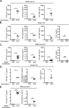
Maternal IgG-allergen immune complex, but not free allergen, are transferred to offspring via breast milk. (A) OVA-specific Igs in mother sera. (B and C) OVA-specific Igs and OVA in breast milk (B) and offspring sera (C). (D) OVA-IgG-ICs and TGF-β1 in breast milk. (E) OVA-IgG-ICs in offspring sera. Groups of animals were compared using the Mann-Whitney U test. Data are mean ± SEM of two independent experiments (A–E). *, P < 0.05; **, P < 0.01; ***, P < 0.001; ****, P < 0.0001; ns, not significant. SAL, saline.
Figure 5.
Figure 5.
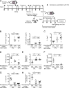
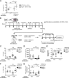
Breastfeeding by allergen-sensitized mothers protects offspring from food allergy. (A) Experimental protocol. (B) Serum OVA-IgG-ICs in weaned offspring. (C) Analysis of OVA-specific Foxp3+ cells expanded from offspring MLN cells. (D) Serum OVA-IgE levels. (E) Core body temperature change. (F) Serum mMCP1 levels. (G and H) Flow cytometric analysis of jejunal mast cell frequencies (G) and numbers (H). Groups of animals were compared using nonparametric one-way ANOVA. Data are mean ± SEM of two independent experiments (B–H). *, P < 0.05; **, P < 0.01; ***, P < 0.001; ns, not significant. SAL, saline.
Figure 6.
Figure 6.
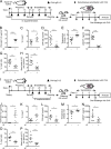
IC supplementation to mothers protects offspring from food allergy. (A–H) IC supplementation during pregnancy and breastfeeding. Experimental protocol (A), serum OVA-IgG1-IC in weaned offspring (B), OVA-specific Foxp3+ cells expanded from offspring MLN cells (C), serum OVA-IgE levels (D), core body temperature change (E), serum mMCP1 levels (F), jejunal mast cell frequencies (G), and numbers (H). (I–P) IC supplementation during breastfeeding. Experimental protocol (I), serum OVA-IgG1-IC in weaned offspring (J), OVA-specific Foxp3+ cells expanded from offspring MLN cells (K), serum OVA-IgE levels (L), core body temperature change (M), serum mMCP1 levels (N), jejunal mast cell frequencies (O), and numbers (P). Groups of animals were compared using the Mann-Whitney U test. Data are mean ± SEM and representative of 2 independent experiments (B–H, J–P). *, P < 0.05; **, P < 0.01; ***, P < 0.001; ****, P < 0.0001.
Figure 7.
Figure 7.
Offspring FcRn is required for protection of offspring from food allergy. (A) Experimental protocol. (B) Serum OVA-IgG1-IC in weaned offspring. (C) Analysis of OVA-specific Foxp3+ cells expanded from offspring MLN cells. (D) Serum OVA-IgE levels. (E) Core body temperature change. (F) Serum mMCP1 levels. (G and H) Flow cytometric analysis of jejunal mast cell frequencies (G) and numbers (H). Groups of animals were compared using nonparametric one-way ANOVA. Data are mean ± SEM of two independent experiments (B–H). *, P < 0.05; **, P < 0.01; ***, P < 0.001; ns, not significant. SAL, saline.
Figure 8.
Figure 8.
Milk-borne IgG-IC induces allergen-specific T reg cells via MLN CD11c+ DCs. (A and B) Flow cytometric analysis (A) and frequencies (B) of CD4+DO11.10+Foxp3EGFP+ T reg cells. MLN CD11c+ cells were isolated from naive WT mice and cultured with CD4+DO11.10+Foxp3EGFP- cells in the presence or absence of breast milk. (C and D) Flow cytometric analysis (C) and frequencies (D) of CD4+DO11.10+Foxp3EGFP+ T reg cells. MLN CD11c+ cells were isolated from offspring of SAL or OVA mothers and cultured with CD4+DO11.10+Foxp3EGFP- cells without exogenous allergens. (E–L) Adoptive transfer of MLN CD11c+ cells from offspring of SAL or OVA mothers. Experimental protocol (E), OVA-IgE (F), core body temperature change (G), serum mMCP1 levels (H), jejunal mast cell frequencies (I) and numbers (J), and flow cytometric analysis of CD4+DO11.10+Foxp3EGFP+ T reg cells in MLN (K) and jejunum (L) of recipients. Representative plots from two independent experiments shown (A and C). Numbers indicate percentages (A and C). Groups of animals were compared using nonparametric one-way ANOVA (B) and the Mann-Whitney U test (D and F–L). Data are mean ± SEM of two independent experiments (B and D) or representative of two independent experiments (F–L). *, P < 0.05; **, P < 0.01; ns, not significant. SAL, saline.
Figure 9.
Figure 9.
FcRn in CD11c+ DCs is critical for induction of tolerance in offspring. (A) Flow cytometric analysis of CD4+DO11.10+Foxp3EGFP+ T reg cells. MLN CD11c+ DCs were isolated from WT or Fcgrt−/− mice and cultured with CD4+DO11.10+Foxp3EGFP- cells with breast milk from OVA-sensitized WT mothers or with exogenous TGF-β1 and OVA323-339 peptide. (B) Experimental protocol. (C) Analysis of OVA-specific Foxp3+ cells expanded from weaned offspring MLN cells. (D) Serum OVA-IgE levels. (E) Core body temperature change. (F and G) Flow cytometric analysis of jejunal mast cell frequencies (F) and numbers (G). Groups of animals were compared using the Mann-Whitney U test (A, C–G). Data are mean ± SEM of two independent experiments (A) or 3 independent experiments (C–G). *, P < 0.05; **, P < 0.01; ***, P < 0.001; ****, P < 0.0001; ns, not significant. SAL, saline.
Figure 10.
Figure 10.
IgG-IC in human breast milk protects humanized FcRn mice from food allergy. (A) Experimental protocol. (B) Analysis of allergen-specific Foxp3+ cells expanded from MLN cells. (C) Serum egg white-IgE levels. (D) Core body temperature change. (E and F) Flow cytometric analysis of jejunal mast cell frequencies (E) and numbers (F). Groups of animals were compared using a nonparametric one-way ANOVA. Data are mean ± SEM and representative of two independent experiments (B–F). *, P < 0.05; **, P < 0.01; ***, P < 0.001; ns, not significant.

Comment in

Similar articles

Cited by

References

    1. Akdis C.A., and Akdis M.. 2011. Mechanisms of allergen-specific immunotherapy. J. Allergy Clin. Immunol. 127:18–27. 10.1016/j.jaci.2010.11.030 - DOI - PubMed
    1. Akilesh S., Huber T.B., Wu H., Wang G., Hartleben B., Kopp J.B., Miner J.H., Roopenian D.C., Unanue E.R., and Shaw A.S.. 2008. Podocytes use FcRn to clear IgG from the glomerular basement membrane. Proc. Natl. Acad. Sci. USA. 105:967–972. 10.1073/pnas.0711515105 - DOI - PMC - PubMed
    1. Antohe F., Rădulescu L., Gafencu A., Gheţie V., and Simionescu M.. 2001. Expression of functionally active FcRn and the differentiated bidirectional transport of IgG in human placental endothelial cells. Hum. Immunol. 62:93–105. 10.1016/S0198-8859(00)00244-5 - DOI - PubMed
    1. Bai Y., Ye L., Tesar D.B., Song H., Zhao D., Björkman P.J., Roopenian D.C., and Zhu X.. 2011. Intracellular neutralization of viral infection in polarized epithelial cells by neonatal Fc receptor (FcRn)-mediated IgG transport. Proc. Natl. Acad. Sci. USA. 108:18406–18411. 10.1073/pnas.1115348108 - DOI - PMC - PubMed
    1. Baker K., Qiao S.W., Kuo T.T., Aveson V.G., Platzer B., Andersen J.T., Sandlie I., Chen Z., de Haar C., Lencer W.I., et al. . 2011. Neonatal Fc receptor for IgG (FcRn) regulates cross-presentation of IgG immune complexes by CD8-CD11b+ dendritic cells. Proc. Natl. Acad. Sci. USA. 108:9927–9932. 10.1073/pnas.1019037108 - DOI - PMC - PubMed

Publication types

MeSH terms