. 2018 Mar;138(3):657-668.
doi: 10.1016/j.jid.2017.09.040. Epub 2017 Oct 17.

Repigmentation of Human Vitiligo Skin by NBUVB Is Controlled by Transcription of GLI1 and Activation of the β-Catenin Pathway in the Hair Follicle Bulge Stem Cells

Affiliations

Repigmentation of Human Vitiligo Skin by NBUVB Is Controlled by Transcription of GLI1 and Activation of the β-Catenin Pathway in the Hair Follicle Bulge Stem Cells

Nathaniel B Goldstein et al. J Invest Dermatol. 2018 Mar.

Abstract

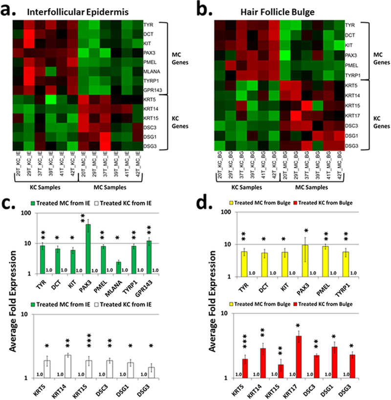
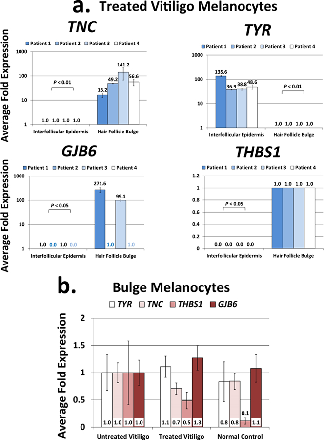
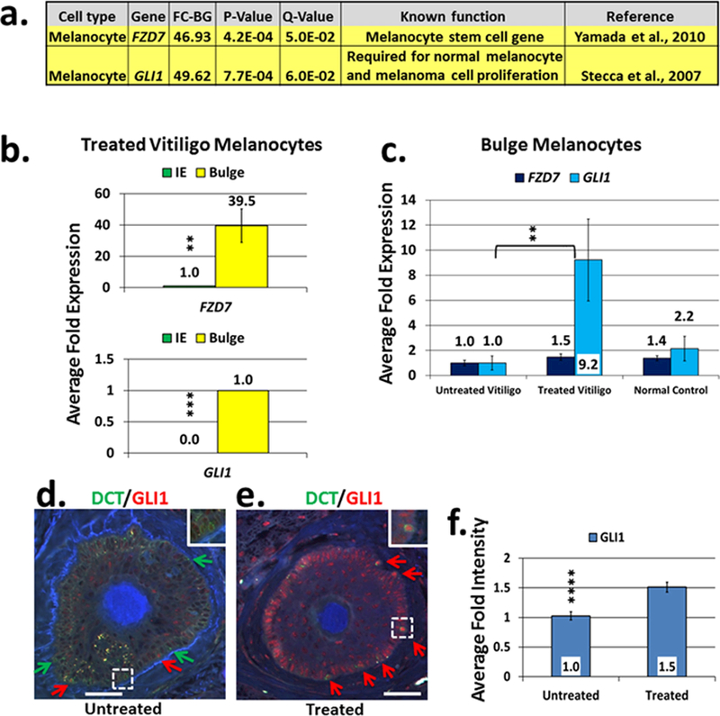
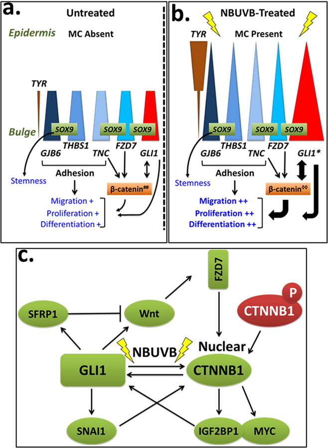
Vitiligo repigmentation is a complex process in which the melanocyte-depleted interfollicular epidermis is repopulated by melanocyte precursors from hair follicle bulge that proliferate, migrate, and differentiate into mature melanocytes on their way to the epidermis. The strongest stimulus for vitiligo repigmentation is narrow-band UVB (NBUVB), but how the hair follicle melanocyte precursors are activated by UV light has not been extensively studied. To better understand this process, we developed an application that combined laser capture microdissection and subsequent whole transcriptome RNA sequencing of hair follicle bulge melanocyte precursors and compared their gene signatures to that of regenerated mature epidermal melanocytes from NBUVB-treated vitiligo skin. Using this strategy, we found up-regulation of TNC, GJB6, and THBS1 in the hair follicle bulge melanocytes and of TYR in the epidermal melanocytes of the NBUVB-treated vitiligo skin. We validated these results by quantitative real-time-PCR using NBUVB-treated vitiligo skin and untreated normal skin. We also identified that GLI1, a candidate stem cell-associated gene, is significantly up-regulated in the melanocytes captured from NBUVB-treated vitiligo bulge compared with untreated vitiligo bulge. These signals are potential key players in the activation of bulge melanocyte precursors during vitiligo repigmentation.


Conflict of interest statement

CONFLICT OF INTEREST

We state no conflict of interest.

Figures

Fig. 1.
Fig. 1.. Heatmaps of melanocyte (MC)-and keratinocyte (KC)-specific genes in the RNA-seq data.
Heatmaps of RNA expression levels of melanocyte-specific and keratinocyte-specific genes in the vitiligo skin of NBUVB-treated IE (Panel a) and of NBUVB-treated bulge (Panel b) (N=6). We found upregulation of melanocyte-specific genes in melanocyte samples (green squares, right-sided upper corner) and keratinocyte-specific genes in keratinocyte samples (green squares, left-sided lower corner). The red squares represent downregulated melanocyte-specific genes. Bar plots represent the gene enrichment in the interfollicular epidermis (Panel c) and bulge (Panel d) +/− standard error of the mean (SEM) (N=6). In both NBUVB-treated IE and NBUVB-treated bulge we found a significant enrichment of melanocyte-specific genes in the melanocyte samples [upper figures of Panel c (green bars) and of Panel d (yellow bars), with expression values set to 1-fold in keratinocytes] and a significant enrichment of keratinocyte-specific genes in the keratinocytes samples [lower figures of Panel c (white bars), and Panel d (red bars), with expression values set to 1-fold in melanocytes]. (*P <0.05; **P <0.01; ***P <0.001). Each bar represents mean and standard deviation (n=3).
Fig. 2.
Fig. 2.. Expression of top genes differentially expressed in the NBUVB-treated vitiligo bulge melanocytes and NBUVB-treated epidermis melanocytes.
Fig.2a. qRT-PCR confirmation of the top differentially expressed genes resulted from the RNA-seq study: TNC, GJB6 and THBS1 were significantly upregulated in the melanocyte samples from the NBUVB-treated bulge, while TYR was significantly upregulated in the melanocyte samples from the NBUVB-treated IE (Paired T-test, Padjusted<0.05) (N=4 NBUVB-treated vitiligo patients). Each bar represents mean and standard deviation (n=3). Expression values were set to 1-fold in the IE for TNC and THBS1, and in the bulge for TYR and GJB6. Fig.2b. qRT-PCR gene expression analysis comparing the expression values of TNC, GJB6, THBS1 and TYR in the bulge melanocyte samples collected from NBUVB-treated vitiligo patients (N=6), untreated vitligo patients (N=4), and normal control subjects (N=6). The expression values of the 4 genes did not vary significantly between the groups tested (one-way ANOVA, Tukey’s Post Hoc Test: adjusted P>0.05 for all pairwise comparisons). Expression values were set to 1-fold in the untreated vitiligo samples. Each bar represents mean and standard deviation (n=3).
Fig. 3.
Fig. 3.. Candidate stem cell associated gene expression analysis in the RNA-Seq data and GLI1 confirmation in the hair follicle bulge of vitiligo skin.
Fig.3a. Candidate stem cell associated gene expression analysis of the RNA-seq data: FZD7 and GLI1 were the top candidate stem cell-associated genes differentially expressed in the melanocyte samples from the bulge versus interfollicular epidermis collected from NBUVB-treated vitiligo patients (N=6). Fig.3b. qRT-PCR confirmation study of FZD7 and GLI1 following the RNA-seq study: both genes were significantly upregulated in the melanocyte samples from the bulge compared to the interfollicular epidermis (IE) of NBUVB-treated vitiligo patients (N=6) (**Padjusted <0.01; ***Padjusted <0.001). RNA analyzed was extracted from melanocyte samples after new rounds of laser capture microdissection. Expression values were set to 1-fold in the IE for FZD7 and in the bulge for GLI1. Fig.3c. Analysis of NBUVB effects on FZD7 and GLI1 in the human HF bulge of vitiligo skin: Expression analysis of FZD7 and GLI1 in melanocyte samples from the bulge of NBUVB-treated vitiligo skin (N=7), untreated vitiligo skin (N=6) and normal control skin (N=6). GLI1 expression showed significant upregulation in the bulge of NBUVB-treated vs untreated vitiligo samples (**Padjusted<0.01), and did not show significant variation in the other comparisons. Expression values were set to 1-fold in the untreated vitiligo samples. Each bar represents mean and standard deviation (n=3). Fig.3d–f. Immunostaining analysis of GLI1 localization in the hair follicle bulge. Figs. 3d and 3e. Double fluorescent immunostaining of FFPE transverse sections using an anti-GLI1 antibody (red) in combination with anti-DCT antibody (green, labels melanocytes) in the untreated vitiligo bulge (3d) and NBUVB-treated vitiligo bulge (3e). White scale bars: 50 μm. Red arrows indicate DCT(+)/GLI1(+) cells; green arrows indicate DCT(+)/GLI1(−) cells. White dotted lines indicate area of higher magnification (inset). GLI1 is also expressed in the bulge keratinocytes which are DCT(−)/GLI1(+) cells. Fig. 3f. Signal intensity analysis for the anti-GLI1 antibody. The average intensity of the anti-GLI1 antibody signal is significantly higher in the bulge melanocytes of the NBUVB-treated vitiligo skin (N=7) as compared with untreated vitiligo skin (N=7) (****P=4.4E-04; +1.5-fold).
Fig. 4.
Fig. 4.. Hypothetical model of the effects of NBUVB on the bulge and epidermis of human vitiligo skin during repigmentation.
Fig 4a, b. Scheme of melanocyte-induced proliferation, migration and differentiation through GLI1 activation. In the human vitiligo skin, bulge-specific genes GJB6, THBS1, TNC, and FZD7 (blue shapes) are expressed in the melanocyte precursors in the bulge at similar levels before (Fig. 4a) and after NBUVB-treatment (Fig. 4b). These genes are proposed to be involved in cellular adhesion and in maintaining stemness in the bulge melanocyte precursors through β-catenin signaling. Epidermal melanocytes are absent in the interfollicular epidermis (IE) of untreated vitiligo skin, illustrated by the decreased height of all triangles, and their absence in the IE in Fig. 4a. In contrast, TYR (brown triangles) is expressed at low levels in both untreated and NBUVB-treated bulge melanocyte precursors, and is expressed at significantly higher levels in the regenerated epidermal melanocytes of NBUVB-treated vitiligo skin, indicating active melanogenesis. GLI1 expression (red triangles) is significantly higher in the NBUVB-treated bulge melanocytes of vitiligo skin compared to untreated vitiligo skin, suggesting a role for GLI1 in the repigmentation process. We identified SOX9 constitutively expressed by the melanocytes precursors in the human bulge of normal and vitiligo skin (Goldstein et al, 2016). Activation of FZD7 (a Wnt receptor) was associated with reduced phosphorylation of β-catenin and its nuclear accumulation (Merle et al., 2005). Gli1 can induce the accumulation of transcriptionally active β-catenin in cell nuclei (Li et al., 2007). β-catenin can enhance the transcriptional activity of GLI1 (Maeda et al., 2006; Varnat et al., 2010; Song et al., 2015). GLI1 induction in the bulge melanocyte and keratinocyte precursors by NBUVB is supposedly influenced by nuclear translocation of β-catenin, and by interaction with SOX9, with a shift in the balance of maintaining stemness to greater differentiation, proliferation, and migration. Fig. 4c. Cross-talk activation between β-catenin and GLI1 under NBUVB. β-catenin can enhance the transcriptional activity of GLI1 (Maeda et al., 2006) likely through regulation of the β-catenin key downstream targets, IGF2BP1 (CRD-BP) and myc (Noubissi et al. 2006). Once activated by β-catenin, IGF2BP1 and myc can activate GLI1 likely by binding to the GLI1 mRNA coding region (Varnat at et al., 2010; Noubissi et al, 2009). GLI1 also regulates β-catenin at the transcription level, through its targets, SNAIL, WNT, and SFRP1 (Song et al., 2015). GLI1 can induce SNAIL expression which then interacts with β-catenin and stimulates its expression activity at transcription level (Song et al., 2015), and at protein level (Li et al., 2007). SFRP1 and Wnt have opposite effects on β-catenin signaling, as a negative feedback, or as a control mechanism after GLI1 induction, to prevent over-activation of WNT/β-catenin pathway. SFRP1 is a melanocyte stem gene that we identified upregulated in the bulge of NBVUB-treated vitiligo skin and normal skin (Goldstein et al., 2016). Experimental evidence used for this figure was generated in either human models, or Gli1 transgenic mice, or on in vitro experiments performed on rat kidney cells, murine LLC-11 hepatocellular carcinoma cells, human stomach, colon, and lung cancer cells. ##, cytoplasmic β-catenin; ◊◊, nuclear β-catenin

Similar articles

Cited by

References

    1. Amoh Y, Aki R, Hamada Y, Niiyama S, Eshima K, Kawahara K, Sato Y, et al. Nestin-positive hair follicle pluripotent stem cells can promote regeneration of impinged peripheral nerve injury. J Dermatol 2012; 39:33–8. - PubMed
    1. Barakat B, Yu L, Lo C, Vu D, De Luca E, Cain JE, et al. Interaction of smoothened with integrin-linked kinase in primary cilia mediates Hedgehog signalling. EMBO Rep 2013;14:837–44. - PMC - PubMed
    1. Bikle DD, Xie Z, Tu CL. Calcium regulation of keratinocyte differentiation. Expert Rev Endocrinol Metab 2012; 7:461–72. - PMC - PubMed
    1. Birlea SA, Costin GE, Roop DR, Norris DA. Trends in Regenerative Medicine: Repigmentation in Vitiligo Through Melanocyte Stem Cell Mobilization. Med Res Rev 2016; [Epub ahead of print]. - PMC - PubMed
    1. Birlea SA, Goldstein NB, Norris DA. Repigmentation through melanocyte regeneration in vitiligo. Dermatol Clin 2017; 35:205–18. - PubMed

Publication types