. 2017 Oct 13;91(21):e00530-17.
doi: 10.1128/JVI.00530-17. Print 2017 Nov 1.

An Epstein-Barr Virus MicroRNA Blocks Interleukin-1 (IL-1) Signaling by Targeting IL-1 Receptor 1

Affiliations

An Epstein-Barr Virus MicroRNA Blocks Interleukin-1 (IL-1) Signaling by Targeting IL-1 Receptor 1

Camille M Skinner et al. J Virol. .

Abstract

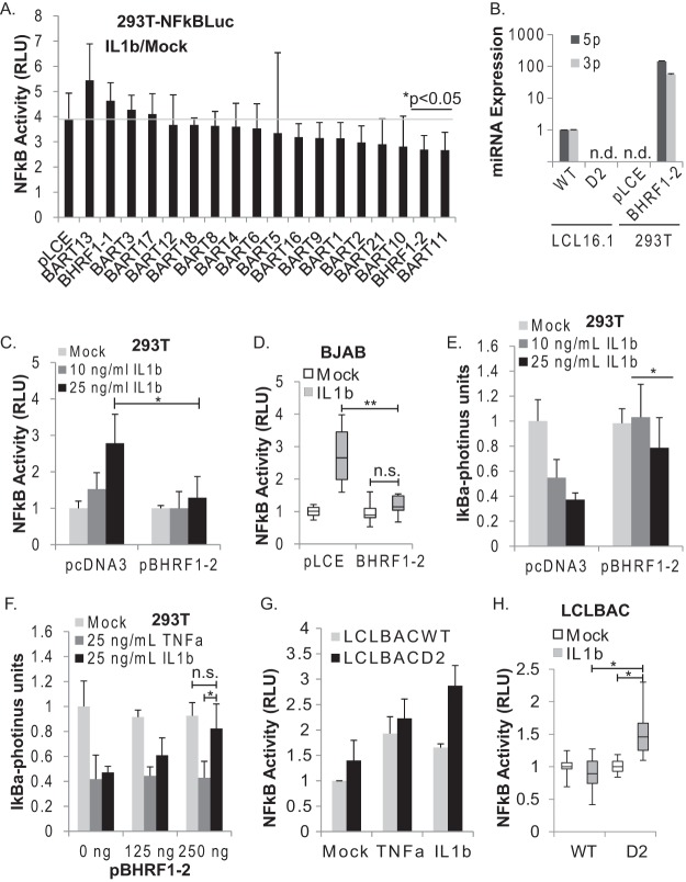
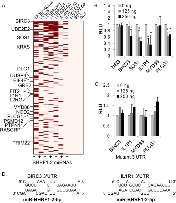
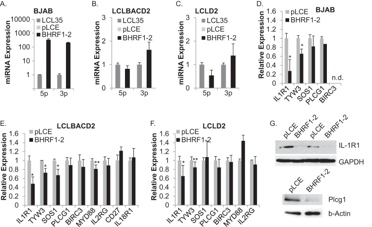
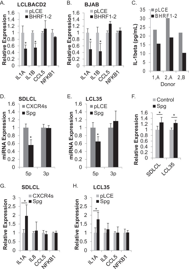
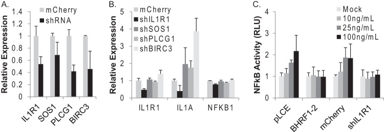
Epstein-Barr virus (EBV) encodes >44 viral microRNAs (miRNAs) that are differentially expressed throughout infection, can be detected in Epstein-Barr virus (EBV)-positive tumors, and manipulate several biological processes, including cell proliferation, apoptosis, and immune responses. Here, we show that EBV BHRF1-2 miRNAs block NF-κB activation following treatment with proinflammatory cytokines, specifically interleukin-1β (IL-1β). Analysis of EBV PAR-CLIP miRNA targetome data sets combined with pathway analysis revealed multiple BHRF1-2 miRNA targets involved in interleukin signaling pathways. By further analyzing changes in cellular gene expression patterns, we identified the IL-1 receptor 1 (IL1R1) as a direct target of miR-BHRF1-2-5p. Targeting the IL1R1 3' untranslated region (UTR) by EBV miR-BHRF1-2-5p was confirmed using 3'-UTR luciferase reporter assays and Western blot assays. Manipulation of EBV BHRF1-2 miRNA activity in latently infected B cells altered steady-state cytokine levels and disrupted IL-1β responsiveness. These studies demonstrate functionally relevant BHRF1-2 miRNA interactions during EBV infection, which is an important step in understanding their roles in pathogenesis.IMPORTANCE IL-1 signaling plays an important role in inflammation and early activation of host innate immune responses following virus infection. Here, we demonstrate that a viral miRNA downregulates the IL-1 receptor 1 during EBV infection, which consequently alters the responsiveness of cells to IL-1 stimuli and changes the cytokine expression levels within infected cell populations. We postulate that this viral miRNA activity not only disrupts IL-1 autocrine and paracrine signaling loops that can alert effector cells to sites of infection but also provides a survival advantage by dampening excessive inflammation that may be detrimental to the infected cell.

Keywords: Epstein-Barr virus; RNA interference; cytokines; herpesviruses; interleukins; microRNA.


Figures

FIG 1
FIG 1
EBV BHRF1-2 miRNAs inhibit IL-1β-mediated NF-κB activity. (A) Functional screen of EBV miRNAs reveals BHRF1-2 and BART11 miRNAs inhibit NF-κB activation by IL-1β. 293T-NF-κBLuc cells were transfected with individual pLCE-based EBV miRNA expression vectors. At 44 h posttransfection, the cells were stimulated with 25 ng/ml IL-1β for 4 h and then lysed and assayed for dual luciferase activity. Reported are the averages of two independent experiments performed in triplicates. *, P < 0.05 (Student t test). (B) Expression of EBV miR-BHRF1-2-5p and miR-BHRF1-2-3p in 293T cells. 293T cells were transfected with pLCE or pLCE-BHRF1-2, and the total RNA was harvested at 48 h posttransfection. miRNA expression was assayed by stem-loop qRT-PCR (normalized to miR-16) and is reported relative to levels in EBV B9-8 wild-type (WT) or BHRF1-2 miRNA KO (D2) LCLs. (C) 293T-NF-κBLuc cells were transfected with 250 ng of pcDNA3 or pBHRF1-2 as indicated. At 44 h posttransfection, the cells were stimulated with increasing amounts of IL-1β for 4 h. Reported are averages and the SD of two independent experiments performed in quadruplicate. *, P < 0.05 (Student t test). (D) BJAB-NF-κBLuc cells were transduced with pLCE or pLCE-BHRF1-2. At 5 or 7 days postransduction, the cells were plated in 96-well black-well plates at 3 × 10⁴ cells/well, treated with 25 ng/ml IL-1β for 18 h, and then lysed and assayed for dual luciferase activity. Box-and-whisker plots represent averages of two independent experiments with five replicates (n = 10). n.s., not significant. **, P < 0.01 (Student t test). (E) 293T-IκBαLuc cells were transfected with 250 ng of pcDNA3 or pBHRF1-2 as indicated. At 44 h posttransfection, the cells were stimulated with increasing amounts of IL-1β for 4 h and then lysed and assayed for luciferase activity. Reported are averages and the SD of two independent experiments performed in triplicates. *, P < 0.01 (Student t test) compared to empty-vector control cells. (F) 293T-IκBαLuc cells were transfected with increasing amounts of pBHRF1-2 as indicated. At 44 h posttransfection, the cells were stimulated with TNF-α or IL-1β for 4 h and then lysed and assayed for luciferase activity. Reported are averages and the SD of two independent experiments performed in triplicates. n.s., not significant. *, P < 0.01 (Student t test) compared to empty-vector control cells. (G) EBV B95-8 wild-type (LCLBACWT) or BHRF1-2 miRNA KO (LCLBACD2) LCLs were cotransduced with a NF-κB reporter (pL-NF-κB-Fluc) and a Renilla luciferase internal control (pL-RSV-Rluc). At 72 h postransduction, the cells were plated in fresh medium and stimulated with 100 ng/ml TNF-α or IL-1β for 3 h. Reported are averages of two experiments. (H) LCLBACWT or BHRF1-2 miRNA KO (LCLBACD2) cells were electroporated with 2.7 μg of pNF-kB-Fluc and 300 ng of pL-RSV-RLuc. At 24 h posttransfection, the cells were plated in 96-well plates at 1.2 × 10⁵ cells per well and then treated for 18 h with 25 ng/ml IL-1β. Box-and-whisker plots represent averages of five independent experiments performed in quadruplicates (n = 20). *, P < 0.01 (Student t test). RLU, relative light units.
FIG 2
FIG 2
Targets of the EBV BHRF1-2 miRNAs are involved in interleukin signaling. (A) Analysis of BHRF1-2 miRNA interaction sites captured in PAR-CLIP data sets. Data sets are from EBV+ LCLs, rhesus LCLs, and KSHV+ PELs. BHRF1-2 miRNA expression is indicated for each cell line. A total of 225 PAR-CLIP'ed, cellular 3′-UTR interaction sites were identified that exhibited ≥7mer canonical seed matches to either miR-BHRF1-2-5p or miR-BHRF1-2-3p. Red boxes indicate presence of the interaction site within each PAR-CLIP sample. The 18 genes highlighted on the left were identified by pathway analysis through the reactome database and are involved in signaling by interleukins (R-HSA-449147). (B and C) 3′-UTR reporter assays confirm BHRF1-2 miRNA target interactions. Select 3′ UTRs were cloned downstream of Renilla in the psiCheck2 dual reporter luciferase vector. For panel C, PAR-CLIP identified seed match sites for the BHRF1-2 miRNAs were mutated using site-directed mutagenesis. 293T cells were cotransfected with 20 ng of 3′-UTR reporter and pcDNA3 or pBHRF1-2 as indicated. Lysates were harvested 48 to 72 h posttransfection and analyzed for dual luciferase activity. Reported are the averages of at least three independent experiments performed in triplicates. *, P < 0.01 (Student t test).
FIG 3
FIG 3
IL1R1 is upregulated in the absence of the BHRF1-2 miRNAs. (A) RNA-Seq analysis of LCLBACWT (WT) and LCLBACD2 (D2) reveals upregulated cellular transcripts in LCLs lacking the BHRF1-2 miRNAs. TPM, transcripts per million mapped. (B) Eight upregulated genes in LCLBACD2 (compared to WT) overlap PAR-CLIP-identified BHRF1-2 miRNA targets. Shown are log2FC values relative to LCLBACWT. (C) qRT-PCR analysis of IL1R1, TYW3, IL2RG, CD27, and IL18R1 transcripts in donor-matched LCL pairs generated with wild-type EBV B95-8 (WT) or BHRF1-2 miRNA KO (D2) viruses. Values are normalized to GAPDH and are reported relative to WT LCLs for each pair. Reported are average expression values with the SD from at least three independent RT experiments; qPCR was performed in duplicate. For IL1R1, n = 5 experiments for donors 1 and 2 and n = 4 experiments for donor 3. n.s., not significant. *, P < 0.01; **, P < 0.05 (Student t test). (D) Western blot analysis of IL-1R1 and β-actin in LCLBACWT and LCLBACD2 (donor 1). Shown are blots from two independent experiments.
FIG 4
FIG 4
Ectopic BHRF1-2 miRNA expression reduces IL1R1 levels. (A) qRT-PCR analysis of BHRF1-2 miRNA expression levels in BJAB cells stably transduced with pLCE (control vector) or pLCE-BHRF1-2 (n = 2 in duplicate). (B and C) qRT-PCR analysis of BHRF1-2 miRNA expression levels in BHRF1-2 miRNA KO LCLs (LCLBACD2 or LCLD2) stably transduced with pLCE (control vector) or pLCE-BHRF1-2 (n = 3 in duplicate for LCLBACD2; n = 4 in duplicate for LCLD2). For panels A to C, miR-BHRF1-2-5p and miR-BHRF1-2-3p expression values are normalized to snoRNA U6 or miR-16 and reported relative to levels in EBV B95-8 LCL35. (D to F) qRT-PCR analysis of IL1R1 transcript levels and other cellular genes in BJAB cells and BHRF1-2 KO LCLs (LCLBACD2 and LCL-D2) ectopically expressing BHRF1-2 miRNAs. Values are normalized to GAPDH and reported relative to control cells (pLCE). Average expression values and standard deviations were calculated from three to five independent experiments. *, P < 0.005; **, P < 0.05 (Student t test). (G) Western blot analysis of IL1R1 or PLCγ1 (Plcg1) in transduced BJAB cells. GAPDH or β-actin levels are shown as loading controls.
FIG 5
FIG 5
BHRF1-2 miRNA activity regulates steady-state IL-1 expression. (A and B) Ectopic BHRF1-2 miRNA expression dampens cytokine expression. Total RNA was harvested from BHRF1-2 KO LCLs (LCLBACD2) (A) or EBV-negative BJAB cells (B) transduced with pLCE or pLCE-BHRF1-2. NF-κB-responsive cytokine genes were measured by TaqMan qRT-PCR. Values are normalized to GAPDH as an internal control and reported relative to cells transduced with pLCE control vector. Shown are the averages of four independent experiments for LCLBACD2 and three independent experiments for BJAB, performed in duplicate. *, P < 0.005 (Student t test). (C) Secreted IL-1β is reduced in the presence of BHRF1-2 miRNAs. BHRF1-2 KO LCLs transduced with pLCE or pLCE-BHRF1-2 were plated at 106 cells/ml, and cell culture supernatants were harvested after 24 h. IL-1β levels were measured by ELISA. Donor 1, LCLBACD2; donor 2, LCLD2. (D and E) Sponge inhibition of miR-BHRF1-2-5p increases cytokine expression. BHRF1-2 miRNA expression was analyzed by qRT-PCR in two EBV B95-8 wild-type LCLs (SDLCL and LCL35) transduced with the miR-BHRF1-2-5p sponge (Spg). 5p and 3p indicate miR-BHRF1-2-5p and miR-BHRF1-2-3p. Values were normalized to U6 and are reported relative to miRNA expression levels in cells transduced with control vector (CXCR4s or pLCE). (F to H) qRT-PCR analysis of IL1R1 and cytokine transcripts in miR-BHRF1-2-5p sponged LCLs. Values were normalized to GAPDH and are reported relative to cells transduced with control vector (pLCE-CXCR4s or pLCE). For panels D to H, reported are the averages and the SD of at least three independent experiments (n = 4 for IL1A) performed in duplicate. *, P < 0.005 (Student t test).
FIG 6
FIG 6
Inhibition of IL1R1 phenocopies miR-BHRF1-2-5p activity. (A) 293T cells were transduced with pLmCherry-shRNAs, and total RNA was harvested at 72 h. Knockdown of individual target genes was assayed by qRT-PCR analysis. (B) Steady-state IL1A transcripts, assayed by qRT-PCR, are reduced by the IL1R1 shRNA in 293T cells. For panels A and B, the expression levels were normalized to GAPDH and are reported relative to control (mCherry) cells. Reported is the average and the SD of two independent experiments performed in duplicate. (C) 293T cells were transfected with 20 ng of NF-κB luciferase reporter, 20 ng of Renilla expression vector, and either 250 ng of control vector (pLCE or pLmCherry) or miRNA or shIL1R1 vector as indicated. At 44 h posttransfection, the cells were stimulated with increasing amounts of IL-1β for 4 h. Lysates were harvested and assayed for dual luciferase activity. Reported is the average and the SD of two independent experiments performed in triplicate.
FIG 7
FIG 7
miR-BHRF1-2-5p regulates IL-1β responsiveness during latent EBV infection. (A and B) SDLCL or LCL35 cells stably transduced with either control vector (pLCE-CXCR4s or pLCE) or miR-BHRF1-2-5p sponge inhibitor (BHRF1-2-5ps) were plated in 12-well plates and treated for 18 h with IL-1β. Total RNA was harvested and assayed using qRT-PCR for expression of cytokine and chemokine transcripts. Shown are the averages of three independent experiments; PCR was performed in duplicate. *, P < 0.05; **, P < 0.005 (Student t test). (C) Schematic model depicting EBV miR-BHRF1-2-5p regulation of IL-1 signaling that occurs primarily through inhibition of the IL-1 receptor. By silencing IL1R1, the viral miRNA disrupts a positive loop that stimulates inflammatory cytokine expression in both infected and neighboring cells through autocrine and paracrine signals.

Similar articles

Cited by

References

    1. Babcock GJ, Decker LL, Volk M, Thorley-Lawson DA. 1998. EBV persistence in memory B cells in vivo. Immunity 9:395–404. doi:10.1016/S1074-7613(00)80622-6. - DOI - PubMed
    1. Hislop AD, Taylor GS, Sauce D, Rickinson AB. 2007. Cellular responses to viral infection in humans: lessons from Epstein-Barr virus. Annu Rev Immunol 25:587–617. doi:10.1146/annurev.immunol.25.022106.141553. - DOI - PubMed
    1. Ressing ME, van Gent M, Gram AM, Hooykaas MJ, Piersma SJ, Wiertz EJ. 2015. Immune evasion by Epstein-Barr virus. Curr Top Microbiol Immunol 391:355–381. doi:10.1007/978-3-319-22834-1_12. - DOI - PubMed
    1. Vockerodt M, Yap LF, Shannon-Lowe C, Curley H, Wei W, Vrzalikova K, Murray PG. 2015. The Epstein-Barr virus and the pathogenesis of lymphoma. J Pathol 235:312–322. doi:10.1002/path.4459. - DOI - PubMed
    1. Tsao SW, Tsang CM, To KF, Lo KW. 2015. The role of Epstein-Barr virus in epithelial malignancies. J Pathol 235:323–333. doi:10.1002/path.4448. - DOI - PMC - PubMed

Publication types

MeSH terms