

Lipid Droplet Biogenesis
- PMID: 28793795
- PMCID: PMC6986389
- DOI: 10.1146/annurev-cellbio-100616-060608
Lipid Droplet Biogenesis
Abstract
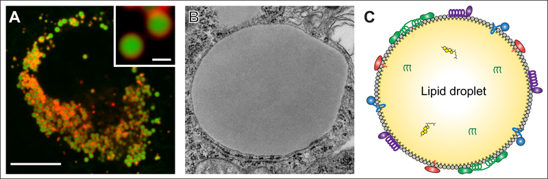
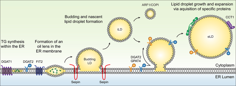
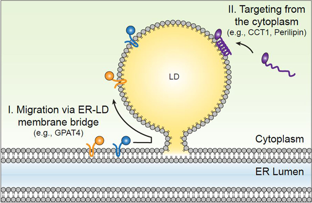
Lipid droplets (LDs) are ubiquitous organelles that store neutral lipids for energy or membrane synthesis and act as hubs for metabolic processes. Cells generate LDs de novo, converting cells to emulsions with LDs constituting the dispersed oil phase in the aqueous cytoplasm. Here we review our current view of LD biogenesis. We present a model of LD formation from the ER in distinct steps and highlight the biology of proteins that govern this biophysical process. Areas of incomplete knowledge are identified, as are connections with physiology and diseases linked to alterations in LD biology.
Keywords: biogenesis; endoplasmic reticulum; lipid droplets; neutral lipids; seipin; sterol esters; triacylglycerol; triglycerides.
Figures
Similar articles
-
Membrane shaping proteins, lipids, and cytoskeleton: Recipe for nascent lipid droplet formation.Bioessays. 2022 Sep;44(9):e2200038. doi: 10.1002/bies.202200038. Epub 2022 Jul 13. Bioessays. 2022. PMID: 35832014
-
Structure and function of lipid droplet assembly complexes.Curr Opin Struct Biol. 2023 Jun;80:102606. doi: 10.1016/j.sbi.2023.102606. Epub 2023 May 5. Curr Opin Struct Biol. 2023. PMID: 37150040 Free PMC article. Review.
-
Lipid droplet biogenesis: A mystery "unmixing"?Semin Cell Dev Biol. 2020 Dec;108:14-23. doi: 10.1016/j.semcdb.2020.03.001. Epub 2020 Mar 17. Semin Cell Dev Biol. 2020. PMID: 32192830 Review.
-
Spatial compartmentalization of lipid droplet biogenesis.Biochim Biophys Acta Mol Cell Biol Lipids. 2020 Jan;1865(1):158499. doi: 10.1016/j.bbalip.2019.07.008. Epub 2019 Jul 25. Biochim Biophys Acta Mol Cell Biol Lipids. 2020. PMID: 31352131 Free PMC article. Review.
-
Mechanisms of lipid droplet biogenesis.Biochem J. 2019 Jul 9;476(13):1929-1942. doi: 10.1042/BCJ20180021. Biochem J. 2019. PMID: 31289128 Review.
Cited by
-
The Plasma Membrane H+ ATPase CsPMA2 Regulates Lipid Droplet Formation, Appressorial Development and Virulence in Colletotrichum siamense.Int J Mol Sci. 2023 Dec 11;24(24):17337. doi: 10.3390/ijms242417337. Int J Mol Sci. 2023. PMID: 38139168 Free PMC article.
-
Triglyceride lipolysis triggers liquid crystalline phases in lipid droplets and alters the LD proteome.J Cell Biol. 2022 Nov 7;221(11):e202205053. doi: 10.1083/jcb.202205053. Epub 2022 Sep 16. J Cell Biol. 2022. PMID: 36112368 Free PMC article.
-
Nazo, the Drosophila homolog of the NBIA-mutated protein-c19orf12, is required for triglyceride homeostasis.PLoS Genet. 2024 Feb 9;20(2):e1011137. doi: 10.1371/journal.pgen.1011137. eCollection 2024 Feb. PLoS Genet. 2024. PMID: 38335241 Free PMC article.
-
TMEM41B and VMP1 are scramblases and regulate the distribution of cholesterol and phosphatidylserine.J Cell Biol. 2021 Jun 7;220(6):e202103105. doi: 10.1083/jcb.202103105. J Cell Biol. 2021. PMID: 33929485 Free PMC article.
-
MOSPD2 is an endoplasmic reticulum-lipid droplet tether functioning in LD homeostasis.J Cell Biol. 2022 Jun 6;221(6):e202110044. doi: 10.1083/jcb.202110044. Epub 2022 Apr 7. J Cell Biol. 2022. PMID: 35389430 Free PMC article.
References
-
- Altmann R 1894. Die Elementarorganismen und ihre Beziehungen zu den Zellen. Leipzig: Viet & Co; 271 pp.
-
- Anderson RA, Joyce C, Davis M, Reagan JW, Clark M, et al. 1998. Identification of a form of acyl- CoA:cholesterol acyltransferase specific to liver and intestine in nonhuman primates. J. Biol. Chem. 273:26747–54 - PubMed
-
- Anstee QM, Day CP. 2013. The genetics of NAFLD. Nat. Rev. Gastroenterol. Hepatol. 10:645–55 - PubMed
Publication types
MeSH terms
Substances
Grants and funding
LinkOut - more resources
Full Text Sources
Other Literature Sources
Research Materials
