

Protocol for Directed Differentiation of Human Induced Pluripotent Stem Cells (iPSCs) to a Hepatic Lineage
- PMID: 28752455
- PMCID: PMC7252494
- DOI: 10.1007/978-1-4939-7163-3_15
Protocol for Directed Differentiation of Human Induced Pluripotent Stem Cells (iPSCs) to a Hepatic Lineage
Abstract
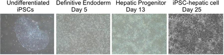
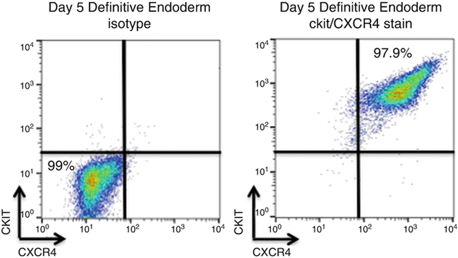
Directed differentiation is a powerful cell culture technique where developmental pathways are applied to a pluripotent progenitor in order to generate specific terminally differentiated cell populations. Here, we describe a serum-free protocol using growth factors in defined concentrations to derive iPSC-hepatic cells starting from both feeder and feeder-free conditions. The generated iPSC-hepatic cells are developmentally similar to fetal stage hepatocytes, and when generated from patients with genetic mutations such as alpha-1 antitrypsin deficiency recapitulate pathologic changes associated with clinical disease, such as protein misfolding, intracellular retention of misfolded proteins, and elevated levels of ER stress.
Keywords: Definitive endoderm; Directed differentiation; Induced pluripotent stem cell (iPSC); iPSC-hepatic cell.
Figures

Similar articles
-
Human Induced Pluripotent Stem Cell-Derived Definitive Endoderm Bulk Culture and Hepatic Differentiation.Methods Mol Biol. 2019;1994:41-53. doi: 10.1007/978-1-4939-9477-9_4. Methods Mol Biol. 2019. PMID: 31124103
-
Generation of functional human hepatic endoderm from human induced pluripotent stem cells.Hepatology. 2010 Jan;51(1):329-35. doi: 10.1002/hep.23335. Hepatology. 2010. PMID: 19877180 Free PMC article.
-
An Efficient Method for the Differentiation of Human iPSC-Derived Endoderm toward Enterocytes and Hepatocytes.Cells. 2021 Apr 6;10(4):812. doi: 10.3390/cells10040812. Cells. 2021. PMID: 33917333 Free PMC article.
-
Continuous human iPSC-macrophage mass production by suspension culture in stirred tank bioreactors.Nat Protoc. 2022 Feb;17(2):513-539. doi: 10.1038/s41596-021-00654-7. Epub 2022 Jan 17. Nat Protoc. 2022. PMID: 35039668 Free PMC article. Review.
-
Applications of patient-specific induced pluripotent stem cells; focused on disease modeling, drug screening and therapeutic potentials for liver disease.Int J Biol Sci. 2010 Dec 14;6(7):796-805. doi: 10.7150/ijbs.6.796. Int J Biol Sci. 2010. PMID: 21179587 Free PMC article. Review.
Cited by
-
A Highly Phenotyped Open Access Repository of Alpha-1 Antitrypsin Deficiency Pluripotent Stem Cells.Stem Cell Reports. 2020 Jul 14;15(1):242-255. doi: 10.1016/j.stemcr.2020.06.006. Epub 2020 Jul 2. Stem Cell Reports. 2020. PMID: 32619491 Free PMC article.
-
Omics-Based Platform for Studying Chemical Toxicity Using Stem Cells.J Proteome Res. 2018 Jan 5;17(1):579-589. doi: 10.1021/acs.jproteome.7b00693. Epub 2017 Dec 20. J Proteome Res. 2018. PMID: 29261316 Free PMC article.
-
Induction and Maturation of Hepatocyte-Like Cells In Vitro: Focus on Technological Advances and Challenges.Front Cell Dev Biol. 2021 Nov 26;9:765980. doi: 10.3389/fcell.2021.765980. eCollection 2021. Front Cell Dev Biol. 2021. PMID: 34901010 Free PMC article. Review.
-
iPSC-derived hepatocytes generated from NASH donors provide a valuable platform for disease modeling and drug discovery.Biol Open. 2020 Dec 16;9(12):bio055087. doi: 10.1242/bio.055087. Biol Open. 2020. PMID: 33268331 Free PMC article.
-
Recombinant Lloviu virus as a tool to study viral replication and host responses.PLoS Pathog. 2022 Feb 4;18(2):e1010268. doi: 10.1371/journal.ppat.1010268. eCollection 2022 Feb. PLoS Pathog. 2022. PMID: 35120176 Free PMC article.
References
-
- Evans MJ, Kaufman MH (1981) Establishment in culture of pluripotent cells from mouse embryos. Nature 292:154–156 - PubMed
-
- Thomson JA, Itskovitz-Eldor J, Shapiro SS et al. (1998) Embryonic stem cell lines derived from human blastocysts. Science:1145–1147 - PubMed
-
- Takahashi K, Yamanaka S (2006) Induction of pluripotent stem cells from mouse embryonic and adult fibroblast cultures by defined factors. Cell 126:663–676 - PubMed
-
- Takahashi K, Tanabe K, Ohnuki M et al. (2007) Induction of pluripotent stem cells from adult human fibroblasts by defined factors. Cell 131:861–872 - PubMed
Publication types
MeSH terms
Grants and funding
LinkOut - more resources
Full Text Sources
Other Literature Sources
