

Antibodies from a Human Survivor Define Sites of Vulnerability for Broad Protection against Ebolaviruses
- PMID: 28525755
- PMCID: PMC5808922
- DOI: 10.1016/j.cell.2017.04.037
Antibodies from a Human Survivor Define Sites of Vulnerability for Broad Protection against Ebolaviruses
Abstract
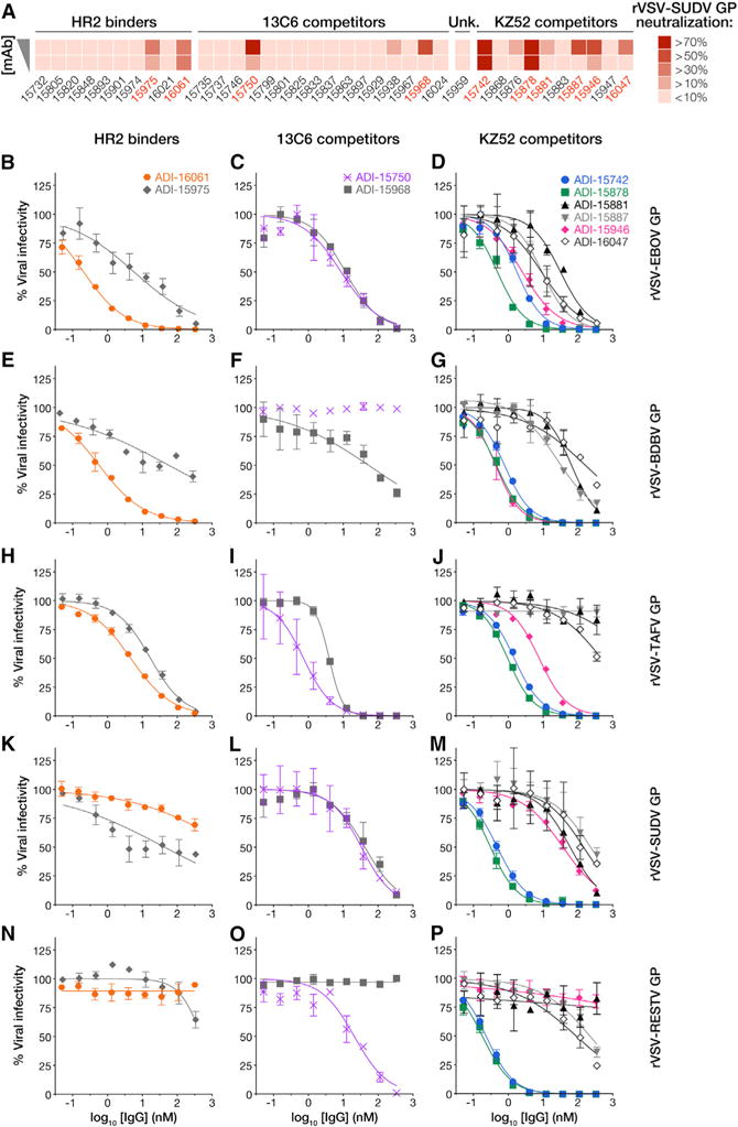
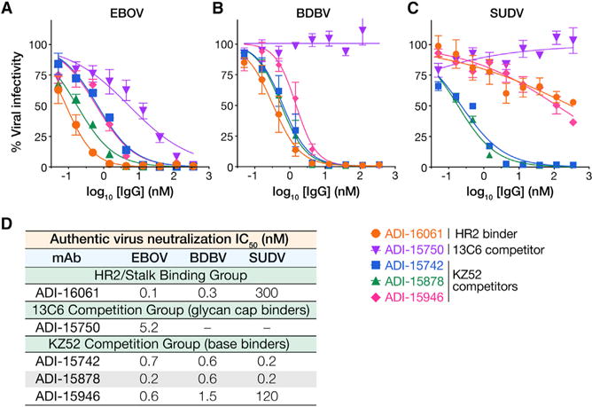
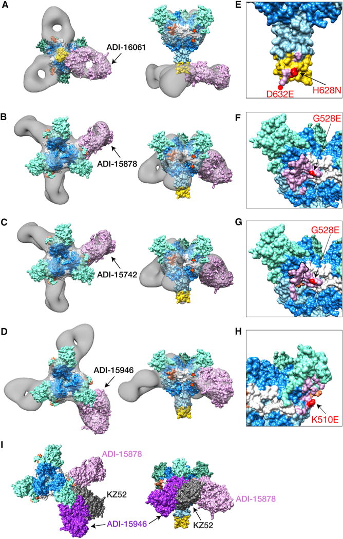
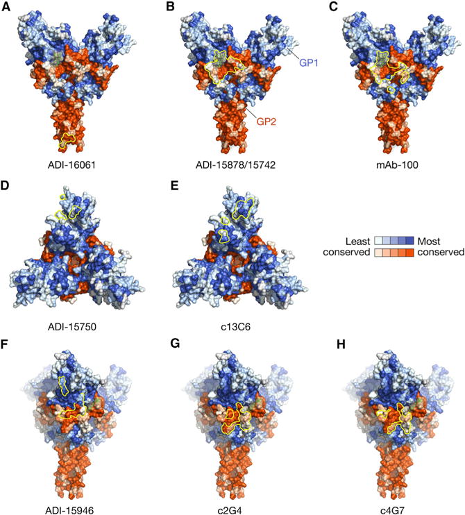
Experimental monoclonal antibody (mAb) therapies have shown promise for treatment of lethal Ebola virus (EBOV) infections, but their species-specific recognition of the viral glycoprotein (GP) has limited their use against other divergent ebolaviruses associated with human disease. Here, we mined the human immune response to natural EBOV infection and identified mAbs with exceptionally potent pan-ebolavirus neutralizing activity and protective efficacy against three virulent ebolaviruses. These mAbs recognize an inter-protomer epitope in the GP fusion loop, a critical and conserved element of the viral membrane fusion machinery, and neutralize viral entry by targeting a proteolytically primed, fusion-competent GP intermediate (GPCL) generated in host cell endosomes. Only a few somatic hypermutations are required for broad antiviral activity, and germline-approximating variants display enhanced GPCL recognition, suggesting that such antibodies could be elicited more efficiently with suitably optimized GP immunogens. Our findings inform the development of both broadly effective immunotherapeutics and vaccines against filoviruses.
Keywords: 15878; EBOV; Ebola virus; ebolavirus; ferret; human broadly neutralizing antibodies; infection; mouse; neutralization; protection.
Copyright © 2017 Elsevier Inc. All rights reserved.
Figures

Comment in
-
Ebolavirus's Foibles.Cell. 2017 May 18;169(5):773-775. doi: 10.1016/j.cell.2017.05.008. Cell. 2017. PMID: 28525748
-
Neutralizing the Threat: Pan-Ebolavirus Antibodies Close the Loop.Trends Mol Med. 2017 Aug;23(8):669-671. doi: 10.1016/j.molmed.2017.06.008. Epub 2017 Jul 8. Trends Mol Med. 2017. PMID: 28697885 Free PMC article.
Similar articles
-
Immunization-Elicited Broadly Protective Antibody Reveals Ebolavirus Fusion Loop as a Site of Vulnerability.Cell. 2017 May 18;169(5):891-904.e15. doi: 10.1016/j.cell.2017.04.038. Cell. 2017. PMID: 28525756 Free PMC article.
-
Multifunctional Pan-ebolavirus Antibody Recognizes a Site of Broad Vulnerability on the Ebolavirus Glycoprotein.Immunity. 2018 Aug 21;49(2):363-374.e10. doi: 10.1016/j.immuni.2018.06.018. Epub 2018 Jul 17. Immunity. 2018. PMID: 30029854 Free PMC article.
-
Antibody-Mediated Protective Mechanisms Induced by a Trivalent Parainfluenza Virus-Vectored Ebolavirus Vaccine.J Virol. 2019 Feb 5;93(4):e01845-18. doi: 10.1128/JVI.01845-18. Print 2019 Feb 15. J Virol. 2019. PMID: 30518655 Free PMC article.
-
Achieving cross-reactivity with pan-ebolavirus antibodies.Curr Opin Virol. 2019 Feb;34:140-148. doi: 10.1016/j.coviro.2019.01.003. Epub 2019 Mar 15. Curr Opin Virol. 2019. PMID: 30884329 Free PMC article. Review.
-
Structural Biology Illuminates Molecular Determinants of Broad Ebolavirus Neutralization by Human Antibodies for Pan-Ebolavirus Therapeutic Development.Front Immunol. 2022 Jan 10;12:808047. doi: 10.3389/fimmu.2021.808047. eCollection 2021. Front Immunol. 2022. PMID: 35082794 Free PMC article. Review.
Cited by
-
A conserved immunogenic and vulnerable site on the coronavirus spike protein delineated by cross-reactive monoclonal antibodies.Nat Commun. 2021 Mar 17;12(1):1715. doi: 10.1038/s41467-021-21968-w. Nat Commun. 2021. PMID: 33731724 Free PMC article.
-
Functional Profiling of Antibody Immune Repertoires in Convalescent Zika Virus Disease Patients.Front Immunol. 2021 Feb 24;12:615102. doi: 10.3389/fimmu.2021.615102. eCollection 2021. Front Immunol. 2021. PMID: 33732238 Free PMC article.
-
Potent germline-like monoclonal antibodies: rapid identification of promising candidates for antibody-based antiviral therapy.Antib Ther. 2021 May 17;4(2):89-98. doi: 10.1093/abt/tbab008. eCollection 2021 Apr. Antib Ther. 2021. PMID: 34104872 Free PMC article.
-
Relationship between Anti-Spike Protein Antibody Titers and SARS-CoV-2 In Vitro Virus Neutralization in Convalescent Plasma.bioRxiv [Preprint]. 2020 Jun 9:2020.06.08.138990. doi: 10.1101/2020.06.08.138990. bioRxiv. 2020. Update in: J Clin Invest. 2020 Dec 1;130(12):6728-6738. doi: 10.1172/JCI141206. PMID: 32577662 Free PMC article. Updated. Preprint.
-
Structural Basis of Pan-Ebolavirus Neutralization by an Antibody Targeting the Glycoprotein Fusion Loop.Cell Rep. 2018 Sep 4;24(10):2723-2732.e4. doi: 10.1016/j.celrep.2018.08.009. Cell Rep. 2018. PMID: 30184505 Free PMC article.
References
-
- Brannan JM, Froude JW, Prugar LI, Bakken RR, Zak SE, Daye SP, Wilhelmsen CE, Dye JM. Interferon α/β receptor-deficient mice as a model for Ebola virus disease. J Infect Dis. 2015;212(Suppl 2):S282–S294. - PubMed
-
- Bray M, Davis K, Geisbert T, Schmaljohn C, Huggins J. A mouse model for evaluation of prophylaxis and therapy of Ebola hemorrhagic fever. J Infect Dis. 1998;178:651–661. - PubMed
MeSH terms
Substances
Grants and funding
LinkOut - more resources
Full Text Sources
Other Literature Sources
Medical
Molecular Biology Databases
