. 2017 Apr 20;169(3):407-421.e16.
doi: 10.1016/j.cell.2017.03.047.

Structural and Functional Analysis of a β2-Adrenergic Receptor Complex with GRK5

Affiliations

Structural and Functional Analysis of a β2-Adrenergic Receptor Complex with GRK5

Konstantin E Komolov et al. Cell. .

Abstract

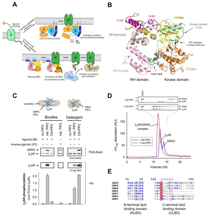
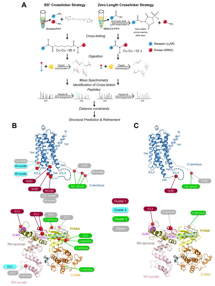
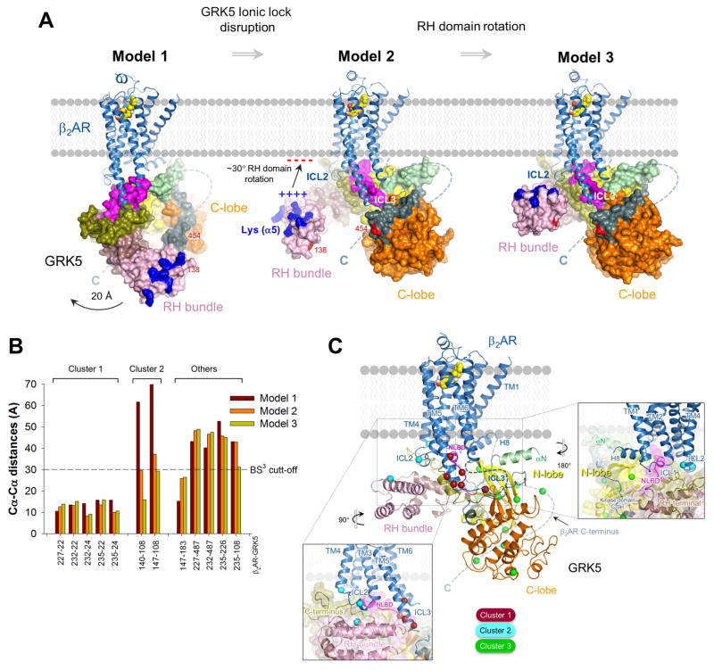
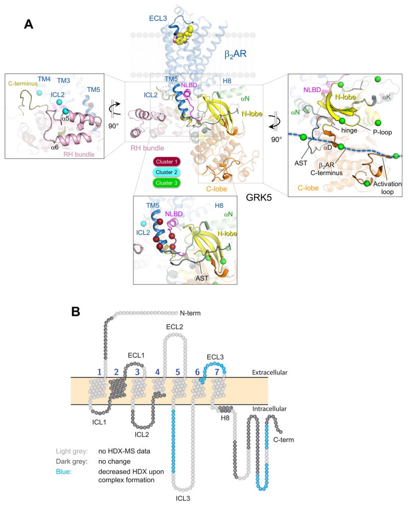
The phosphorylation of agonist-occupied G-protein-coupled receptors (GPCRs) by GPCR kinases (GRKs) functions to turn off G-protein signaling and turn on arrestin-mediated signaling. While a structural understanding of GPCR/G-protein and GPCR/arrestin complexes has emerged in recent years, the molecular architecture of a GPCR/GRK complex remains poorly defined. We used a comprehensive integrated approach of cross-linking, hydrogen-deuterium exchange mass spectrometry (MS), electron microscopy, mutagenesis, molecular dynamics simulations, and computational docking to analyze GRK5 interaction with the β2-adrenergic receptor (β2AR). These studies revealed a dynamic mechanism of complex formation that involves large conformational changes in the GRK5 RH/catalytic domain interface upon receptor binding. These changes facilitate contacts between intracellular loops 2 and 3 and the C terminus of the β2AR with the GRK5 RH bundle subdomain, membrane-binding surface, and kinase catalytic cleft, respectively. These studies significantly contribute to our understanding of the mechanism by which GRKs regulate the function of activated GPCRs. PAPERCLIP.

Keywords: G-protein-coupled receptor; G-protein-coupled receptor kinases; cross-linking; mass spectrometry; molecular dynamics; phosphorylation; β(2)-adrenergic receptor.


Figures

Figure 1
Figure 1. Receptor agonist and anionic lipids are required for functional interaction between GRK5 and the β2AR in bicelles and detergent solution
(A) Schematic of G protein, GRK and arrestin interaction with GPCRs. (B) Crystal structure of GRK5 bound to AMP-PNP (PDB ID 4TND). The RH bundle and terminal subdomains, catalytic C-lobe and N-lobe subdomains, N-terminal lipid binding domain (NLBD) and an ionic lock between the RH and kinase domains are highlighted. Disordered αN-helix (green) and C-terminal lipid binding domain (CLBD) (magenta) were computationally modeled. (C) Direct binding (pull-down) and phosphorylation assays showing GRK5 coupling to β2AR reconstituted into bicelles or solubilized in MNG. Values represent mean ± SEM from three independent experiments. (D) Analytical gel filtration of GRK5 and MNG-solubilized BI-bound β2AR. (E) Sequence alignment of NLBD and CLBD of human GRKs (residues 22–29 and 546–565 in GRK5, respectively). Identical residues are boxed in red while residues showing similarity are in red and grouped in a blue frame. See also Figure S1.
Figure 2
Figure 2. Validation of GRK5/β2AR complex formation
(A) The crystal structure of nanobody Nb6B9 complex with β2AR (PDB ID 4LDE). (B) Nb6B9 inhibits GRK5-mediated phosphorylation of agonist-bound β2AR (BI-β2AR). Values represent mean ± SEM from three independent experiments. (C) Ribbon representation of the active (PDB 3SN6) and inactive (PDB 4GBR) structures of the β2AR with TM6 and the position of Cys265 (yellow) highlighted. The cytoplasmic end of TM6 moves outward following β2AR activation, increasing solvent exposure of a bimane probe covalently bound to Cys265. (D) Fluorescence emission spectra for monobromobimane-labeled β2AR in rHDL in the presence or absence of agonist (ISO), GRK5 and Gs. See also Figure S1.
Figure 3
Figure 3. Electrostatic contact between the RH and kinase domains regulates GRK5 plasticity and catalytic activity
(A) The network of electrostatic interactions between the kinase and RH domains of GRK5 (“ionic lock”). (B) Effect of ionic lock mutations on Michaelis-Menten kinetics for ATP. The data represent the mean ± SEM from three independent experiments and were fit using GraphPad Prism. (C) Interdomain distances in MD simulations of GRK5 wild type (blue) and the ionic lock mutant (red) in which the ionic lock was disrupted by alanine mutations. Distances are measured between alpha carbons on residues 92 and 455 (see panel D) and smoothed using an 8 ns moving average. Three of the six simulations performed under each condition are shown. (D) A snapshot of a representative elongated conformation of GRK5 (colors), as observed in MD simulations of the ionic lock mutant, superimposed on the crystallographic conformation from which the simulations started (gray). (E) Representative 2D class averages of GRK5 ionic lock mutant and a GRK5 mutant that contains C138–C454 disulfide bond stabilizing RH/kinase interface (GRK5-DC; described in Figure 4). (F) Thermal unfolding of wild type and the ionic lock mutant of GRK5 was monitored by measuring the ellipticity at 222 nm as a function of temperature. The cooperativity index, n, was calculated as described in the STAR Methods. See also Figures S2 and S3 and Movies S1 and S2.
Figure 4
Figure 4. Effective coupling of GRK5 to β2AR requires disruption of the ionic lock and RH/kinase domain separation
(A) Oxidation using K3Fe(CN)6 causes an upward shift in migration of GRK5 double-cysteine mutant (DC) in a gel under non-reducing conditions, suggesting disulfide bond formation. (B) Identification of C138–C454 disulfide bond in DC mutant by tandem MS. Inset shows relative positions of the residues in GRK5/AMP-PNP atomic structure (4TND) that were expected to cross-link (E91C/K454C) and were found to cross-link (C138/K454C) in DC mutant upon oxidation. The Cα-Cα distances are indicated. (C) A time course of C138–C454 disulfide bond formation in GRK5-DC under different conditions (domain proximity assay). The samples were run in SDS-PAGE under non-reducing conditions and stained by Coomassie blue. Positions of cross-linked (GRK5-DCS-S) and non cross-linked (GRK5-DC) species are indicated. (D) Kinetics of rhodopsin phosphorylation in reducing (+DTT) and non-reducing (-DTT) conditions. Values represent mean ± SEM from three independent experiments. (E) Conformational changes in GRK5 involving RH/kinase domain separation upon binding to active GPCR while association with phospholipids favors a compact conformation.
Figure 5
Figure 5. Identification of structural restraints for the β2AR/GRK5 complex using XL-MS
(A) Overview of BS3 and zero-length XL-MS analysis for mapping the β2AR/GRK5 binding interface. (B) Location of identified BS3 cross-linked Lys residues (red spheres around Cα) on the crystal structure of β2AR (PDB ID 3SN6) and GRK5 (PDB ID 4TND). Three main clusters of inter-chain cross-links map structural proximity of ICL3 and GRK5 lipid binding domains (deep red), ICL2 and the RH bundle subdomain (cyan), and the β2AR C-terminus and kinase catalytic cleft (green). Identified cross-links that are not assigned to the three main clusters are highlighted in grey. (C) Location of identified zero-length cross-linked residues (red spheres around Cα) on the crystal structure of β2AR (PDB ID 3SN6) and GRK5 (PDB ID 4TND). The color code for the cluster assignment is the same as in panel B. See also Figure S4 and Table S1.
Figure 6
Figure 6. Topology of β2AR/GRK5 complex as suggested by computational modeling and docking guided by cross-linking structural restraints
(A) Three models of the GRK5/β2AR complex, showing a possible progression of GRK5 from initial binding pose to the final complex through a series of conformational changes. GRK5 was docked on the β2AR in compact conformation (Model 1), elongated based on MD simulations of the ionic lock disruption (Model 2) and the RH bundle subdomain rotated (Model 3). (B) Cα-Cα distances calculated for BS3 cross-linked residues in three β2AR/GRK5 docking models depicted in panel A. The distances for some cross-links were not measured due to the absence of atomic coordinates for the regions where they are located (Table S1). Expected distance cut-off for BS3 cross-linker is ~30 Å. (C) Ribbon representation of Model 3. ICL2 of β2AR is aligned against RH bundle subdomain of GRK5 (pink) (left box), and ICL1/helix 8 of β2AR is aligned against N-lobe and NLBD of GRK5 (right box). Cross-linked residues are shown as spheres around Cα with colors according to the clusters they belong to. See also Figure S5 and Table S1.
Figure 7
Figure 7. Mapping binding interface and allosteric conformational changes in β2AR/GRK5 complex using HDX-MS
(A) Protein regions with HDX rate decrease in the complex as compared to individual proteins are shown as non-transparent elements on the ribbon diagram of Model 3 (Figure 6A) while transparent elements map regions of either no change in HDX rate or not covered in the analysis. Boxed regions with significant HDX rate decrease are enlarged and compared to cross-linking data for cluster 1 (bottom box), cluster 2 (left box) and cluster 3 cross-links (right box). Cross-linked residues are shown as spheres around Cα with colors according to the clusters they belong to. (B) Snake map showing differential deuterium uptake for the β2AR in complex with GRK5 as compared to the absence of GRK5. See also Figures S6 and S7.

Similar articles

Cited by

References

    1. Best RB, Zhu X, Shim J, Lopes PEM, Mittal J, Feig M, MacKerell AD. Optimization of the additive CHARMM all-atom protein force field targeting improved sampling of the backbone ϕ, ψ and side-chain χ 1 and χ 2 dihedral angles. J Chem Theory Comput. 2012;8:3257–3273. - PMC - PubMed
    1. Boguth CA, Singh P, Huang C, Tesmer JJG. Molecular basis for activation of G protein-coupled receptor kinases. EMBO J. 2010;29:3249–3259. - PMC - PubMed
    1. Case DA, Betz RM, Cerutti DS, Cheatham TE, III , Darden TA, Duke RE, Giese TJ, Gohlke H, Goetz AW, Homeyer N, et al. AMBER 2016. University of California; San Francisco: 2016.
    1. Cox J, Mann M. MaxQuant enables high peptide identification rates, individualized p. p.b.-range mass accuracies and proteome-wide protein quantification. Nat Biotechnol. 2008;26:1367–1372. - PubMed
    1. Duc NM, Du Y, Thorsen TS, Lee SY, Zhang C, Kato H, Kobilka BK, Chung KY. Effective application of bicelles for conformational analysis of G protein-coupled receptors by hydrogen/deuterium exchange mass spectrometry. J Am Soc Mass Spectrom. 2015;26:808–817. - PMC - PubMed

Publication types

Substances