

Large Intragenic Deletion in DSTYK Underlies Autosomal-Recessive Complicated Spastic Paraparesis, SPG23
- PMID: 28157540
- PMCID: PMC5294675
- DOI: 10.1016/j.ajhg.2017.01.014
Large Intragenic Deletion in DSTYK Underlies Autosomal-Recessive Complicated Spastic Paraparesis, SPG23
Abstract
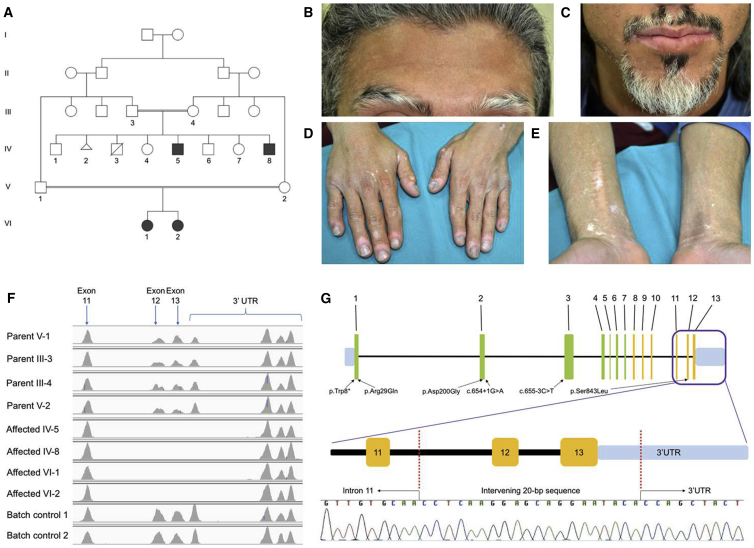
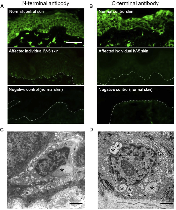
SPG23 is an autosomal-recessive neurodegenerative subtype of lower limb spastic paraparesis with additional diffuse skin and hair dyspigmentation at birth followed by further patchy pigment loss during childhood. Previously, genome-wide linkage in an Arab-Israeli pedigree mapped the gene to an approximately 25 cM locus on chromosome 1q24-q32. By using whole-exome sequencing in a further Palestinian-Jordanian SPG23 pedigree, we identified a complex homozygous 4-kb deletion/20-bp insertion in DSTYK (dual serine-threonine and tyrosine protein kinase) in all four affected family members. DSTYK is located within the established linkage region and we also found the same mutation in the previously reported pedigree and another Israeli pedigree (total of ten affected individuals from three different families). The mutation removes the last two exons and part of the 3' UTR of DSTYK. Skin biopsies revealed reduced DSTYK protein levels along with focal loss of melanocytes. Ultrastructurally, swollen mitochondria and cytoplasmic vacuoles were also noted in remaining melanocytes and some keratinocytes and fibroblasts. Cultured keratinocytes and fibroblasts from an affected individual, as well as knockdown of Dstyk in mouse melanocytes, keratinocytes, and fibroblasts, were associated with increased cell death after ultraviolet irradiation. Keratinocytes from an affected individual showed loss of kinase activity upon stimulation with fibroblast growth factor. Previously, dominant mutations in DSTYK were implicated in congenital urological developmental disorders, but our study identifies different phenotypic consequences for a recurrent autosomal-recessive deletion mutation in revealing the genetic basis of SPG23.
Keywords: DSTYK; Spastic Paraplegia 23; autosomal-recessive; deletion; gene; hereditary spastic paraplegia; mutation; pigmentation; vitiligo; whole-exome sequencing.
Copyright © 2017 American Society of Human Genetics. Published by Elsevier Inc. All rights reserved.
Figures

Similar articles
-
Mutations in DSTYK and dominant urinary tract malformations.N Engl J Med. 2013 Aug 15;369(7):621-9. doi: 10.1056/NEJMoa1214479. Epub 2013 Jul 17. N Engl J Med. 2013. PMID: 23862974 Free PMC article.
-
A locus for complicated hereditary spastic paraplegia maps to chromosome 1q24-q32.Ann Neurol. 2003 Dec;54(6):796-803. doi: 10.1002/ana.10768. Ann Neurol. 2003. PMID: 14681889
-
Stop-gain mutations in UBAP1 cause pure autosomal-dominant spastic paraplegia.Brain. 2019 Aug 1;142(8):2238-2252. doi: 10.1093/brain/awz158. Brain. 2019. PMID: 31203368
-
Exon 8-17 deletions of SPAST in a Chinese family with hereditary spastic paraplegia: a case report and literature review.J Neurol Sci. 2015 Oct 15;357(1-2):282-4. doi: 10.1016/j.jns.2015.07.003. Epub 2015 Jul 3. J Neurol Sci. 2015. PMID: 26165777 Review.
-
Molecular genetics of familial spastic paraplegia: a multitude of responsible genes.J Neurol Sci. 1996 May;137(2):131-8. doi: 10.1016/0022-510x(95)00349-7. J Neurol Sci. 1996. PMID: 8782167 Review.
Cited by
-
Functions of the RIP kinase family members in the skin.Cell Mol Life Sci. 2023 Sep 9;80(10):285. doi: 10.1007/s00018-023-04917-2. Cell Mol Life Sci. 2023. PMID: 37688617 Free PMC article. Review.
-
Exploring the possibility of predicting human head hair greying from DNA using whole-exome and targeted NGS data.BMC Genomics. 2020 Aug 5;21(1):538. doi: 10.1186/s12864-020-06926-y. BMC Genomics. 2020. PMID: 32758128 Free PMC article.
-
Exome sequencing implicates a novel heterozygous missense variant in DSTYK in autosomal dominant lower urinary tract dysfunction and mild hereditary spastic paraparesis.Mol Cell Pediatr. 2021 Oct 4;8(1):13. doi: 10.1186/s40348-021-00122-y. Mol Cell Pediatr. 2021. PMID: 34608560 Free PMC article.
-
DSTYK Promotes Metastasis and Chemoresistance via EMT in Colorectal Cancer.Front Pharmacol. 2020 Sep 2;11:1250. doi: 10.3389/fphar.2020.01250. eCollection 2020. Front Pharmacol. 2020. PMID: 32982725 Free PMC article.
-
Copy Number Variations in Hereditary Spastic Paraplegia-Related Genes: Evaluation of an Iranian Hereditary Spastic Paraplegia Cohort and Literature Review.Mol Syndromol. 2023 Dec;14(6):477-484. doi: 10.1159/000531507. Epub 2023 Jul 7. Mol Syndromol. 2023. PMID: 38058755 Free PMC article.
References
-
- Lison M., Kornbrut B., Feinstein A., Hiss Y., Boichis H., Goodman R.M. Progressive spastic paraparesis, vitiligo, premature graying, and distinct facial appearance: a new genetic syndrome in 3 sibs. Am. J. Med. Genet. 1981;9:351–357. - PubMed
-
- Mukamel M., Weitz R., Metzker A., Varsano I. Spastic paraparesis, mental retardation, and cutaneous pigmentation disorder. A new syndrome. Am. J. Dis. Child. 1985;139:1090–1092. - PubMed
-
- Bamforth J.S. Vitiligo-spasticity syndrome: new case. Clin. Dysmorphol. 2003;12:137–139. - PubMed
Publication types
MeSH terms
Substances
Supplementary concepts
Grants and funding
LinkOut - more resources
Full Text Sources
Other Literature Sources
Medical
Molecular Biology Databases
Miscellaneous
