

Targeted Fcγ Receptor (FcγR)-mediated Clearance by a Biparatopic Bispecific Antibody
- PMID: 28100773
- PMCID: PMC5354496
- DOI: 10.1074/jbc.M116.770628
Targeted Fcγ Receptor (FcγR)-mediated Clearance by a Biparatopic Bispecific Antibody
Abstract
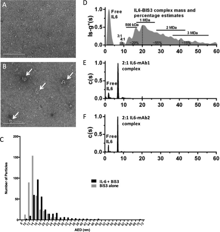
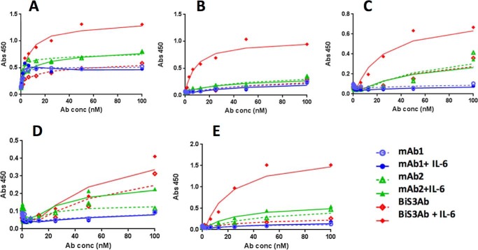
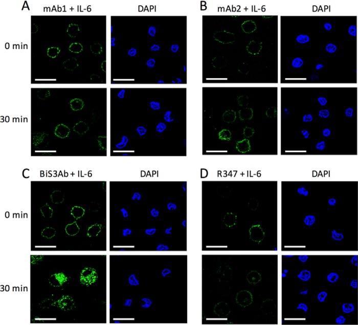
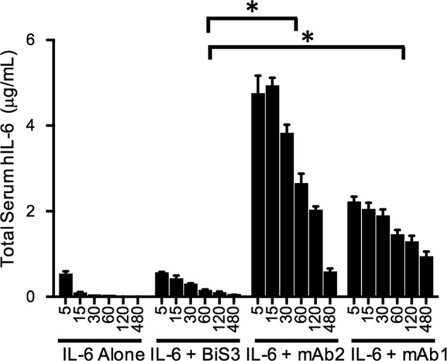
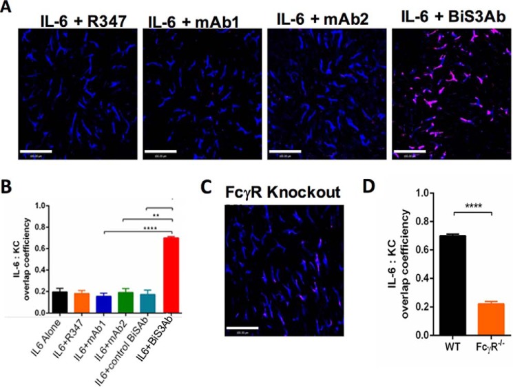
Soluble ligands have commonly been targeted by antibody therapeutics for cancers and other diseases. Although monoclonal antibodies targeting such ligands can block their interactions with their cognate receptors, they can also significantly increase the half-life of their ligands by FcRn-mediated antibody recycling, thereby evading ligand renal clearance and requiring increasingly high antibody doses to neutralize the increasing pool of target. To overcome this issue, we generated a bispecific/biparatopic antibody (BiSAb) that targets two different epitopes on IL-6 to block IL-6-mediated signaling. The BiSAb formed large immune complexes with IL-6 that can bind Fcγ receptors on phagocytic cells and are rapidly internalized. In addition, rapid clearance of the BiSAb·IL-6 complex was observed in mice while the parental antibodies prolonged the serum half-life of IL-6. Intravital imaging of the liver in mice confirmed that the rapid clearance of these large immune complexes was associated with Fcγ receptor-dependent binding to Kupffer cells in the liver. The approach described here provides a general strategy for therapeutic antibodies with the ability to not only neutralize but also actively drive clearance of their soluble antigens.
Keywords: Fc receptor; IL-6; antibody; antibody engineering; electron microscopy (EM); macrophage; microscopic imaging; pharmacokinetics; protein complex.
© 2017 by The American Society for Biochemistry and Molecular Biology, Inc.
Conflict of interest statement
S. K., G. J. R., C. G., L. X., X. W., A. P., T. C., C. F., J. B., H. Z., and H. W. own stock in AstraZeneca
Figures


Similar articles
-
Multi-Angle Effector Function Analysis of Human Monoclonal IgG Glycovariants.PLoS One. 2015 Dec 11;10(12):e0143520. doi: 10.1371/journal.pone.0143520. eCollection 2015. PLoS One. 2015. PMID: 26657484 Free PMC article.
-
Human monoclonal antibodies targeting nonoverlapping epitopes on insulin-like growth factor II as a novel type of candidate cancer therapeutics.Mol Cancer Ther. 2012 Jul;11(7):1400-10. doi: 10.1158/1535-7163.MCT-12-0172. Epub 2012 May 2. Mol Cancer Ther. 2012. PMID: 22553356 Free PMC article.
-
Field flow fractionation for assessing neonatal Fc receptor and Fcγ receptor binding to monoclonal antibodies in solution.Anal Biochem. 2011 Jul 1;414(1):88-98. doi: 10.1016/j.ab.2011.03.001. Epub 2011 Mar 6. Anal Biochem. 2011. PMID: 21385563
-
Current strategies in antibody engineering: Fc engineering and pH-dependent antigen binding, bispecific antibodies and antibody drug conjugates.Biotechnol J. 2012 Dec;7(12):1444-50. doi: 10.1002/biot.201200250. Epub 2012 Nov 1. Biotechnol J. 2012. PMID: 23125076 Review.
-
The Neonatal Fc Receptor (FcRn): A Misnomer?Front Immunol. 2019 Jul 10;10:1540. doi: 10.3389/fimmu.2019.01540. eCollection 2019. Front Immunol. 2019. PMID: 31354709 Free PMC article. Review.
Cited by
-
Multiple Variables at the Leukocyte Cell Surface Impact Fc γ Receptor-Dependent Mechanisms.Front Immunol. 2019 Feb 14;10:223. doi: 10.3389/fimmu.2019.00223. eCollection 2019. Front Immunol. 2019. PMID: 30837990 Free PMC article. Review.
-
Expanding the Boundaries of Biotherapeutics with Bispecific Antibodies.BioDrugs. 2018 Oct;32(5):441-464. doi: 10.1007/s40259-018-0299-9. BioDrugs. 2018. PMID: 30132211 Free PMC article. Review.
-
Factors Affecting the Pharmacology of Antibody-Drug Conjugates.Antibodies (Basel). 2018 Feb 7;7(1):10. doi: 10.3390/antib7010010. Antibodies (Basel). 2018. PMID: 31544862 Free PMC article. Review.
-
Plasma cell biology: Foundations for targeted therapeutic development in transplantation.Immunol Rev. 2021 Sep;303(1):168-186. doi: 10.1111/imr.13011. Epub 2021 Jul 12. Immunol Rev. 2021. PMID: 34254320 Free PMC article. Review.
-
PET imaging of Aspergillus infection using Zirconium-89 labeled anti-β-glucan antibody fragments.Eur J Nucl Med Mol Imaging. 2024 Sep;51(11):3223-3234. doi: 10.1007/s00259-024-06760-4. Epub 2024 May 24. Eur J Nucl Med Mol Imaging. 2024. PMID: 38787397 Free PMC article.
References
-
- Pandey M., and Mahadevan D. (2014) Monoclonal antibodies as therapeutics in human malignancies. Future Oncol. 10, 609–636 - PubMed
-
- Rehlaender B. N., and Cho M. J. (2001) Anti-drug antibodies as drug carriers: I: for small molecules. Pharm. Res. 18, 745–752 - PubMed
-
- Gessner J. E., Heiken H., Tamm A., and Schmidt R. E. (1998) The IgG Fc receptor family. Ann. Hematol. 76, 231–248 - PubMed
-
- Mihara M., Hashizume M., Yoshida H., Suzuki M., and Shiina M. (2012) IL-6/IL-6 receptor system and its role in physiological and pathological conditions. Clin. Sci. 122, 143–159 - PubMed
-
- Mihara M., Nishimoto N., and Ohsugi Y. (2005) The therapy of autoimmune diseases by anti-interleukin-6 receptor antibody. Expert Opin. Biol. Ther. 5, 683–690 - PubMed
MeSH terms
Substances
LinkOut - more resources
Full Text Sources
Other Literature Sources
