Randomized trial of calcipotriol combined with 5-fluorouracil for skin cancer precursor immunotherapy
- PMID: 27869649
- PMCID: PMC5199703
- DOI: 10.1172/JCI89820
Randomized trial of calcipotriol combined with 5-fluorouracil for skin cancer precursor immunotherapy
Abstract
Background: Actinic keratosis is a precursor to cutaneous squamous cell carcinoma. Long treatment durations and severe side effects have limited the efficacy of current actinic keratosis treatments. Thymic stromal lymphopoietin (TSLP) is an epithelium-derived cytokine that induces a robust antitumor immunity in barrier-defective skin. Here, we investigated the efficacy of calcipotriol, a topical TSLP inducer, in combination with 5-fluorouracil (5-FU) as an immunotherapy for actinic keratosis.
Methods: The mechanism of calcipotriol action against skin carcinogenesis was examined in genetically engineered mouse models. The efficacy and safety of 0.005% calcipotriol ointment combined with 5% 5-FU cream were compared with Vaseline plus 5-FU for the field treatment of actinic keratosis in a randomized, double-blind clinical trial involving 131 participants. The assigned treatment was self-applied to the entirety of the qualified anatomical sites (face, scalp, and upper extremities) twice daily for 4 consecutive days. The percentage of reduction in the number of actinic keratoses (primary outcome), local skin reactions, and immune activation parameters were assessed.
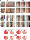
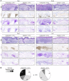
Results: Calcipotriol suppressed skin cancer development in mice in a TSLP-dependent manner. Four-day application of calcipotriol plus 5-FU versus Vaseline plus 5-FU led to an 87.8% versus 26.3% mean reduction in the number of actinic keratoses in participants (P < 0.0001). Importantly, calcipotriol plus 5-FU treatment induced TSLP, HLA class II, and natural killer cell group 2D (NKG2D) ligand expression in the lesional keratinocytes associated with a marked CD4+ T cell infiltration, which peaked on days 10-11 after treatment, without pain, crusting, or ulceration.
Conclusion: Our findings demonstrate the synergistic effects of calcipotriol and 5-FU treatment in optimally activating a CD4+ T cell-mediated immunity against actinic keratoses and, potentially, cancers of the skin and other organs.
Trial registration: ClinicalTrials.gov NCT02019355.
Funding: Not applicable (investigator-initiated clinical trial).
Conflict of interest statement
The authors have declared that no conflict of interest exists.
Figures


Similar articles
-
Skin cancer precursor immunotherapy for squamous cell carcinoma prevention.JCI Insight. 2019 Mar 21;4(6):e125476. doi: 10.1172/jci.insight.125476. eCollection 2019 Mar 21. JCI Insight. 2019. PMID: 30895944 Free PMC article. Clinical Trial.
-
Topical Calcipotriol Plus 5-Fluorouracil in the Treatment of Actinic Keratosis, Bowen's Disease, and Squamous Cell Carcinoma: A Systematic Review.J Cutan Med Surg. 2024 Jul-Aug;28(4):375-380. doi: 10.1177/12034754241256347. Epub 2024 May 23. J Cutan Med Surg. 2024. PMID: 38783539 Review.
-
Use of Topical Calcipotriol Plus 5-Fluorouracil in the Treatment of Actinic Keratosis: A Systematic Review.J Drugs Dermatol. 2022 Jan 1;21(1):60-65. doi: 10.36849/JDD.2022.6632. J Drugs Dermatol. 2022. PMID: 35005863
-
Calcipotriol and 5-Fluorouracil Combination Therapy for the Treatment of Actinic Keratosis in the Clinic: A Review Article.Clin Drug Investig. 2024 Oct;44(10):733-737. doi: 10.1007/s40261-024-01392-w. Epub 2024 Sep 28. Clin Drug Investig. 2024. PMID: 39342018 Free PMC article. Review.
-
T helper 2 cell-directed immunotherapy eliminates precancerous skin lesions.J Clin Invest. 2025 Jan 2;135(1):e183274. doi: 10.1172/JCI183274. J Clin Invest. 2025. PMID: 39744942 Free PMC article.
Cited by
-
Genome-wide association study of actinic keratosis identifies new susceptibility loci implicated in pigmentation and immune regulation pathways.Commun Biol. 2022 Apr 21;5(1):386. doi: 10.1038/s42003-022-03301-3. Commun Biol. 2022. PMID: 35449187 Free PMC article.
-
Alarmin Cytokines as Central Regulators of Cutaneous Immunity.Front Immunol. 2022 Mar 30;13:876515. doi: 10.3389/fimmu.2022.876515. eCollection 2022. Front Immunol. 2022. PMID: 35432341 Free PMC article. Review.
-
Case report of novel combination of anthralin and calcipotriene leading to trichologic response in alopecia areata.JAAD Case Rep. 2019 Mar 1;5(3):258-260. doi: 10.1016/j.jdcr.2019.01.006. eCollection 2019 Mar. JAAD Case Rep. 2019. PMID: 30891474 Free PMC article. No abstract available.
-
Overview of popular cosmeceuticals in dermatology.Skin Health Dis. 2024 Feb 7;4(2):e340. doi: 10.1002/ski2.340. eCollection 2024 Apr. Skin Health Dis. 2024. PMID: 38577050 Free PMC article. Review.
-
Tumor initiation and early tumorigenesis: molecular mechanisms and interventional targets.Signal Transduct Target Ther. 2024 Jun 19;9(1):149. doi: 10.1038/s41392-024-01848-7. Signal Transduct Target Ther. 2024. PMID: 38890350 Free PMC article. Review.
References
Publication types
MeSH terms
Substances
Associated data
Grants and funding
LinkOut - more resources
Full Text Sources
Other Literature Sources
Medical
Research Materials

