. 2016 Jul 12;113(28):7876-81.
doi: 10.1073/pnas.1600587113. Epub 2016 Jun 24.

A viral kinase mimics S6 kinase to enhance cell proliferation

Affiliations

A viral kinase mimics S6 kinase to enhance cell proliferation

Aadra Prashant Bhatt et al. Proc Natl Acad Sci U S A. .

Abstract

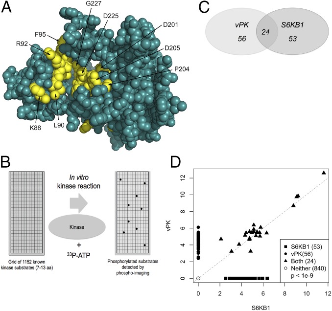
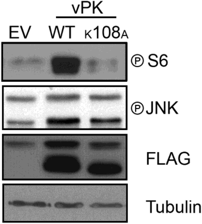
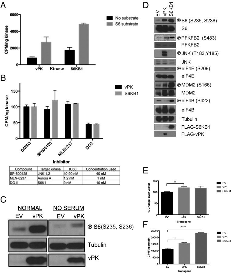
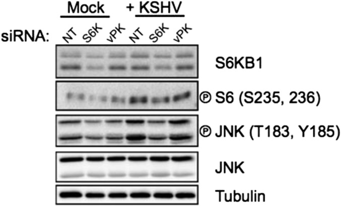
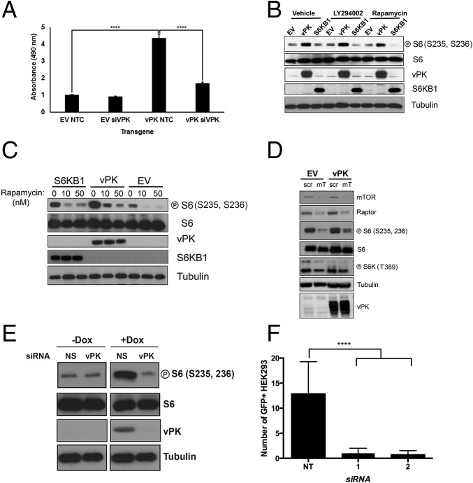
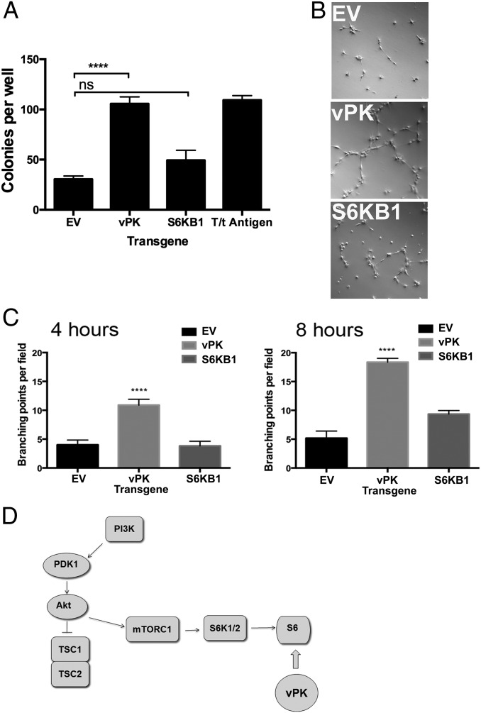
Viruses depend upon the host cell for manufacturing components of progeny virions. To mitigate the inextricable dependence on host cell protein synthesis, viruses can modulate protein synthesis through a variety of mechanisms. We report that the viral protein kinase (vPK) encoded by open reading frame 36 (ORF36) of Kaposi's sarcoma-associated herpesvirus (KSHV) enhances protein synthesis by mimicking the function of the cellular protein S6 kinase (S6KB1). Similar to S6KB1, vPK phosphorylates the ribosomal S6 protein and up-regulates global protein synthesis. vPK also augments cellular proliferation and anchorage-independent growth. Furthermore, we report that both vPK and S6KB1 phosphorylate the enzyme 6-phosphofructo-2-kinase/fructose-2, 6-bisphosphatase 2 (PFKFB2) and that both kinases promote endothelial capillary tubule formation.

Keywords: KSHV; ORF36; S6K; cell signaling; viral protein kinase.


Conflict of interest statement

The authors declare no conflict of interest.

Figures

Fig. 1.
Fig. 1.
KSHV vPK displays limited homology to cellular S6KB1. (A) In silico model of vPK based on the partially activated state of S6KB1 is rendered in spheres. Residues highly conserved between S6KB1 and vPK are colored yellow, and the rest are teal. Most of the conserved residues between the two kinases occur in three motifs that cross the active site pocket. The first motif—KrLGRGaFG (uppercase residues are conserved and in yellow)—consists of residues K88 to G96. The second and third motifs—DvsPDNI and LTDFG—consist of residues D201 to I207 and L223 to G227. (B) A PepChip array was used to identify targets of both vPK and S6KB1. Recombinant kinases were incubated with radiolabeled ATP on a glass slide arrayed with >1,000 kinase substrate peptides. (C) Twenty-four peptides are phosphorylated by both vPK and S6KB1. vPK and S6KB1 were found to uniquely phosphorylate an additional 56 and 53 peptides, respectively. (D) Scatter plot analysis of spot intensities of peptides phosphorylated either uniquely by vPK (y axis; closed circles) or S6KB1 (x axis; closed squares), dually phosphorylated by both (closed triangles), or spots phosphorylated by neither kinase (open circles).
Fig. S1.
Fig. S1.
Immunoblot of 293 cells transfected with indicated plasmids. Transfection of WT vPK elevates phospho-S6 compared with the K108A mutant vPK. As previously reported, increased phosphorylation in phospho-JNK is also observed with vPK transfection. Expression of vPK constructs is confirmed by FLAG immunoblot, and tubulin is used as a loading control.
Fig. 2.
Fig. 2.
KSHV vPK phosphorylates several S6KB1 substrates. (A) An in vitro kinase assay was performed with recombinant vPK and S6K using a synthetic S6 substrate. Data are representative of four independent experiments; error bars denote SEM. (B) Impact of S6KB1-specific and nonspecific kinase inhibitors on recombinant vPK and S6KB1 phosphorylation of S6 peptide substrate in an in vitro kinase assay. Data are representative of three independent experiments; error bars are ±SEM. (C) Ectopic expression of vPK in 293 cells increases phosphorylated levels of S6. Total S6 and tubulin are shown as loading controls. Construct expression is verified using a vPK-specific antibody. These images are representative of three independent experiments. ℗, phospho. (D) Stable HUVECs were first plated in normal media; subsequently, serum was withdrawn for 16 h, and immunoblots were performed with harvested lysates for the indicated phosphorylated and corresponding total proteins. Tubulin is shown as loading control. These images are representative of four independent experiments. (E) Metabolic labeling of de novo protein synthesis using 35S-labeled methionine and cysteine was quantified in 293 cells transiently transfected with vector, vPK, or S6KB1. Counts are normalized to total protein content. Data are representative of three independent experiments; error bars are ±SEM. (F) Metabolic labeling of de novo protein synthesis using 35S-labeled methionine and cysteine was quantified in stable HUVECs expressing vPK, S6KB1, or matched vector control. Counts are normalized to total protein content. Data are representative of three independent experiments; error bars are ±SEM, **P < 0.01, ****P < 0.0001.
Fig. S2.
Fig. S2.
Immunoblot of HUVECs transfected for 24 h with indicated siRNAs and then infected with KSHV for 90 min. Infection was carried out as described by West and Damania (24). One hour postinfection (HPI), cells were harvested, and immunoblots were performed. KSHV infection induces S6 phosphorylation in an S6KB1-independent manner.
Fig. 3.
Fig. 3.
KSHV vPK enhances cellular proliferation. (A) Stable HUVECs transfected with indicated siRNAs were plated in a 96-well plate, and their basal metabolic rate was measured 72 h afterward using the Cell Titer Aqueous One Assay. Data are representative of three independent experiments; error bars are ±SEM, ****P < 0.001. NTC, nontargeting control. (B) Immunoblot analyses of indicated proteins with lysates prepared from 293 cells transiently transfected with empty vector, vPK, or S6KB1 and then treated with either LY294002 or rapamycin (10 nM), each for 1 h. Transgene expression was confirmed using vPK- and FLAG-specific antibodies. Tubulin is shown as a loading control. Images represent one of three independent experiments. (C) Stable HUVECs were treated with increasing doses of rapamycin (10, 50 nM) for 1 h, and harvested lysates were subject to immunoblotting with indicated phospho-specific and total antibodies. Although phospho-S6 remains elevated after rapamycin treatment in HUVEC-vPK, it is diminished in vector- and S6KB1-expressing HUVECs. Images are representative of three independent experiments. (D) Stable 293 cells were transfected with indicated siRNAs for 48 h, and immunoblots were performed after 12-h serum withdrawal. mT, pooled Raptor- and mTOR-specific siRNA; Scr, scrambled. (E) Immunoblot analysis of both latent (No Dox) and reactivated (Dox) iSLK.219 cells transfected with either vPK-directed or a nonspecific (NS) siRNA. Representative of four independent experiments. (F) Infectivity of 293 cells infected with KSHV derived from the experiment described in E; **** P < 0.0001.
Fig. S3.
Fig. S3.
Immunoblot analysis of lysates prepared from stable HUVECs transfected with either nontargeting control (NTC) or vPK-specific siRNA (siVPK). Blot is probed with vPK-specific antibody, and shows significant knockdown in HUVEC-vPK transfected with vPK-specific siRNA (siVPK). Ku70 is shown as a loading control.
Fig. 4.
Fig. 4.
KSHV vPK augments tubule formation. (A) Rat1 fibroblasts stably expressing the indicated transgenes were plated in soft agar for 3 wk. Colonies were stained with crystal violet and quantified to measure anchorage independence. Data are representative of four independent experiments; error bars are ±SEM; ****P < 0.0001. (B) When placed in growth factor-reduced Matrigel, vPK-stable HUVECs form tubular networks within 4 h. Also shown are EV- and S6KB1-expressing cells. (C) Significantly increased branching was observed at 4 h (Left) and 8 h (Right) in vPK-expressing HUVECs, compared with EV- and S6KB1-HUVEC. Tubule formation data are representative of three independent experiments. (D) Proposed model for vPK’s mechanism of action.

Similar articles

Cited by

References

    1. Manning G, Whyte DB, Martinez R, Hunter T, Sudarsanam S. The protein kinase complement of the human genome. Science. 2002;298(5600):1912–1934. - PubMed
    1. Ma XM, Blenis J. Molecular mechanisms of mTOR-mediated translational control. Nat Rev Mol Cell Biol. 2009;10(5):307–318. - PubMed
    1. Fenton TR, Gout IT. Functions and regulation of the 70kDa ribosomal S6 kinases. Int J Biochem Cell Biol. 2011;43(1):47–59. - PubMed
    1. Shahbazian D, et al. The mTOR/PI3K and MAPK pathways converge on eIF4B to control its phosphorylation and activity. EMBO J. 2006;25(12):2781–2791. - PMC - PubMed
    1. Lai KP, et al. S6K1 is a multifaceted regulator of Mdm2 that connects nutrient status and DNA damage response. EMBO J. 2010;29(17):2994–3006. - PMC - PubMed

Publication types

MeSH terms

Substances