

TRPA1 channels: molecular sentinels of cellular stress and tissue damage
- PMID: 27079970
- PMCID: PMC4967735
- DOI: 10.1113/JP270935
TRPA1 channels: molecular sentinels of cellular stress and tissue damage
Abstract
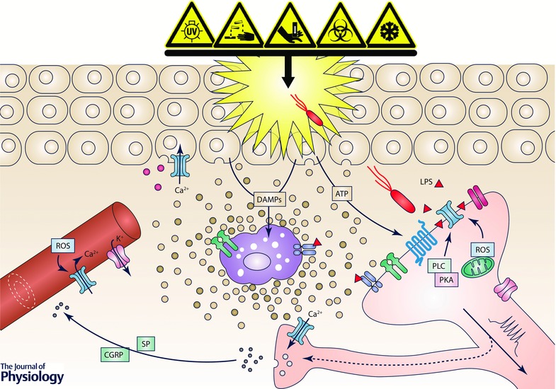
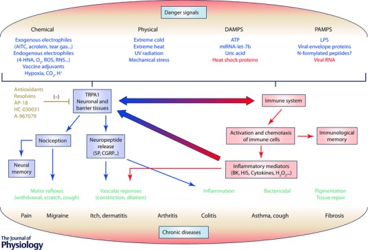
TRPA1 is a non-selective cation channel expressed in mammalian peripheral pain receptors, with a major role in chemonociception. TRPA1 has also been implicated in noxious cold and mechanical pain sensation. TRPA1 has an ancient origin and plays important functions in lower organisms, including thermotaxis, mechanotransduction and modulation of lifespan. Here we highlight the role of TRPA1 as a multipurpose sensor of harmful signals, including toxic bacterial products and UV light, and as a sensor of stress and tissue damage. Sensing roles span beyond the peripheral nervous system to include major barrier tissues: gut, skin and lung. Tissue injury, environmental irritants and microbial pathogens are danger signals that can threaten the health of organisms. These signals lead to the coordinated activation of the nociceptive and the innate immune system to provide a homeostatic response trying to re-establish physiological conditions including tissue repair. Activation of TRPA1 participates in protective neuroimmune interactions at multiple levels, sensing ROS and bacterial products and triggering the release of neuropeptides. However, an exaggerated response to danger signals is maladaptive and can lead to the development of chronic inflammatory conditions.
© 2016 The Authors. The Journal of Physiology © 2016 The Physiological Society.
Figures
Similar articles
-
Recent advances in the biology and medicinal chemistry of TRPA1.Future Med Chem. 2010 May;2(5):843-58. doi: 10.4155/fmc.10.29. Future Med Chem. 2010. PMID: 21426205 Review.
-
TRPA1: A gatekeeper for inflammation.Annu Rev Physiol. 2013;75:181-200. doi: 10.1146/annurev-physiol-030212-183811. Epub 2012 Sep 27. Annu Rev Physiol. 2013. PMID: 23020579 Free PMC article. Review.
-
The TRPA1 channel in inflammatory and neuropathic pain and migraine.Rev Physiol Biochem Pharmacol. 2014;167:1-43. doi: 10.1007/112_2014_18. Rev Physiol Biochem Pharmacol. 2014. PMID: 24668446 Review.
-
Emerging roles of TRPA1 in sensation of oxidative stress and its implications in defense and danger.Arch Pharm Res. 2013 Jul;36(7):783-91. doi: 10.1007/s12272-013-0098-2. Epub 2013 Apr 5. Arch Pharm Res. 2013. PMID: 23558672 Review.
-
TRPV1, TRPA1, and TRPM8 channels in inflammation, energy redirection, and water retention: role in chronic inflammatory diseases with an evolutionary perspective.J Mol Med (Berl). 2014 Sep;92(9):925-37. doi: 10.1007/s00109-014-1175-9. Epub 2014 May 29. J Mol Med (Berl). 2014. PMID: 24871046 Review.
Cited by
-
Renal Tubular Epithelial TRPA1 Acts as An Oxidative Stress Sensor to Mediate Ischemia-Reperfusion-Induced Kidney Injury through MAPKs/NF-κB Signaling.Int J Mol Sci. 2021 Feb 25;22(5):2309. doi: 10.3390/ijms22052309. Int J Mol Sci. 2021. PMID: 33669091 Free PMC article.
-
Effects of norepinephrine on colonic tight junction protein expression during heat stress.Exp Ther Med. 2021 May;21(5):421. doi: 10.3892/etm.2021.9865. Epub 2021 Feb 25. Exp Ther Med. 2021. PMID: 33747161 Free PMC article.
-
Non-Analgesic Symptomatic or Disease-Modifying Potential of TRPA1.Med Sci (Basel). 2019 Sep 23;7(10):99. doi: 10.3390/medsci7100099. Med Sci (Basel). 2019. PMID: 31547502 Free PMC article. Review.
-
A natural agonist of mosquito TRPA1 from the medicinal plant Cinnamosma fragrans that is toxic, antifeedant, and repellent to the yellow fever mosquito Aedes aegypti.PLoS Negl Trop Dis. 2018 Feb 9;12(2):e0006265. doi: 10.1371/journal.pntd.0006265. eCollection 2018 Feb. PLoS Negl Trop Dis. 2018. PMID: 29425195 Free PMC article.
-
H2O2 gel bleaching induces cytotoxicity and pain conduction in dental pulp stem cells via intracellular reactive oxygen species on enamel/dentin disc.PLoS One. 2021 Sep 10;16(9):e0257221. doi: 10.1371/journal.pone.0257221. eCollection 2021. PLoS One. 2021. PMID: 34506603 Free PMC article.
References
-
- Abdullah H, Heaney LG, Cosby SL & McGarvey LP (2014). Rhinovirus upregulates transient receptor potential channels in a human neuronal cell line: implications for respiratory virus‐induced cough reflex sensitivity. Thorax 69, 46–54. - PubMed
-
- Akopian AN (2011). Regulation of nociceptive transmission at the periphery via TRPA1‐TRPV1 interactions. Curr Pharm Biotechnol 12, 89–94. - PubMed
-
- Andersson DA, Gentry C, Alenmyr L, Killander D, Lewis SE, Andersson A, Bucher B, Galzi JL, Sterner O, Bevan S, Högestätt ED & Zygmunt PM (2011). TRPA1 mediates spinal antinociception induced by acetaminophen and the cannabinoid Δ9‐tetrahydrocannabiorcol. Nat Commun 2, 551. - PubMed
Publication types
MeSH terms
Substances
LinkOut - more resources
Full Text Sources
Other Literature Sources