. 2016 Apr 8;12(4):e1005546.
doi: 10.1371/journal.ppat.1005546. eCollection 2016 Apr.

cGAS Senses Human Cytomegalovirus and Induces Type I Interferon Responses in Human Monocyte-Derived Cells

Affiliations

cGAS Senses Human Cytomegalovirus and Induces Type I Interferon Responses in Human Monocyte-Derived Cells

Jennifer Paijo et al. PLoS Pathog. .

Abstract

Human cytomegalovirus (HCMV) infections of healthy individuals are mostly unnoticed and result in viral latency. However, HCMV can also cause devastating disease, e.g., upon reactivation in immunocompromised patients. Yet, little is known about human immune cell sensing of DNA-encoded HCMV. Recent studies indicated that during viral infection the cyclic GMP/AMP synthase (cGAS) senses cytosolic DNA and catalyzes formation of the cyclic di-nucleotide cGAMP, which triggers stimulator of interferon genes (STING) and thus induces antiviral type I interferon (IFN-I) responses. We found that plasmacytoid dendritic cells (pDC) as well as monocyte-derived DC and macrophages constitutively expressed cGAS and STING. HCMV infection further induced cGAS, whereas STING expression was only moderately affected. Although pDC expressed particularly high levels of cGAS, and the cGAS/STING axis was functional down-stream of STING, as indicated by IFN-I induction upon synthetic cGAMP treatment, pDC were not susceptible to HCMV infection and mounted IFN-I responses in a TLR9-dependent manner. Conversely, HCMV infected monocyte-derived cells synthesized abundant cGAMP levels that preceded IFN-I production and that correlated with the extent of infection. CRISPR/Cas9- or siRNA-mediated cGAS ablation in monocytic THP-1 cells and primary monocyte-derived cells, respectively, impeded induction of IFN-I responses following HCMV infection. Thus, cGAS is a key sensor of HCMV for IFN-I induction in primary human monocyte-derived DC and macrophages.


Conflict of interest statement

The authors have declared that no competing interests exist.

Figures
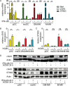
Fig 1
Fig 1. pDC and monocyte-derived cells show constitutive cGAS expression that is enhanced upon IFN-α or HCMV stimulation.
Primary human pDC (yellow bars), moDC (green bars), GM-CSF MΦ (GM-MΦ, red bars), and M-CSF MΦ (M-MΦ, blue bars) were (A) stimulated for 6 h with recombinant IFN-α2b and analyzed for relative MxA, cGAS, and STING mRNA expression. Furthermore, the cells were infected with HCMV at MOI 3 for 24 h and analyzed for (B) cGAS and (C) STING mRNA expression relative to HPRT1 mRNA by qPCR. (D) Protein levels of cGAS and STING upon HCMV infection for 24 h or recombinant IFN-α2b treatment for 6 h (upper panel) or 24 h (lower panel) were determined by western blot analysis, while actin was used as loading control. Mean ± SEM of 5–14 (A), 5–15 (B, C) or representative for 3–4 (D) different donors. ns = not significant, *: p ≤ 0.032, **: p ≤ 0.0078 one-tailed Wilcoxon signed rank test.
Fig 2
Fig 2. HCMV infection induces IFN-I responses in pDC as well as monocyte-derived DC and MΦ.
pDC, moDC, GM-CSF MΦ, and M-CSF MΦ were infected with HCMV at MOI 3 and 24 hours post infection (hpi) (A) IFN-α and (B) IFN-β mRNA expression was determined relative to HPRT1 mRNA by qPCR (mean ± SEM of 7–9 different donors). (C, D) IFN-α expressing cells were analyzed by flow cytometry and (E) the IFN-α content in cell-free supernatants was determined by an ELISA method upon infection with three independently produced HCMV preparations (#1, #2, and #3) at MOI 3. (F) Upon HCMV infection with varying MOI between 0.1 to 30 IFN-α concentrations in cell-free supernatants (y-axis) were blotted against percentages of IFN-α+ cells (x-axis) of the same culture (Values from 12–28 different donors. Black line: confidence interval of 95%). Median of 9–26 (D) and 12–23 (E) different donors. ns = not significant, *: p ≤ 0.031, **: p ≤ 0.0078, ***: p ≤ 0.0002 one-tailed paired Wilcoxon signed rank test was used for statistical analyses between monocyte-derived cell subsets, because the subsets were derived from monocytes of the same donors. In contrast, pDC were derived from different donors. Therefore, statistical analysis between pDC and one of the monocyte-derived cell subsets was performed using one-tailed unpaired Mann-Whitney test.
Fig 3
Fig 3. HCMV infection induces cGAMP formation in monocyte-derived DC and MΦ, but not in pDC.
cGAMP synthesis was analyzed and quantified using a HPLC-MS/MS method. Chromatograms (blue line: quantifier, green/red lines: identifiers) of (A) synthetic cGAS-derived cGAMP (2´-5´/3´-5´) and bacterial cGAMP (3´-5´/3´-5´) (upper panel) or lysed, HCMV stimulated M-CSF MΦ (lower panel) as well as (B) lysates of 24 h HCMV infected pDC, moDC, GM-CSF MΦ, and M-CSF MΦ (enlarged visualization of grey area shown in (A)). (C) Quantification of the detected cGAMP shown in (B). (D) cGAMP (filled circles) synthesis and IFN-α contents in cell-free supernatants (open triangles) were monitored in unstimulated (0 h) moDC, GM-CSF MΦ, and M-CSF MΦ or at indicated time points after HCMV treatment. pDC, moDC, GM-CSF MΦ, and M-CSF MΦ were infected with MVA at MOI 1 and 24 hpi (E) cGAMP synthesis as well as (F) IFN-α contents of cell-free supernatants were quantified. moDC, GM-CSF MΦ, and M-CSF MΦ were infected with VSV-M2 at MOI 1 and 24 hpi (G) cGAMP synthesis as well as (H) IFN-α contents of cell-free supernatants were quantified. Mean ± SEM of 4–7 (C), 4 (D), 3–7 (E, F), or 4 (G, H) different donors. ns = not significant, *: p ≤ 0.032, **: p ≤ 0.0078 one-tailed Wilcoxon signed rank test.
Fig 4
Fig 4. HCMV-derived genomic DNA efficiently activates recombinant human cGAS.
Recombinant human cGAS was incubated for 2 h in the presence or absence of ATP and GTP with (A) a 50 bp long control dsDNA, (B) purified HCMV containing approximately 6.5 x 10⁶ infectious virus particles, or purified HCMV treated for 10 minutes at 95°C, or (C, D) genomic DNA isolated from purified HCMV. All samples were tested with and without DNA digestion prior to incubation with recombinant cGAS. cGAMP formation was quantified using a HPLC-MS/MS method. Mean ± SEM of 5–6 (A) and 5–7 (B, C, D) data points from 3 independent experiments. **: p ≤ 0.005, ***: p ≤ 0.0003 one-tailed Mann-Whitney test.
Fig 5
Fig 5. pDC are stimulated by cGAMP to mount IFN-I responses, whereas they sense HCMV in a TLR9-dependent manner.
(A) IFN-α responses of pDC were monitored by an ELISA method in supernatants of untreated pDC (Ø), and pDC stimulated with HCMV at MOI 3, or the TLR7/8 agonist R848, in the presence or absence of the TLR9 inhibitory oligonucleotide IRS869. (B) pDC were left untreated (Ø), or mock transfected, or transfected with synthetic cGAMP, and IFN-α expression was monitored by an ELISA method. Mean ± SEM of 5–6 (A) and 7 (B) different donors. ns = not significant, *: p ≤ 0.016, **: p ≤ 0.0078 one-tailed Wilcoxon signed rank test.
Fig 6
Fig 6. Upon HCMV stimulation PMA-matured THP-1 cells mount IFN-β responses in a cGAS-dependent manner.
WT (wild-type), and 2 clones (#1 and #2) of cGAS, IFI16, or STING deficient THP-1 cells differentiated with PMA for 3 days were stimulated with (A) HCMV at MOI 10, (B) MVA at MOI 1, or (C) VSV at MOI 1 for 24 h. Cell-free supernatant was tested for IFN-β by an ELISA method (A, B), or cell lysates were tested for IFN-β mRNA expression relative to HPRT1 mRNA by qPCR (C). Lysates of virus infected cells were analyzed for phosphorylated IRF3 (P-IRF3) and IRF3 by western blot (A, B, C). Mean ± SEM of 3–6 (A), 3 (B), and 4–5 (C) data points from 3 (A, C) and 2 (B) independent experiments. *: p ≤ 0.05, **: p ≤ 0.0076 one-tailed Mann-Whitney test.
Fig 7
Fig 7. HCMV infected monocyte-derived DC and MΦ mount IFN-α responses in a cGAS-dependent manner.
(A) moDC, GM-CSF MΦ, and M-CSF MΦ were transfected with siRNA directed against cGAS or control siRNA and cGAS or IFI16 expression was monitored by western blot analysis, while actin was used as loading control. Untreated or siRNA-mediated cGAS knock-down moDC, GM-CSF MΦ, and M-CSF MΦ were infected with HCMV-GFP at MOI 3 for 24 h and (B) IFN-α contents of cell-free supernatants were monitored by ELISA or (C) IFN-α+ and/or HCMV-GFP+ cells were analyzed by flow cytometry. (D) cGAS knock-down moDC, GM-CSF MΦ, and M-CSF MΦ were mock transfected or transfected with synthetic cGAMP and analyzed for IFN-β mRNA expression relative to HPRT1 mRNA by qPCR. Mean ± SEM of 6 (B) and 5 (D) or representative for 6 (A, C) different donors from 3 independent experiments. ns = not significant, *: p ≤ 0.04 one-tailed Wilcoxon signed rank test.
Fig 8
Fig 8. Upon HCMV-GFP infection, GFP+ cells are more abundant in monocyte-derived cells than in pDC.
(A, B) pDC, moDC, GM-CSF MΦ, and M-CSF MΦ were infected with three independently produced HCMV-GFP preparations (#1, #2, and #3) at MOI 3 for 24 h and percentages of HCMV-GFP+ cells were analyzed by flow cytometry. (C) pDC, moDC, GM-CSF MΦ, and M-CSF MΦ were infected with HCMV-GFP and percentages of GFP+ cells as well as cGAMP synthesis were determined by FACS analysis and a HPLC-MS/MS method, respectively. Percentages of GFP+ cells (x-axis) were blotted against the amount of cGAMP synthesized (y-axis) from the same cultures for all different cell subsets in one blot and the Spearman correlation (rS) between the factors was determined. (D) pDC and M-CSF MΦ were infected with HCMV-GFP at varying MOI between 0.1 to 30 and percentages of GFP+ cells (x-axis) were blotted against percentages of IFN-α+ cells or IFN-α contents of cell-free supernatants of the same cultures (y-axis). Median of 8–30 (B) or values from 3–7 (C) and 14–42 (D) different donors. **: p ≤ 0.0013, ***: p ≤ 0.00031 one-tailed paired Wilcoxon signed rank test was used for statistical analyses between monocyte-derived cell subsets, because the subsets were derived from monocytes of the same donors. In contrast, pDC were derived from different donors. Therefore, statistical analysis between pDC and one of the monocyte-derived cell subsets was performed using one-tailed unpaired Mann-Whitney test.

Similar articles

Cited by

References

    1. Hengel H, Brune W, Koszinowski UH (1998) Immune evasion by cytomegalovirus—survival strategies of a highly adapted opportunist. Trends Microbiol 6: 190–197. - PubMed
    1. Loewendorf A, Benedict CA (2010) Modulation of host innate and adaptive immune defenses by cytomegalovirus: timing is everything. J Intern Med 267: 483–501. 10.1111/j.1365-2796.2010.02220.x - DOI - PMC - PubMed
    1. Griffiths P, Baraniak I, Reeves M (2015) The pathogenesis of human cytomegalovirus. J Pathol 235: 288–297. 10.1002/path.4437 - DOI - PubMed
    1. Revello MG, Gerna G (2002) Diagnosis and Management of Human Cytomegalovirus Infection in the Mother, Fetus, and Newborn Infant. Clin Microbiol Rev 15: 680–715. - PMC - PubMed
    1. Sinclair J, Sissons P (2006) Latency and reactivation of human cytomegalovirus. J Gen Virol 87: 1763–1779. - PubMed

Publication types

MeSH terms

Grants and funding

This study was supported by funding from the Helmholtz Virtual Institute (VH-VI-424 Viral Strategies of Immune Evasion, VISTRIE) to MM, and UK and by funding from the Helmholtz-Alberta Initiative, Infectious Diseases Research (HAI-IDR SO-073) to UK. GW was funded by the Deutsche Forschungsgemeinschaft (DFG, grant GRK1721) and JS and MD were supported by the Hannover Biomedical Research School (HBRS) and the Center for Infection Biology (ZIB). The funders had no role in study design, data collection and analysis, decision to publish, or preparation of the manuscript.