

Advanced brain aging: relationship with epidemiologic and genetic risk factors, and overlap with Alzheimer disease atrophy patterns
- PMID: 27045845
- PMCID: PMC4872397
- DOI: 10.1038/tp.2016.39
Advanced brain aging: relationship with epidemiologic and genetic risk factors, and overlap with Alzheimer disease atrophy patterns
Abstract
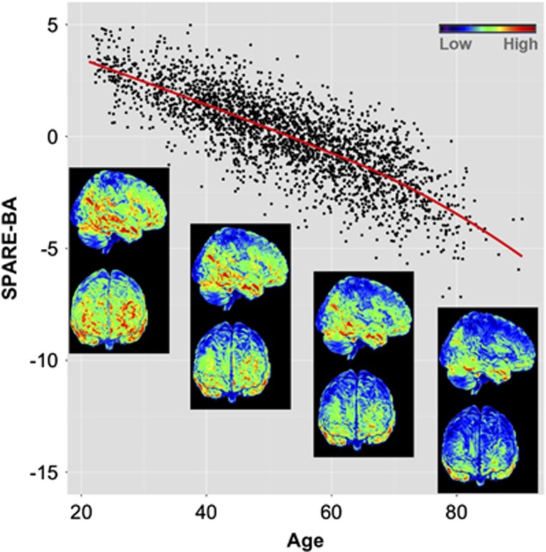
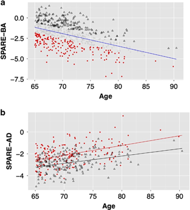
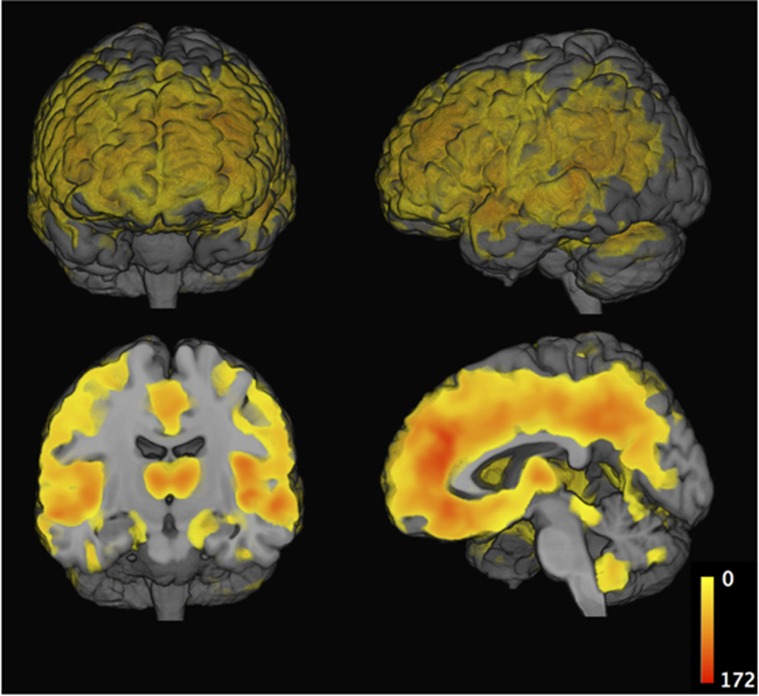
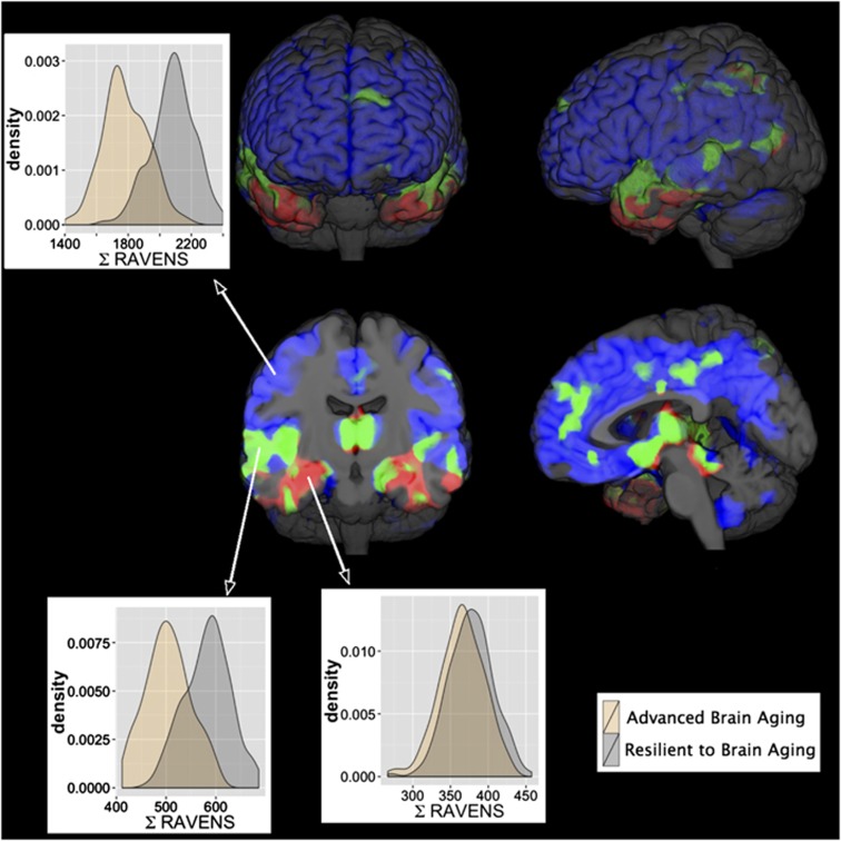
We systematically compared structural imaging patterns of advanced brain aging (ABA) in the general-population, herein defined as significant deviation from typical BA to those found in Alzheimer disease (AD). The hypothesis that ABA would show different patterns of structural change compared with those found in AD was tested via advanced pattern analysis methods. In particular, magnetic resonance images of 2705 participants from the Study of Health in Pomerania (aged 20-90 years) were analyzed using an index that captures aging atrophy patterns (Spatial Pattern of Atrophy for Recognition of BA (SPARE-BA)), and an index previously shown to capture atrophy patterns found in clinical AD (Spatial Patterns of Abnormality for Recognition of Early Alzheimer's Disease (SPARE-AD)). We studied the association between these indices and risk factors, including an AD polygenic risk score. Finally, we compared the ABA-associated atrophy with typical AD-like patterns. We observed that SPARE-BA had significant association with: smoking (P<0.05), anti-hypertensive (P<0.05), anti-diabetic drug use (men P<0.05, women P=0.06) and waist circumference for the male cohort (P<0.05), after adjusting for age. Subjects with ABA had spatially extensive gray matter loss in the frontal, parietal and temporal lobes (false-discovery-rate-corrected q<0.001). ABA patterns of atrophy were partially overlapping with, but notably deviating from those typically found in AD. Subjects with ABA had higher SPARE-AD values; largely due to the partial spatial overlap of associated patterns in temporal regions. The AD polygenic risk score was significantly associated with SPARE-AD but not with SPARE-BA. Our findings suggest that ABA is likely characterized by pathophysiologic mechanisms that are distinct from, or only partially overlapping with those of AD.
Figures
Similar articles
-
White matter hyperintensities and imaging patterns of brain ageing in the general population.Brain. 2016 Apr;139(Pt 4):1164-79. doi: 10.1093/brain/aww008. Epub 2016 Feb 24. Brain. 2016. PMID: 26912649 Free PMC article.
-
Inflammatory markers and imaging patterns of advanced brain aging in the general population.Brain Imaging Behav. 2020 Aug;14(4):1108-1117. doi: 10.1007/s11682-019-00058-y. Brain Imaging Behav. 2020. PMID: 30820858 Free PMC article.
-
Hippocampal Sclerosis of Aging, a Common Alzheimer's Disease 'Mimic': Risk Genotypes are Associated with Brain Atrophy Outside the Temporal Lobe.J Alzheimers Dis. 2016;52(1):373-83. doi: 10.3233/JAD-160077. J Alzheimers Dis. 2016. PMID: 27003218 Free PMC article.
-
Comparisons between Alzheimer disease, frontotemporal lobar degeneration, and normal aging with brain mapping.Top Magn Reson Imaging. 2005 Dec;16(6):409-25. doi: 10.1097/01.rmr.0000245457.98029.e1. Top Magn Reson Imaging. 2005. PMID: 17088691 Review.
-
Imaging and CSF studies in the preclinical diagnosis of Alzheimer's disease.Ann N Y Acad Sci. 2007 Feb;1097:114-45. doi: 10.1196/annals.1379.012. Ann N Y Acad Sci. 2007. PMID: 17413016 Review.
Cited by
-
Alzheimer's disease genetic risk and changes in brain atrophy and white matter hyperintensities in cognitively unimpaired adults.Brain Commun. 2024 Aug 14;6(5):fcae276. doi: 10.1093/braincomms/fcae276. eCollection 2024. Brain Commun. 2024. PMID: 39229494 Free PMC article.
-
Visual and Auditory Sensory Impairments Differentially Relate with Alzheimer's Pathology.Clin Psychopharmacol Neurosci. 2024 Nov 30;22(4):610-623. doi: 10.9758/cpn.24.1169. Epub 2024 Aug 16. Clin Psychopharmacol Neurosci. 2024. PMID: 39420608 Free PMC article.
-
Ten Years of BrainAGE as a Neuroimaging Biomarker of Brain Aging: What Insights Have We Gained?Front Neurol. 2019 Aug 14;10:789. doi: 10.3389/fneur.2019.00789. eCollection 2019. Front Neurol. 2019. PMID: 31474922 Free PMC article. Review.
-
ADCoC: Adaptive Distribution Modeling Based Collaborative Clustering for Disentangling Disease Heterogeneity from Neuroimaging Data.IEEE Trans Emerg Top Comput Intell. 2023 Apr;7(2):308-318. doi: 10.1109/tetci.2021.3136587. Epub 2022 Jan 5. IEEE Trans Emerg Top Comput Intell. 2023. PMID: 36969108 Free PMC article.
-
The personalized Alzheimer's disease cortical thickness index predicts likely pathology and clinical progression in mild cognitive impairment.Alzheimers Dement (Amst). 2018 Mar 17;10:301-310. doi: 10.1016/j.dadm.2018.02.007. eCollection 2018. Alzheimers Dement (Amst). 2018. PMID: 29780874 Free PMC article.
References
-
- Buckner RL. Memory and executive function in aging and ad: multiple factors that cause decline and reserve factors that compensate. Neuron 2004; 44: 195–208. - PubMed
-
- Rachael IS, Chris F, Rhian J, Jennifer LW, Martin NR, Nick CF. A longitudinal study of brain volume changes in normal aging using serial registered magnetic resonance imaging. Arch Neurol 2003; 60: 989–994. - PubMed
Publication types
MeSH terms
Grants and funding
LinkOut - more resources
Full Text Sources
Other Literature Sources
Medical
