Thymic stromal lymphopoietin (TSLP) inhibits human colon tumor growth by promoting apoptosis of tumor cells
- PMID: 26919238
- PMCID: PMC4941354
- DOI: 10.18632/oncotarget.7614
Thymic stromal lymphopoietin (TSLP) inhibits human colon tumor growth by promoting apoptosis of tumor cells
Abstract
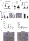
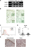
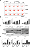
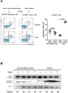
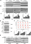
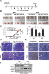
Thymic stromal lymphopoietin (TSLP) has recently been suggested in several epithelial cancers, either pro-tumor or anti-tumor. However, the role of TSLP in colon cancer remains unknown. We here found significantly decreased TSLP levels in tumor tissues compared with tumor-surrounding tissues of patients with colon cancer and TSLP levels negatively correlated with the clinical staging score of colon cancer. TSLPR, the receptor of TSLP, was expressed in all three colon cancer cell lines investigated and colon tumor tissues. The addition of TSLP significantly enhanced apoptosis of colon cancer cells in a TSLPR-dependent manner. Interestingly, TSLP selectively induced the apoptosis of colon cancer cells, but not normal colonic epithelial cells. Furthermore, we demonstrated that TSLP induced JNK and p38 activation and initiated apoptosis mainly through the extrinsic pathway, as caspase-8 inhibitor significantly reversed the apoptosis-promoting effect of TSLP. Finally, using a xenograft mouse model, we demonstrated that peritumoral administration of TSLP greatly reduced tumor growth accompanied with extensive tumor apoptotic response, which was abolished by tumor cell-specific knockdown of TSLPR. Collectively, our study reveals a novel anti-tumor effect of TSLP via direct promotion of the apoptosis of colon cancer cells, and suggests that TSLP could be of value in treating colon cancer.
Keywords: TSLP; TSLPR; apoptosis; caspase3; colon cancer.
Conflict of interest statement
There is no conflict of interest with any financial organization regarding the material discussed in the manuscript.
Figures
Similar articles
-
Expression and Regulation of Thymic Stromal Lymphopoietin and Thymic Stromal Lymphopoietin Receptor Heterocomplex in the Innate-Adaptive Immunity of Pediatric Asthma.Int J Mol Sci. 2018 Apr 18;19(4):1231. doi: 10.3390/ijms19041231. Int J Mol Sci. 2018. PMID: 29670037 Free PMC article. Review.
-
Thymic stromal lymphopoietin protects in a model of airway damage and inflammation via regulation of caspase-1 activity and apoptosis inhibition.Mucosal Immunol. 2020 Jul;13(4):584-594. doi: 10.1038/s41385-020-0271-0. Epub 2020 Feb 26. Mucosal Immunol. 2020. PMID: 32103153 Free PMC article.
-
Role of thymic stromal lymphopoietin in the pathogenesis of lumbar disc degeneration.Medicine (Baltimore). 2017 Jul;96(30):e7516. doi: 10.1097/MD.0000000000007516. Medicine (Baltimore). 2017. PMID: 28746197 Free PMC article.
-
Cervical carcinoma cells stimulate the angiogenesis through TSLP promoting growth and activation of vascular endothelial cells.Am J Reprod Immunol. 2013 Jul;70(1):69-79. doi: 10.1111/aji.12104. Epub 2013 Mar 18. Am J Reprod Immunol. 2013. PMID: 23495958
-
Thymic stromal lymphopoietin.Ann N Y Acad Sci. 2010 Jan;1183:13-24. doi: 10.1111/j.1749-6632.2009.05128.x. Ann N Y Acad Sci. 2010. PMID: 20146705 Free PMC article. Review.
Cited by
-
TSLP enhances progestin response in endometrial cancer via androgen receptor signal pathway.Br J Cancer. 2024 Mar;130(4):585-596. doi: 10.1038/s41416-023-02545-y. Epub 2024 Jan 3. Br J Cancer. 2024. PMID: 38172534 Free PMC article.
-
Expression and Regulation of Thymic Stromal Lymphopoietin and Thymic Stromal Lymphopoietin Receptor Heterocomplex in the Innate-Adaptive Immunity of Pediatric Asthma.Int J Mol Sci. 2018 Apr 18;19(4):1231. doi: 10.3390/ijms19041231. Int J Mol Sci. 2018. PMID: 29670037 Free PMC article. Review.
-
Expression and allele frequencies of Thymic stromal lymphopoietin are a key factor of breast cancer risk.Mol Genet Genomic Med. 2019 Aug;7(8):e813. doi: 10.1002/mgg3.813. Epub 2019 Jun 17. Mol Genet Genomic Med. 2019. PMID: 31210014 Free PMC article.
-
Targeting the Epithelium-Derived Innate Cytokines: From Bench to Bedside.Immune Netw. 2022 Feb 22;22(1):e11. doi: 10.4110/in.2022.22.e11. eCollection 2022 Feb. Immune Netw. 2022. PMID: 35291657 Free PMC article. Review.
-
Attenuated Dengue virus PV001-DV induces oncolytic tumor cell death and potent immune responses.J Transl Med. 2023 Jul 19;21(1):483. doi: 10.1186/s12967-023-04344-8. J Transl Med. 2023. PMID: 37468934 Free PMC article.
References
-
- Park LS, Martin U, Garka K, Gliniak B, Di Santo JP, Muller W, Largaespada DA, Copeland NG, Jenkins NA, Farr AG, Ziegler SF, Morrissey PJ, Paxton R, Sims JE. Cloning of the murine thymic stromal lymphopoietin (TSLP) receptor: Formation of a functional heteromeric complex requires interleukin 7 receptor. J Exp Med. 2000;192:659–670. - PMC - PubMed
-
- Pandey A, Ozaki K, Baumann H, Levin SD, Puel A, Farr AG, Ziegler SF, Leonard WJ, Lodish HF. Cloning of a receptor subunit required for signaling by thymic stromal lymphopoietin. Nat Immunol. 2000;1:59–64. - PubMed
MeSH terms
Substances
LinkOut - more resources
Full Text Sources
Other Literature Sources
Research Materials

