

Anti-Microbial Dendrimers against Multidrug-Resistant P. aeruginosa Enhance the Angiogenic Effect of Biological Burn-wound Bandages
- PMID: 26912450
- PMCID: PMC4766566
- DOI: 10.1038/srep22020
Anti-Microbial Dendrimers against Multidrug-Resistant P. aeruginosa Enhance the Angiogenic Effect of Biological Burn-wound Bandages
Erratum in
-
Corrigendum: Anti-Microbial Dendrimers against Multidrug-Resistant P. aeruginosa Enhance the Angiogenic Effect of Biological Burn-wound Bandages.Sci Rep. 2016 Apr 7;6:23872. doi: 10.1038/srep23872. Sci Rep. 2016. PMID: 27052685 Free PMC article. No abstract available.
Abstract
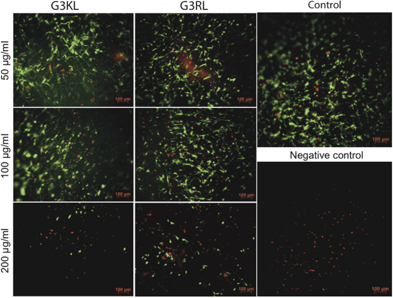
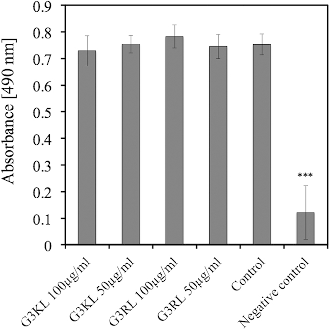
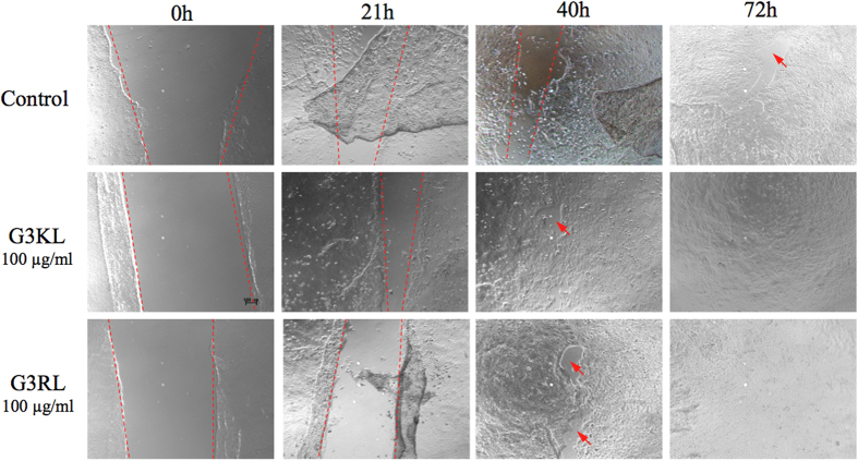
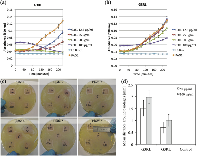
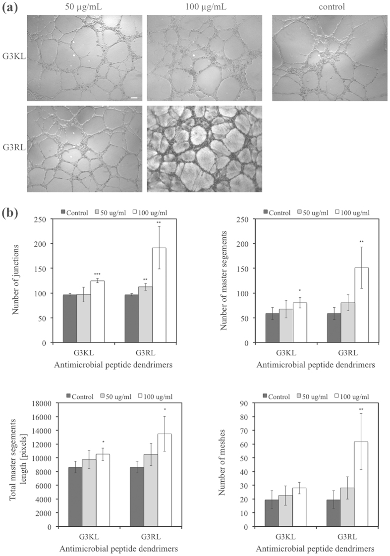
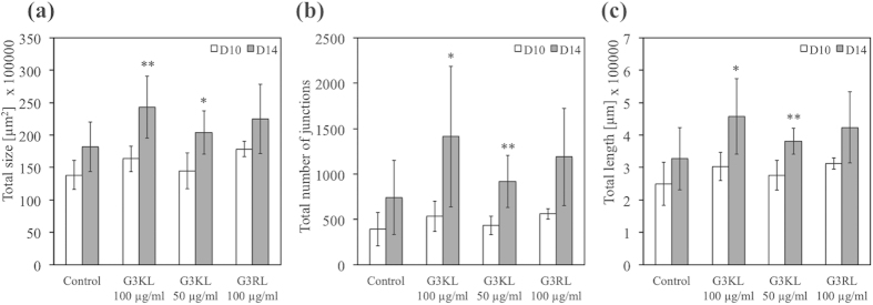
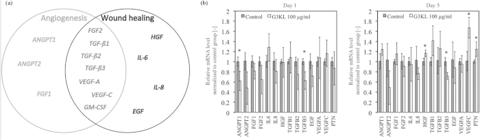
Multi-drug resistant Pseudomonas aeruginosa has increased progressively and impedes further regression in mortality in burn patients. Such wound infections serve as bacterial reservoir for nosocomial infections and are associated with significant morbidity and costs. Anti-microbial polycationic dendrimers G3KL and G3RL, able to kill multi-drug resistant P. aeruginosa, have been previously developed. The combination of these dendrimers with a class of biological bandages made of progenitor skin cells, which secrete growth factors, could positively impact wound-healing processes. However, polycations are known to be used as anti-angiogenic agents for tumor suppression. Since, neovascularization is pivotal in the healing of deep burn-wounds, the use of anti-microbial dendrimers may thus hinder the healing processes. Surprisingly, we have seen in this study that G3KL and G3RL dendrimers can have angiogenic effects. Moreover, we have shown that a dendrimer concentration ranging between 50 and 100 μg/mL in combination with the biological bandages can suppress bacterial growth without altering cell viability up to 5 days. These results show that antimicrobial dendrimers can be used in combination with biological bandages and could potentially improve the healing process with an enhanced angiogenesis.
Figures
Similar articles
-
Multifunctional Antimicrobial Nanofiber Dressings Containing ε-Polylysine for the Eradication of Bacterial Bioburden and Promotion of Wound Healing in Critically Colonized Wounds.ACS Appl Mater Interfaces. 2020 Apr 8;12(14):15989-16005. doi: 10.1021/acsami.9b21683. Epub 2020 Mar 27. ACS Appl Mater Interfaces. 2020. PMID: 32172559
-
Time-kill Kinetics of a Novel Antimicrobial Silver Wound Gel Against Select Wound Pathogens.Wounds. 2015 Dec;27(12):336-46. Wounds. 2015. PMID: 27447106
-
Impact of a novel, antimicrobial dressing on in vivo, Pseudomonas aeruginosa wound biofilm: quantitative comparative analysis using a rabbit ear model.Wound Repair Regen. 2014 Nov-Dec;22(6):712-9. doi: 10.1111/wrr.12232. Epub 2015 Jan 8. Wound Repair Regen. 2014. PMID: 25230854
-
A review of the scientific evidence for biofilms in wounds.Wound Repair Regen. 2012 Sep-Oct;20(5):647-57. doi: 10.1111/j.1524-475X.2012.00836.x. Wound Repair Regen. 2012. PMID: 22985037 Review.
-
New Nanotechnologies for the Treatment and Repair of Skin Burns Infections.Int J Mol Sci. 2020 Jan 8;21(2):393. doi: 10.3390/ijms21020393. Int J Mol Sci. 2020. PMID: 31936277 Free PMC article. Review.
Cited by
-
Synthesis and Characterization of Dendronized Gold Nanoparticles Bearing Charged Peripheral Groups with Antimicrobial Potential.Nanomaterials (Basel). 2022 Jul 29;12(15):2610. doi: 10.3390/nano12152610. Nanomaterials (Basel). 2022. PMID: 35957042 Free PMC article.
-
Stereorandomization as a Method to Probe Peptide Bioactivity.ACS Cent Sci. 2021 Jan 27;7(1):126-134. doi: 10.1021/acscentsci.0c01135. Epub 2021 Jan 19. ACS Cent Sci. 2021. PMID: 33532575 Free PMC article.
-
Are Antimicrobial Peptide Dendrimers an Escape from ESKAPE?Adv Wound Care (New Rochelle). 2020 May 19;9(7):378-95. doi: 10.1089/wound.2019.1113. Online ahead of print. Adv Wound Care (New Rochelle). 2020. PMID: 32320368 Free PMC article.
-
Shaping the Future of Antimicrobial Therapy: Harnessing the Power of Antimicrobial Peptides in Biomedical Applications.J Funct Biomater. 2023 Nov 2;14(11):539. doi: 10.3390/jfb14110539. J Funct Biomater. 2023. PMID: 37998108 Free PMC article. Review.
-
Nanotechnology-Driven Therapeutic Interventions in Wound Healing: Potential Uses and Applications.ACS Cent Sci. 2017 Mar 22;3(3):163-175. doi: 10.1021/acscentsci.6b00371. Epub 2017 Feb 27. ACS Cent Sci. 2017. PMID: 28386594 Free PMC article. Review.
References
Publication types
MeSH terms
Substances
LinkOut - more resources
Full Text Sources
Other Literature Sources
Medical
