. 2016 Mar;26(3):288-303.
doi: 10.1038/cr.2016.16. Epub 2016 Jan 29.

The RNA-binding protein Mex3B is a coreceptor of Toll-like receptor 3 in innate antiviral response

Affiliations

The RNA-binding protein Mex3B is a coreceptor of Toll-like receptor 3 in innate antiviral response

Yan Yang et al. Cell Res. 2016 Mar.

Abstract

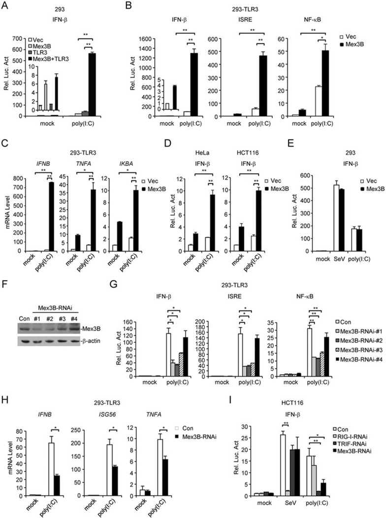
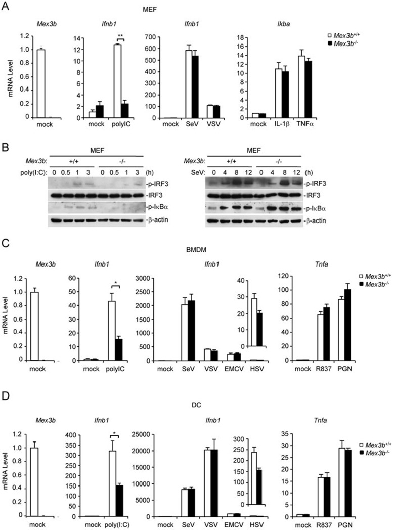
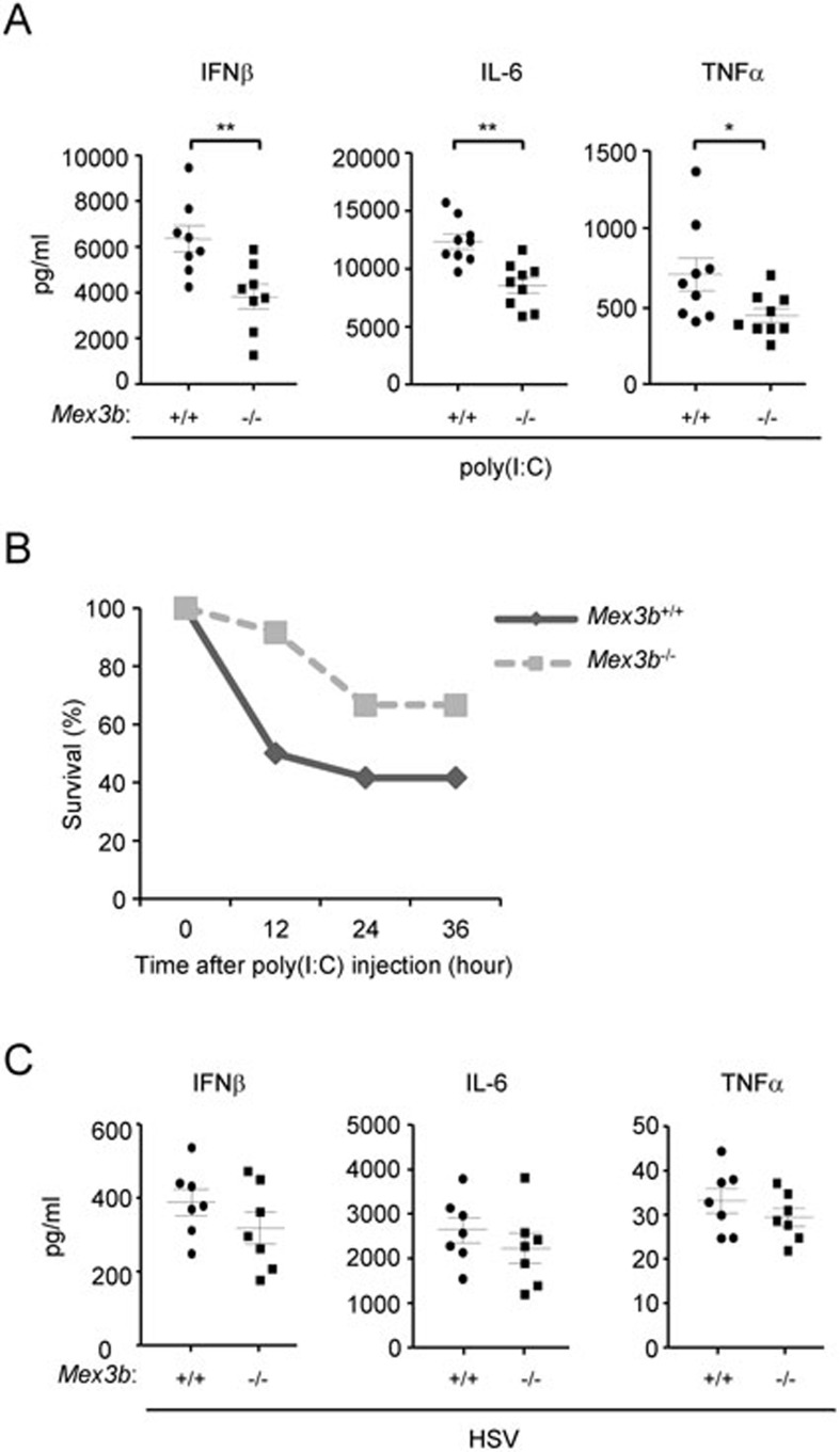
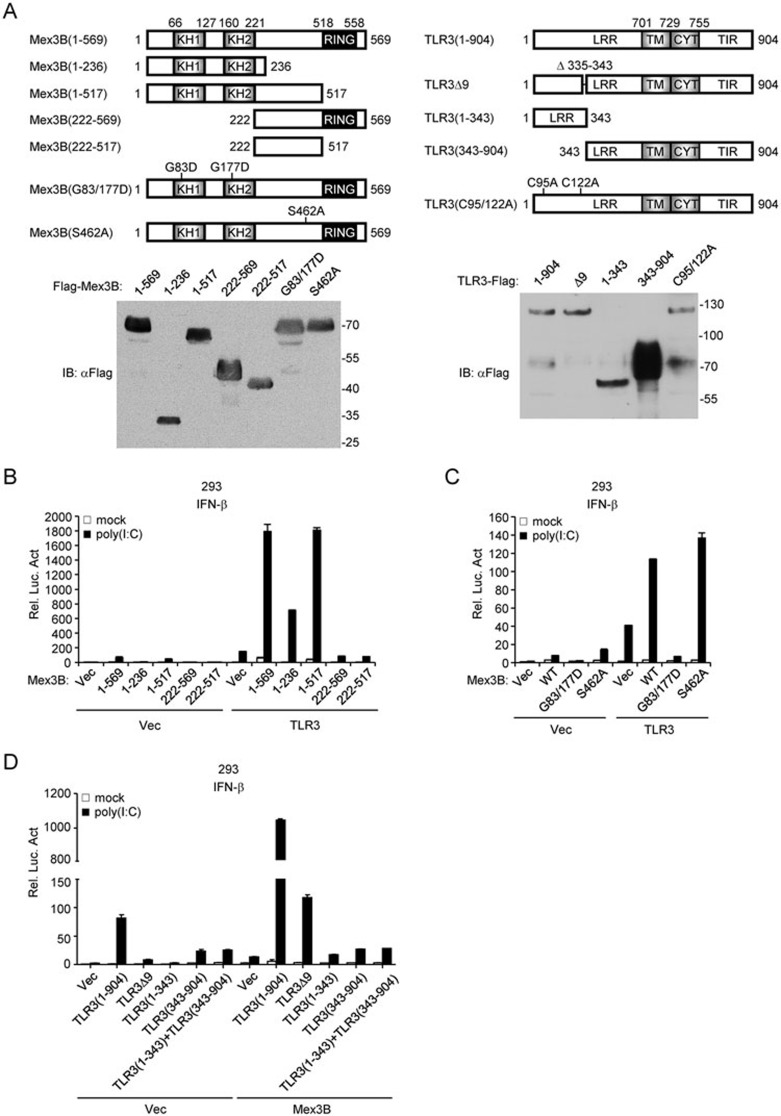
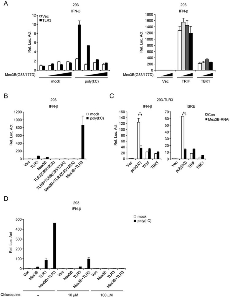
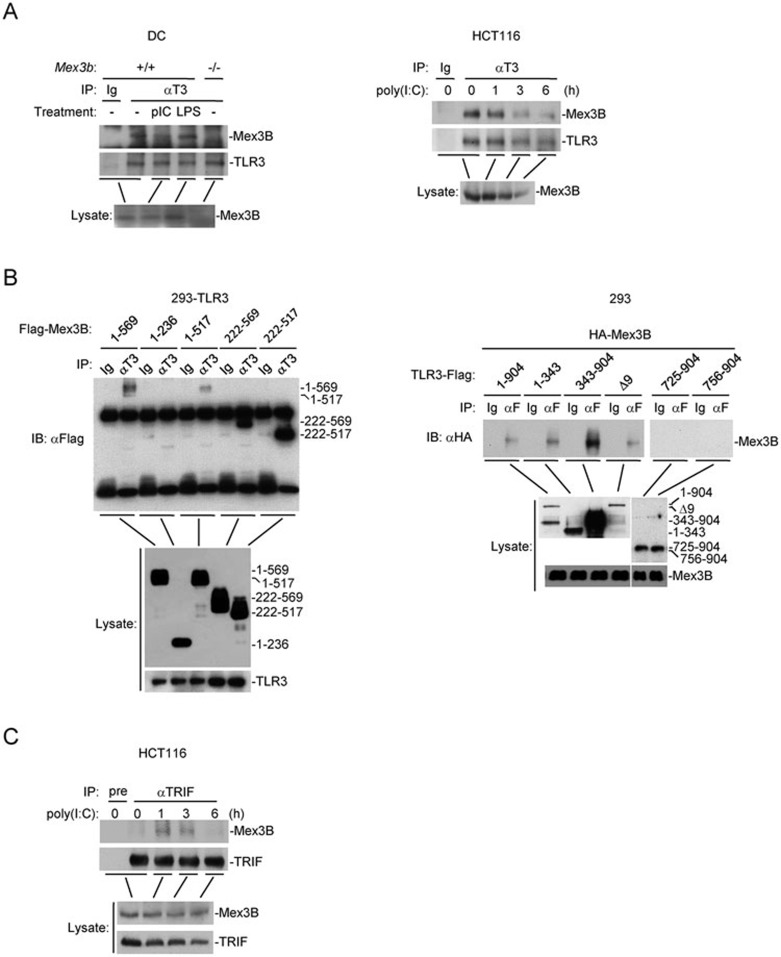
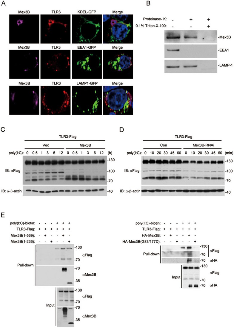
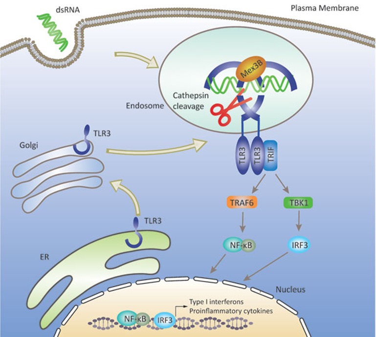
Recognition of viral dsRNA by Toll-like receptor 3 (TLR3) leads to induction of interferons (IFNs) and proinflammatory cytokines, and innate antiviral response. Here we identified the RNA-binding protein Mex3B as a positive regulator of TLR3-mediated signaling by expression cloning screens. Cells from Mex3b(-/-) mice exhibited reduced production of IFN-β in response to the dsRNA analog poly(I:C) but not infection with RNA viruses. Mex3b(-/-) mice injected with poly(I:C) was more resistant to poly(I:C)-induced death. Mex3B was associated with TLR3 in the endosomes. It bound to dsRNA and increased the dsRNA-binding activity of TLR3. Mex3B also promoted the proteolytic processing of TLR3, which is critical for its activation. Mutants of Mex3B lacking its RNA-binding activity inhibited TLR3-mediated IFN-β induction. These findings suggest that Mex3B acts as a coreceptor of TLR3 in innate antiviral response.


Figures

Figure 1
Figure 1
Mex3B is a positive regulator of TLR3-mediated signaling. (A) Mex3B potentiates TLR3-mediated signaling in 293 cells. The cells were transfected with the indicated plasmids for 24 h, and then treated with poly(I:C) for 6 h before luciferase assays. (B) Mex3B potentiates poly(I:C)-induced signaling in 293-TLR3 cells. Reporter assays were similarly performed as in A. (C) Mex3B potentiates poly(I:C)-induced expression of downstream genes in 293-TLR3 cells. The cells were transfected with the indicated plasmids for 24 h, and then treated with poly(I:C) for 3 h before qPCR analysis. (D) Mex3B potentiates poly(I:C)-induced activation of the IFN-β promoter in HeLa and HCT116 cells. Reporter assays were similarly performed as in A. (E) Overexpression of Mex3B has no marked effects on SeV- or cytoplasmic poly(I:C)-induced activation of the IFN-β promoter. The 293 cells were transfected with the indicated plasmids for 24 h, and then infected with SeV or re-transfected with poly(I:C) for 12 h before luciferase assays. (F) Effects of Mex3B-RNAi on expression of Mex3B. The 293 cells were transfected with a Mex3B-RNAi plasmid for 24 h before immunoblot analysis. (G) Effects of Mex3B-RNAi on TLR3-mediated signaling. Reporter assays were similarly performed as in A. (H) Effects of Mex3B-RNAi on poly(I:C)-induced transcription of IFNB1, ISG56 and TNFA genes. The experiments were similarly performed as in C. (I) Effects of Mex3B-RNAi on poly(I:C)- and SeV-induced activation of the IFN-β promoter in HCT116 cells. The cells were transfected with the indicated plasmids for 24 h, and then infected with SeV for 12 h or treated with poly(I:C) for 6 h before luciferase assays. Data are represented as mean ± SEM. *P < 0.05, **P < 0.01 (Student's t-test).
Figure 2
Figure 2
Mex3B is essential for TLR3-mediated signaling in MEFs, BMDMs and DCs. (A) Mex3B deficiency inhibits poly(I:C)-induced transcription of Ifnb1 gene in MEFs. MEFs (2 × 10⁵) were left untreated, treated with poly(I:C) (20 μg/ml), TNFα (10 ng/ml) or IL-1β (10 ng/ml) for 3 h, or infected with SeV or VSV for 12 h. Total RNA was then extracted for qPCR analysis. (B) Effects of Mex3B deficiency on poly(I:C)-induced phosphorylation of IRF3 and IκBα in MEFs. MEFs (2 × 10⁵) were left untreated, infected with SeV or treated with poly(I:C) for the indicated times. Cells were lysed and immunoblot was performed with the indicated antibodies. (C, D) Mex3B deficiency inhibits poly(I:C)-induced transcription of Ifnb1 gene in BMDMs (C) and DCs (D). The cells (2 × 10⁵) were left untreated, treated with poly(I:C) (20 μg/ml), R837 (20 μg/ml) or PGN (10 μg/ml) for 3 h or infected with SeV, VSV, EMCV or HSV for 12 h before qPCR analysis. Data are represented as mean ± SEM. *P < 0.05, **P < 0.01 (Student's t-test).
Figure 3
Figure 3
Mex3b−/− mice are resistant to poly(I:C)-induced death. (A) Sex- and age-matched Mex3b+/+ and Mex3b−/− mice (n = 8 or 9) were injected intraperitoneally with poly(I:C) for 2 h. Serum concentrations of the indicated cytokines were measured by ELISA. Data are represented as mean ± SEM. *P < 0.05, **P < 0.01 (Student's t-test). (B) Sex- and age-matched Mex3b+/+ and Mex3b−/− mice (n = 12) were injected intraperitoneally with poly(I:C) plus D-galactosamine. The survival of injected mice was monitored every 12 h for 4 days. (C) Mex3b+/+ and Mex3b−/− mice were infected i.p. with HSV-1 at 1 × 10⁷ p.f.u. per mouse (n = 7) for 6 h. Serum concentrations of the indicated cytokines were measured by ELISA.
Figure 4
Figure 4
The RNA-binding activity of Mex3B and proteolytic cleavage of TLR3 are indispensable for Mex3B to potentiate TLR3-mediated signaling. (A) A schematic presentation and expression of full-length Mex3B, TLR3 and their mutants. (B-D) Effects of Mex3B mutants on TLR3-mediated signaling. The 293 cells (1 × 10⁵) were transfected with the IFN-β promoter reporter (0.1 μg) and the indicated plasmids (0.1 μg) for 24 h, and then left untreated or treated with poly(I:C) (20 μg/ml) for 6 h before luciferase assays.
Figure 5
Figure 5
Mex3B regulates poly(I:C)-triggered signaling through TLR3. (A) Mex3B(G83/177D) inhibits poly(I:C)- but not TRIF-induced signaling. The 293 cells (1 × 10⁵) were transfected with the indicated plasmids and increased amounts of a Mex3B plasmid. Twenty-four hours after transfection, cells were treated with poly(I:C) (20 μg/ml) for 6 h before luciferase assays were performed. (B) TLR3(C95/122A) inhibits poly(I:C)-induced, TLR3/Mex3B-mediated signaling. The experiments were similarly performed as in A. (C) Knockdown of Mex3B inhibits poly(I:C)- but not TRIF- or TBK1-mediated activation of the IFN-β promoter. The 293-TLR3 cells (1 × 10⁵) were transfected with the IFN-β promoter reporter (0.1 μg) and either a control or Mex3B-RNAi plasmid (0.5 μg). Transfected cells were selected with puromycin (1 μg/ml) for 24 h followed by either treatment with poly(I:C) (20 μg/ml) or retransfection with TRIF or TBK-1 plasmids (0.1 μg). Luciferase assays were performed 6 h after treatment or 18 h after retransfection. Data are represented as mean ± SEM. *P < 0.05, **P < 0.01 (Student's t-test). (D) Effects of chloroquine on TLR3- and Mex3B-mediated signaling. The 293 cells (1 × 10⁵) were transfected with the indicated plasmids (0.1 μg each) for 24 h. The cells were pretreated with chloroquine for 30 min, and then treated with poly(I:C) (20 μg /ml) for 6 h before luciferase assays.
Figure 6
Figure 6
Mex3B is associated with TLR3 and facilitates the recruitment of TRIF upon poly(I:C) stimulation. (A) Mex3B interacts with TLR3. HCT116(Mex3B-3×Flag) cells (1 × 10⁸), in which a cDNA encoding three copies of Flag tag was inserted immediately before the stop codon of Mex3b gene, or mouse DCs (1 × 10⁸) were treated with poly(I:C) (20 μg /ml) or LPS (20 ng/ml) for the indicated times. Coimmunoprecipitation and immunoblots were performed with the indicated antibodies. pIC, poly(I:C). (B) Domain mapping of the Mex3B-TLR3 interaction. The 293-TLR3 cells (2 × 10⁶) were transfected with either the full-length or the indicated Mex3B truncation plasmids (5 μg). Coimmunoprecipitation and immunoblot were performed with the indicated antibodies. Ig, mouse IgG; αT3, anti-TLR3. (C) Mex3B interacts with TRIF upon poly(I:C) stimulation. HCT116 (Mex3B-3×Flag) cells (1 × 10⁸) were left untreated or treated with poly(I:C) (20 μg/ml) for the indicated times. Coimmunoprecipitation and immunoblots were performed with the indicated antibodies.
Figure 7
Figure 7
Mex3B colocalizes with TLR3 in the endosome and promotes the processing, stability and ligand binding of TLR3. (A) Mex3B colocalizes with TLR3 in the endosomes. The 293 cells (1 × 10⁵) were cotransfected with Mex3B-HA and TLR3-Flag plus KDEL-GFP (ER marker), EEA1-GFP (early endosome marker) or LAMP-1-GFP (lysosome marker). Twenty hours after transfection, cells were fixed with 4% paraformaldehyde, stained with anti-Flag and anti-HA antibodies, and subjected to confocal microscopy. (B) Mex3B is sorted into the lumen of endosomes. HCT116 (Mex3B-3×Flag) cells (4 × 10⁸) were collected and washed twice with ice-cold PBS. Endosome/lysosome fractions were isolated and either left untreated, treated with proteinase K (5 ng/ml) or treated with proteinase plus 0.1% Triton X-100 for 15 min at room temperature. The samples were then analyzed with the indicated antibodies. (C) Overexpression of Mex3B increases the abundance of processed C-terminal fragment of TLR3. The 293 cells (2 × 10⁵) were transfected with the indicated plasmids. Twenty-four hours after transfection, cells were treated with poly(I:C) (20 μg/ml) for the indicated times before immunoblot. The relative amount of total TLR3 was quantitated by the Bio-Rad Quantity One program. (D) Knockdown of Mex3B affects the stability of TLR3. The 293 cells (2 × 10⁵) were transfected with TLR3 (0.5 μg) and Mex3B-RNAi plasmid (1 μg). Transfected cells were selected with puromycin (1 μg/ml) for 24 h and then treated with poly(I:C) for the indicated times before immunoblot. (E) Mex3B enhances binding of poly(I:C) to TLR3. The 293 cells (2 × 10⁶) were transfected with the indicated plasmids (5 μg each). Cell lysates were mixed at 1:1 ratio as indicated and incubated with biotinylated-poly(I:C) for 1 h before pull-down assays were performed with streptavidin agarose. Precipitates and cell lysates were analyzed by immunoblot with the indicated antibodies.
Figure 8
Figure 8
A working model on the involvement of Mex3B in TLR3-mediated signaling. Upon viral infection, TLR3 transports from the ER to endosomes where it is proteolytically cleaved, resulting in the formation of a very stable “cleaved/associated” TLR3 that represents the primary form of the signaling receptor. The RNA-binding protein Mex3B localizes in the lumen of endosomes, functions as a coreceptor of TLR3 by promoting ligand binding and proteolytic processing of TLR3, resulting in full activation of TLR3.

Comment in

Similar articles

Cited by

References

    1. Kawai T, Akira S. Toll-like receptors and their crosstalk with other innate receptors in infection and immunity. Immunity 2011; 34:637–650. - PubMed
    1. Jin MS, Lee JO. Structures of the toll-like receptor family and its ligand complexes. Immunity 2008; 29:182–191. - PubMed
    1. Blasius AL, Beutler B. Intracellular toll-like receptors. Immunity 2010; 32:305–315. - PubMed
    1. McGettrick AF, O'Neill LA. Localisation and trafficking of Toll-like receptors: an important mode of regulation. Curr Opin Immunol 2010; 22:20–27. - PubMed
    1. Matsumoto M, Funami K, Tanabe M, et al. Subcellular localization of Toll-like receptor 3 in human dendritic cells. J Immunol 2003; 171:3154–3162. - PubMed

Publication types