

Adult Stem Cell Therapies for Wound Healing: Biomaterials and Computational Models
- PMID: 26793702
- PMCID: PMC4707872
- DOI: 10.3389/fbioe.2015.00206
Adult Stem Cell Therapies for Wound Healing: Biomaterials and Computational Models
Abstract
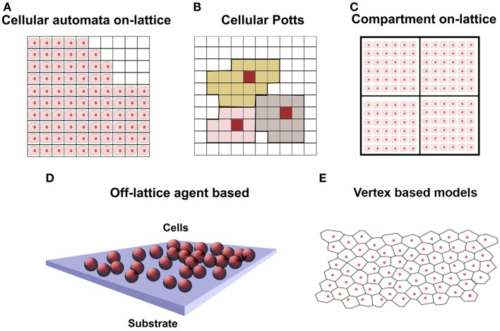
The increased incidence of diabetes and tumors, associated with global demographic issues (aging and life styles), has pointed out the importance to develop new strategies for the effective management of skin wounds. Individuals affected by these diseases are in fact highly exposed to the risk of delayed healing of the injured tissue that typically leads to a pathological inflammatory state and consequently to chronic wounds. Therapies based on stem cells (SCs) have been proposed for the treatment of these wounds, thanks to the ability of SCs to self-renew and specifically differentiate in response to the target bimolecular environment. Here, we discuss how advanced biomedical devices can be developed by combining SCs with properly engineered biomaterials and computational models. Examples include composite skin substitutes and bioactive dressings with controlled porosity and surface topography for controlling the infiltration and differentiation of the cells. In this scenario, mathematical frameworks for the simulation of cell population growth can provide support for the design of bioconstructs, reducing the need of expensive, time-consuming, and ethically controversial animal experimentation.
Keywords: Chaste; FLAME; adipose stem cells; cell-based modeling approaches; mesenchymal stem cells; wound healing.
Figures
Similar articles
-
[Effects of adipose-derived mesenchymal stem cells from type 2 diabetes mellitus patients on wound healing of pressure ulcers in mice].Zhonghua Shao Shang Za Zhi. 2019 Jan 20;35(1):40-47. doi: 10.3760/cma.j.issn.1009-2587.2019.01.008. Zhonghua Shao Shang Za Zhi. 2019. PMID: 30678400 Chinese.
-
Engineered Biopolymeric Scaffolds for Chronic Wound Healing.Front Physiol. 2016 Aug 5;7:341. doi: 10.3389/fphys.2016.00341. eCollection 2016. Front Physiol. 2016. PMID: 27547189 Free PMC article. Review.
-
Adipose Tissue-Derived Stromal Cells for Wound Healing.Adv Exp Med Biol. 2018;1119:133-149. doi: 10.1007/5584_2018_220. Adv Exp Med Biol. 2018. PMID: 29858972
-
[Effects of allogeneic mouse adipose-derived mesenchymal stem cell-microporous sheep acellular dermal matrix on healing of wound with full-thickness skin defect in mouse and the related mechanism].Zhonghua Shao Shang Za Zhi. 2018 Dec 20;34(12):901-906. doi: 10.3760/cma.j.issn.1009-2587.2018.12.015. Zhonghua Shao Shang Za Zhi. 2018. PMID: 30585055 Chinese.
-
Advanced Therapeutic Dressings for Effective Wound Healing--A Review.J Pharm Sci. 2015 Nov;104(11):3653-3680. doi: 10.1002/jps.24610. Epub 2015 Aug 26. J Pharm Sci. 2015. PMID: 26308473 Review.
Cited by
-
Biomembrane-Based Nanostructure- and Microstructure-Loaded Hydrogels for Promoting Chronic Wound Healing.Int J Nanomedicine. 2023 Jan 19;18:385-411. doi: 10.2147/IJN.S387382. eCollection 2023. Int J Nanomedicine. 2023. PMID: 36703725 Free PMC article. Review.
-
The Ethical Implications of Tissue Engineering for Regenerative Purposes: A Systematic Review.Tissue Eng Part B Rev. 2023 Apr;29(2):167-187. doi: 10.1089/ten.TEB.2022.0033. Epub 2022 Oct 20. Tissue Eng Part B Rev. 2023. PMID: 36112697 Free PMC article. Review.
-
The recent advances in the mathematical modelling of human pluripotent stem cells.SN Appl Sci. 2020;2(2):276. doi: 10.1007/s42452-020-2070-3. Epub 2020 Jan 27. SN Appl Sci. 2020. PMID: 32803125 Free PMC article. Review.
-
Hydrogel Scaffolds to Deliver Cell Therapies for Wound Healing.Front Bioeng Biotechnol. 2021 May 3;9:660145. doi: 10.3389/fbioe.2021.660145. eCollection 2021. Front Bioeng Biotechnol. 2021. PMID: 34012956 Free PMC article. Review.
-
Human adipose-derived stem cells promote seawater-immersed wound healing via proangiogenic effects.Aging (Albany NY). 2021 Mar 26;13(13):17118-17136. doi: 10.18632/aging.202773. Epub 2021 Mar 26. Aging (Albany NY). 2021. PMID: 33819183 Free PMC article.
References
-
- Byrne D. P., Lacroix D., Planell J. A., Kelly D. J., Prendergast P. J. (2007). Simulation of tissue differentiation in a scaffold as a function of porosity, Young’s modulus and dissolution rate: application of mechanobiological models in tissue engineering. Biomaterials 28, 5544–5554.10.1016/j.biomaterials.2007.09.003 - DOI - PubMed
Publication types
LinkOut - more resources
Full Text Sources
Other Literature Sources