

Enteroendocrine cells: a review of their role in brain-gut communication
- PMID: 26691223
- PMCID: PMC4842178
- DOI: 10.1111/nmo.12754
Enteroendocrine cells: a review of their role in brain-gut communication
Abstract
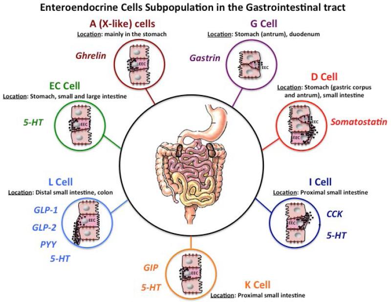
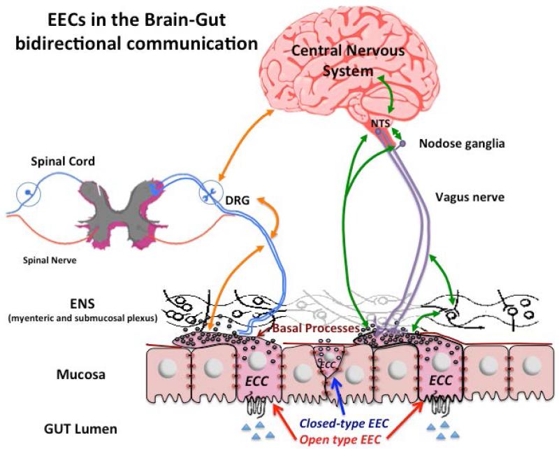
Background: Specialized endoderm-derived epithelial cells, that is, enteroendocrine cells (EECs), are widely distributed throughout the gastrointestinal (GI) tract. Enteroendocrine cells form the largest endocrine organ in the body and play a key role in the control of GI secretion and motility, the regulation of food intake, postprandial glucose levels and metabolism. EECs sense luminal content and release signaling molecules that can enter the circulation to act as classic hormones on distant targets, act locally on neighboring cells and on distinct neuronal pathways including enteric and extrinsic neurons. Recent studies have shed light on EEC sensory transmission by showing direct connections between EECs and the nervous system via axon-like processes that form a well-defined neuroepithelial circuits through which EECs can directly communicate with the neurons innervating the GI tract to initiate appropriate functional responses.
Purpose: This review will highlight the role played by the EECs in the complex and integrated sensory information responses, and discuss the new findings regarding EECs in the brain-gut axis bidirectional communication.
Keywords: afferent neurons; gut chemosensing; peptides.
© 2015 John Wiley & Sons Ltd.
Figures
Similar articles
-
Development and Anatomy of the Enteroendocrine System in Humans.Endocr Dev. 2017;32:20-37. doi: 10.1159/000475729. Epub 2017 Aug 15. Endocr Dev. 2017. PMID: 28873382
-
Enteroendocrine Regulation of Nutrient Absorption.J Nutr. 2020 Jan 1;150(1):10-21. doi: 10.1093/jn/nxz191. J Nutr. 2020. PMID: 31504661
-
Gut chemosensing mechanisms.J Clin Invest. 2015 Mar 2;125(3):908-17. doi: 10.1172/JCI76309. Epub 2015 Feb 9. J Clin Invest. 2015. PMID: 25664852 Free PMC article. Review.
-
Neuropods.Cell Mol Gastroenterol Hepatol. 2019;7(4):739-747. doi: 10.1016/j.jcmgh.2019.01.006. Epub 2019 Jan 30. Cell Mol Gastroenterol Hepatol. 2019. PMID: 30710726 Free PMC article. Review.
-
Gut chemosensing: interactions between gut endocrine cells and visceral afferents.Auton Neurosci. 2010 Feb 16;153(1-2):41-6. doi: 10.1016/j.autneu.2009.07.007. Epub 2009 Aug 11. Auton Neurosci. 2010. PMID: 19674941 Free PMC article. Review.
Cited by
-
Enteroendocrine cell regulation of the gut-brain axis.Front Neurosci. 2023 Nov 7;17:1272955. doi: 10.3389/fnins.2023.1272955. eCollection 2023. Front Neurosci. 2023. PMID: 38027512 Free PMC article. Review.
-
The complex involvement of the digestive tract in human defense behavior - structural and functional arguments.J Med Life. 2022 Sep;15(9):1081-1089. doi: 10.25122/jml-2022-0096. J Med Life. 2022. PMID: 36415517 Free PMC article. Review.
-
Novel Noninvasive Approaches to the Treatment of Obesity: From Pharmacotherapy to Gene Therapy.Endocr Rev. 2022 May 12;43(3):507-557. doi: 10.1210/endrev/bnab034. Endocr Rev. 2022. PMID: 35552683 Free PMC article. Review.
-
Lipopolysaccharides modulate intestinal epithelial permeability and inflammation in a species-specific manner.Gut Microbes. 2020 May 3;11(3):421-432. doi: 10.1080/19490976.2019.1629235. Epub 2019 Jun 16. Gut Microbes. 2020. PMID: 31203717 Free PMC article.
-
Gut microbes and food reward: From the gut to the brain.Front Neurosci. 2022 Jul 25;16:947240. doi: 10.3389/fnins.2022.947240. eCollection 2022. Front Neurosci. 2022. PMID: 35958993 Free PMC article. Review.
References
-
- Rehfeld JF. A centenary of gastrointestinal endocrinology. Horm Metab Res. 2004;36(11-12):735–741. - PubMed
-
- Janssen S, Depoortere I. Nutrient sensing in the gut: new road to therapeutics? Trends Endocrinol Metab. 2013;24(2):92–100. - PubMed
-
- Larsson LI, Goltermann N, De Magistris L, Rehfeld JF, Schwarz TW. Somatostatin cell processes as pathways for paracrine secretion. Science. 1979;205(4413):1393–1395. - PubMed
-
- Böttcher G, Sjölund K, Ekblad E, Håkanson R, Schwartz TW, Sundler F. Coexistence of peptide YY and glicentin immunoreactivity in endocrine cells of the gut. Regul Pept. 1984;8(4):261–266. - PubMed
Publication types
MeSH terms
Substances
Grants and funding
LinkOut - more resources
Full Text Sources
Other Literature Sources
