. 2015 Dec 14;36(5):678-92.
doi: 10.1128/MCB.00586-15.

SIRT3 Blocks Aging-Associated Tissue Fibrosis in Mice by Deacetylating and Activating Glycogen Synthase Kinase 3β

Affiliations

SIRT3 Blocks Aging-Associated Tissue Fibrosis in Mice by Deacetylating and Activating Glycogen Synthase Kinase 3β

Nagalingam R Sundaresan et al. Mol Cell Biol. .

Abstract

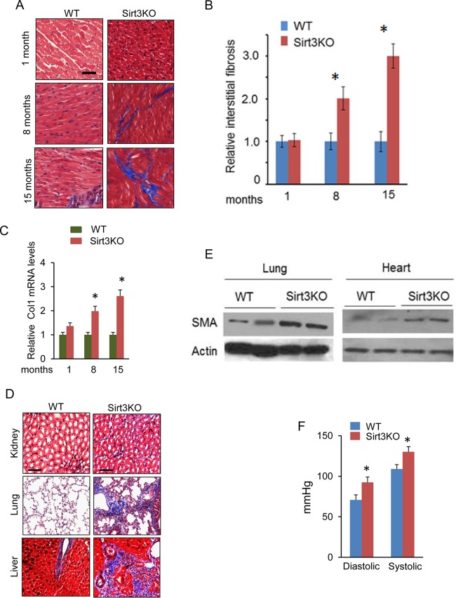
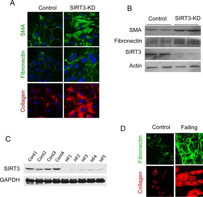
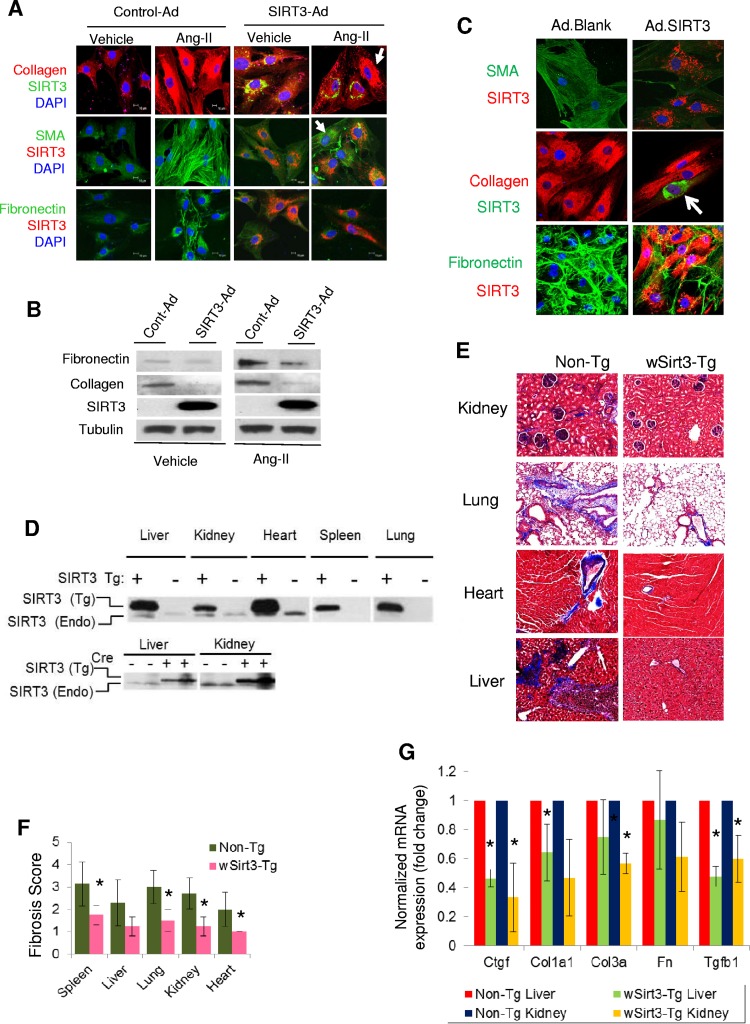
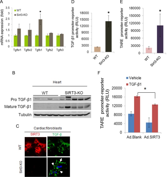
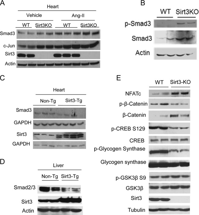
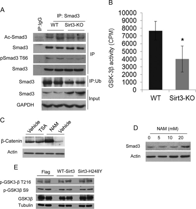
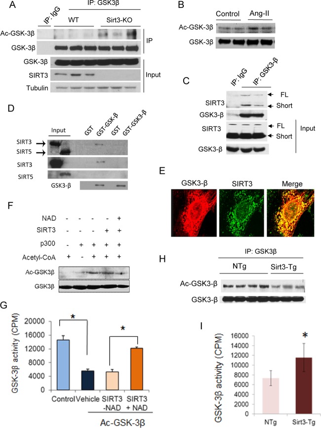
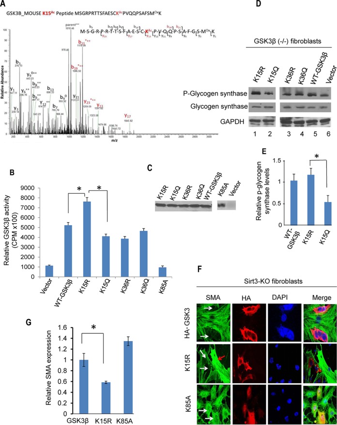
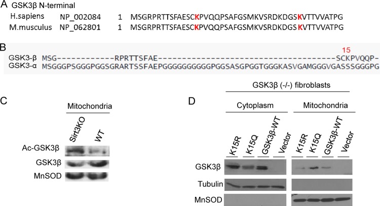
Tissue fibrosis is a major cause of organ dysfunction during chronic diseases and aging. A critical step in this process is transforming growth factor β1 (TGF-β1)-mediated transformation of fibroblasts into myofibroblasts, cells capable of synthesizing extracellular matrix. Here, we show that SIRT3 controls transformation of fibroblasts into myofibroblasts via suppressing the profibrotic TGF-β1 signaling. We found that Sirt3 knockout (KO) mice with age develop tissue fibrosis of multiple organs, including heart, liver, kidney, and lungs but not whole-body SIRT3-overexpressing mice. SIRT3 deficiency caused induction of TGF-β1 expression and hyperacetylation of glycogen synthase kinase 3β (GSK3β) at residue K15, which negatively regulated GSK3β activity to phosphorylate the substrates Smad3 and β-catenin. Reduced phosphorylation led to stabilization and activation of these transcription factors regulating expression of the profibrotic genes. SIRT3 deacetylated and activated GSK3β and thereby blocked TGF-β1 signaling and tissue fibrosis. These data reveal a new role of SIRT3 to negatively regulate aging-associated tissue fibrosis and discloses a novel phosphorylation-independent mechanism controlling the catalytic activity of GSK3β.


Figures

FIG 1
FIG 1
Reduced SIRT3 levels contribute to tissue fibrosis. (A) Comparative study of fibrosis in hearts, stained with Masson's trichrome stain (blue), of Sirt3-KO mice of different age groups and their age-matched wild-type (WT) controls. (B and C) Quantification of cardiac fibrosis and Col1 mRNA levels in Sirt3-KO and WT mice of different age groups. Values are means ± standard errors (n = 4). *, P < 0.01. (D) Tissue sections of 15-month-old WT and Sirt3-KO mice, stained to detect fibrosis (sections are representative of 5 mice per group). (E) Expression of the fibrotic marker SMA in the lung and heart tissue samples collected from 15-month-old Sirt3-KO and WT mice. (F) Systemic arterial pressure (femoral artery) in 15-months-old WT and Sirt3-KO mice. Values are means ± standard errors (n = 10). *, P < 0.001.
FIG 2
FIG 2
SIRT3 deficiency promotes transformation of human cardiac fibroblasts into myofibroblasts. (A and B) Expression of myofibroblast markers in control and SIRT3-depleted (SIRT3-KD) human cardiac fibroblasts as analyzed by confocal microscopy and Western blotting. (C) Western blot analysis showing SIRT3 levels in human cardiac fibroblasts isolated from controls and patients with heart failure (HF). (D) Confocal microscopy showing expression of myofibroblast markers in human cardiac fibroblasts isolated from control and failing hearts.
FIG 3
FIG 3
SIRT3 overexpression blocks aging-associated tissue fibrosis. (A) Mouse cardiac fibroblasts were overexpressed with control or adenovirus (Ad) vector synthesizing SIRT3. The next day, cells were stimulated with Ang-II (200 nM) for 48 h. The expression of myofibroblast markers was analyzed by confocal microscopy. Note the reduced expression of collagen, SMA, and fibronectin in SIRT3-overexpressing cells but not in cells lacking SIRT3 overexpression (arrows). DAPI, 4′,6′-diamidino-2-phenylindole. (B) Western blot analysis showing expression of the indicated proteins in mouse cardiac fibroblasts subjected to SIRT3 overexpression and stimulated with Ang-II. All four group samples were run in the same gel. (C) Cardiac myofibroblasts isolated from patients with heart failure were infected with control (blank) or SIRT3 adenovirus. The expression of myofibroblast markers (SMA, collagen, and fibronectin) was analyzed by confocal microscopy. SIRT3 expression was verified by immunostaining for SIRT3 (red) in SMA and fibronectin panels and in collagen panels (green). Note that SIRT3 overexpression (arrow) blocked the expression of collagen. (D) The Cre-recombinase-dependent overexpression of SIRT3-FLAG in different organs was detected by Western blotting using a specific antiserum for mouse SIRT3. The endogenous (Endo) SIRT3 protein (lacking a tag) is present as a lower-abundance and lower-molecular-weight protein. (E and F) Tissue fibrosis was analyzed in 15-month-old nontransgenic (Non-Tg) and whole-body Sirt3-transgenic (wSirt3-Tg) mice (C57BL/6). Tissue sections were stained with Masson's trichrome stain (E), and relative fibrosis was scored (F) in a blinded fashion (n = 4 to 6 mice per group). (G) Real-time PCR analysis of mRNA of fibrosis-related genes in the liver and kidney samples of non-Tg and wSirt3-Tg mice. Values are means ± standard errors (n = 4 to 7 mice per group). *, P < 0.001.
FIG 4
FIG 4
SIRT3 deficiency activates TGF-β1 synthesis. (A) Reverse transcription-PCR analysis showing expression of different members of the TGF-β family ligands and receptors (r) in the hearts of adult (8 months old) WT and Sirt3-KO mice. Values are means ± standard errors (n = 5 mice per group). *, P < 0.001. (B) Western blot analysis of pro-TGF-β1 and mature TGF-β1 in mouse hearts (n = 4 or 5 mice per group). (C) Cells were immunostained for SIRT3 and TGF-β1 and imaged by confocal microscopy. Note the increased amount of membrane-bound TGF-β1 (arrows) in Sirt3-KO fibroblasts. (D) A luciferase reporter assay showing activity of the TGF-β1 natural promoter in cardiac fibroblast of WT and Sirt3-KO hearts. Values are means ± standard errors (n = 3). *, P < 0.001). (E) A luciferase reporter assay was performed with a synthetic promoter containing multiple TGF-β/activin response elements (TAREs) in cardiac fibroblasts of WT and Sirt3-KO mice. Values are means ± standard errors (n = 6). *, P < 0.001). (F) The TARE promoter/reporter plasmid was transfected into control and SIRT3-overexpressing cardiac fibroblasts. Cells were stimulated with vehicle or 10 nM recombinant TGF-β1 for 12 h, and the luciferase activity was measured. Values are means ± standard errors (n = 3). *, P < 0.001. RLU, relative light units.
FIG 5
FIG 5
SIRT3 regulates expression of profibrotic transcription factors. (A) Western blot analysis of the indicated proteins in adult mouse hearts stimulated to develop cardiac hypertrophy and fibrosis by Ang-II infusion (n = 5 in each group). (B) Western blot analysis showing Smad3 levels in WT and Sirt3-KO cardiac fibroblasts. (C and D) Western blot analysis showing Smad3 levels in the heart and liver of nontransgenic (Non-Tg) and whole-body Sirt3-transgenic mice. Blots are representative of 5 animals in each group. (E) Western blot analysis showing expression levels of different GSK3β targets in WT and Sirt3-KO hearts (n = 5 mice in each group).
FIG 6
FIG 6
Sirt3 deficiency reduces GSK3β activity and stabilizes Smad3 levels. (A) Smad3 was immunoprecipitated (IP) from WT and Sirt3-KO hearts and analyzed for acetylation and T66 phosphorylation using specific antibodies. For this analysis, Smad3 levels were normalized. In the same lysate, ubiquitinated Smad3 was analyzed by immunoprecipitation with antiubiquitin (Ub) antibody, followed by Western blotting. (B) GSK3β prepared from heart lysates of WT and Sirt3-KO mice was tested for its ability to phosphorylate the glycogen synthase recombinant peptide. Values are mean ± standard errors (n = 7). *, P < 0.001. (C) Cardiac fibroblasts were treated with the HDAC inhibitor trichostatin A (TSA; 1 μM) or the pansirtuin inhibitor nicotinamide (NAM; 20 mM) overnight. The β-catenin levels were determined by Western blotting. (D) Western blot analysis showing Smad3 levels in fibroblasts treated with increasing concentrations of NAM. (E) Western analysis showing expression of phosphorylated GSK3β in cardiac fibroblasts subjected to overexpression of WT Sirt3 or an Sirt3 mutant.
FIG 7
FIG 7
SIRT3 deacetylates and activates GSK3β. (A and B) GSK3β was immunoprecipitated from heart lysates of WT and Sirt3-KO mice (A) or mice stimulated to develop hypertrophy by Ang-II infusion (B). The precipitate was analyzed by Western blot analysis using antiacetyllysine antibody. (C) Coimmunoprecipitation experiments showing GSK3β binding to both full-length (FL) and short forms of SIRT3 in human cardiac fibroblasts (n = 3). (D) Flag-tagged [35S]SIRT3 or [35S]SIRT5 were synthesized using a TNT-Quick Coupled transcription/translation system, and their binding to GSK3β was analyzed by a GST pulldown assay. Autoradiograms in the upper panel show binding of [35S]SIRT3 to GST-GSK3β. Expression levels of SIRT3, SIRT5, and GSK3β were detected by Western blotting. Part of this figure was adapted from our previous work (input lanes 1 and 2 from Fig. 2A in reference 18). (E) Confocal microscopy showing colocalization of GSK3-β (red) and SIRT3 (green) in human cardiac fibroblasts (n = 5). (F and G) Recombinant HA-GSK3β was incubated with recombinant p300 and cofactor acetyl coenzyme A (acetyl-CoA). Following completion of the reaction, Ac-GSK3β was incubated with the recombinant SIRT3 in the presence or absence of NAD for 2 h in a deacetylation reaction buffer. GSK3β acetylation was analyzed by Western blotting (F), and enzymatic activity was determined against a glycogen synthase peptide (G). (H) GSK3β immunoprecipitated from heart lysates of non-Tg (NTg) and SIRT3-Tg mice was probed with antiacetyllysine antibody. (I) GSK3β was immunoprecipitated from heart lysates of non-Tg and Sirt3-Tg mice and analyzed for catalytic activity against the glycogen synthase peptide. Values are means ± standard error (n = 4). *, P < 0.001.
FIG 8
FIG 8
Lysine 15 (K15) acetylation regulates GSK3β activity. (A) Annotation of representative tandem mass spectra of trypsin-digested GSK3β, depicting K15 acetylation (in red). (B) HA-tagged wild type and K15R, K15Q, K36R, K36Q, and K85A mutants of GSK3β were purified from GSK3β null mouse embryonic fibroblasts using HA antibody-conjugated agarose beads. The enzymatic activity of GSK3β mutants was determined against a glycogen synthase peptide. Values are means ± standard errors (n = 5). *, P < 0.001. (C) Western analysis showing expression levels of different GSK3β mutants. (D) GSK3β null fibroblasts were overexpressed with different forms of GSK3β. The catalytic activity of GSK3β was assayed by measuring the phosphorylation of glycogen synthase by Western blotting. (E) Quantification of glycogen synthase phosphorylation by GSK3β-K15 mutants. Note the increased activity of GSK3β-K15R in phosphorylation of the substrate compared to that of GSK3β-K15Q. Values are means plus standard errors (n = 3). *, P < 0.01. (F) Sirt3-KO fibroblasts were overexpressed with different GSK3β constructs. Expression of SMA was determined by confocal imaging. White arrows indicate reduced SMA expression in GSK3β-WT- and GSK3β-K15R-expressing cells but not in GSK3β mutant (K85A) cells. The red arrow indicates that cells negative for GSK3β-K15R expression robustly express SMA. (G) Quantification of SMA levels in cells expressing different GSK3β mutants. Values are means ± standard errors (n = 3).
FIG 9
FIG 9
GSK3β acetylation at residue K15 regulates its mitochondrial localization. (A) N-terminal regions of GSK3β from different vertebrate species, showing acetylated K15 and K36 residues (in red). (B) Comparison of the N-terminal regions of GSK3β and GSK3α showing that residue K15 is present only in the β-isoform. (C) GSK3β prepared from the mitochondrial fraction of the heart lysate was assayed by Western blotting using antiacetyllysine and antibodies against the indicated proteins. (D) GSK3β null mouse embryonic fibroblasts were subjected to overexpression of different versions of GSK3β. The cytoplasmic and mitochondrial fractions of cells were prepared and analyzed by Western blotting using antibodies against the indicated proteins. Blots are representative of three separate experiments. Note that K15Q is mostly present in mitochondria and that K15R is located in the cytoplasm. H. sapiens, Homo sapiens; M. musculus, Mus musculus.
FIG 10
FIG 10
TGF-β inhibitors block fibrotic changes of Sirt3-KO cells/tissues. (A) Confocal microscopy showing SMA expression in wild-type and Sirt3-KO cardiac fibroblasts treated with the TGF-β inhibitor follistatin (200 nM) or SB-505124 (1 μM) for 48 h. DAPI, 4′,6′-diamidino-2-phenylindole. (B) Western analysis showing expression levels of SMA and Smad3 in the same cells as used for the experiment described for panel A. (C) WT and Sirt3-KO mice (10 months old) were treated with vehicle or SB-505124 at a dose of 10 mg kg−1 for 2 months (three intraperitoneal injections/week). Tissue fibrosis was analyzed by Masson's trichrome staining of tissue sections (n = 7 mice per group). (D) Real-time PCR analysis of collagen 1 gene expression in different groups of mice. Values are means ± standard errors (n = 4 or 5 mice per group). *, P < 0.001. (E) A simplified scheme illustrating the role of SIRT3 in regulating GSK3β–TGFβ–Smad3 signaling and development of fibrosis. SIRT3 deacetylates GSK3β at mitochondria. In SIRT3-deficient cells GSK3β is acetylated, inhibiting its ability to phosphorylate the substrates. Decreased GSK3β-dependent phosphorylation causes stabilization of substrates like Smad3, c-Jun, and β-catenin, leading to their increased import into the nucleus, where they regulate the expression of profibrotic genes and transformation of fibroblasts into myofibroblasts.

Similar articles

Cited by

References

    1. Mehal WZ, Iredale J, Friedman SL. 2011. Scraping fibrosis: expressway to the core of fibrosis. Nat Med 17:552–553. doi:10.1038/nm0511-552. - DOI - PMC - PubMed
    1. Wynn TA, Ramalingam TR. 2012. Mechanisms of fibrosis: therapeutic translation for fibrotic disease. Nat Med 18:1028–1040. doi:10.1038/nm.2807. - DOI - PMC - PubMed
    1. Krenning G, Zeisberg EM, Kalluri R. 2010. The origin of fibroblasts and mechanism of cardiac fibrosis. J Cell Physiol 225:631–637. doi:10.1002/jcp.22322. - DOI - PMC - PubMed
    1. Frame S, Cohen P. 2001. GSK3 takes centre stage more than 20 years after its discovery. Biochem J 359:1–16. doi:10.1042/0264-6021:3590001. - DOI - PMC - PubMed
    1. Chiara F, Rasola A. 2013. GSK-3 and mitochondria in cancer cells. Front Oncol 3:16. doi:10.3389/fonc.2013.00016. - DOI - PMC - PubMed

Publication types

MeSH terms