. 2016 Feb;356(2):354-65.
doi: 10.1124/jpet.115.230052. Epub 2015 Nov 19.

Dinaciclib, a Cyclin-Dependent Kinase Inhibitor Promotes Proteasomal Degradation of Mcl-1 and Enhances ABT-737-Mediated Cell Death in Malignant Human Glioma Cell Lines

Affiliations

Dinaciclib, a Cyclin-Dependent Kinase Inhibitor Promotes Proteasomal Degradation of Mcl-1 and Enhances ABT-737-Mediated Cell Death in Malignant Human Glioma Cell Lines

Esther P Jane et al. J Pharmacol Exp Ther. 2016 Feb.

Abstract

The prognosis for malignant glioma, the most common brain tumor, is still poor, underscoring the need to develop novel treatment strategies. Because glioma cells commonly exhibit genomic alterations involving genes that regulate cell-cycle control, there is a strong rationale for examining the potential efficacy of strategies to counteract this process. In this study, we examined the antiproliferative effects of the cyclin-dependent kinase inhibitor dinaciclib in malignant human glioma cell lines, with intact, deleted, or mutated p53 or phosphatase and tensin homolog on chromosome 10; intact or deleted or p14ARF or wild-type or amplified epidermal growth factor receptor. Dinaciclib inhibited cell proliferation and induced cell-cycle arrest at the G2/M checkpoint, independent of p53 mutational status. In a standard 72-hour 3-[4,5-dimethylthiazol- 2yl]-5-[3-carboxymethoxyphenyl]-2-[4-sulfophenyl]-2H, tetrazolium (MTS) assay, at clinically relevant concentrations, dose-dependent antiproliferative effects were observed, but cell death was not induced. Moreover, the combination of conventional chemotherapeutic agents and various growth-signaling inhibitors with dinaciclib did not yield synergistic cytotoxicity. In contrast, combination of the Bcl-2/Bcl-xL inhibitors ABT-263 (4-[4-[[2-(4-chlorophenyl)-5,5-dimethylcyclohexen-1-yl]methyl]piperazin-1-yl]-N-[4-[[(2R)-4-morpholin-4-yl-1-phenylsulfanylbutan-2-yl]amino]-3-(trifluoromethylsulfonyl)phenyl]sulfonylbenzamide) or ABT-737 (4-[4-[[2-(4-chlorophenyl)phenyl]methyl]piperazin-1-yl]-N-[4-[[(2R)-4-(dimethylamino)-1-phenylsulfanylbutan-2-yl]amino]-3-nitrophenyl]sulfonylbenzamide) with dinaciclib potentiated the apoptotic response induced by each single drug. The synergistic killing by ABT-737 with dinaciclib led to cell death accompanied by the hallmarks of apoptosis, including an early loss of the mitochondrial transmembrane potential; the release of cytochrome c, smac/DIABLO, and apoptosis-inducing factor; phosphatidylserine exposure on the plasma membrane surface and activation of caspases and poly ADP-ribose polymerase. Mechanistic studies revealed that dinaciclib promoted proteasomal degradation of Mcl-1. These observations may have important clinical implications for the design of experimental treatment protocols for malignant human glioma.


Figures

Fig. 1.
Fig. 1.
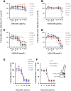
Differential response to CDK inhibitors in malignant human glioma cell lines. Established malignant human glioma cell lines were grown on 96-well plates in growth medium and, after an overnight attachment period, were exposed to selected concentrations of ribociclib (A), palbociclib (B), AZD-5438 (C), AMG-925 (D), or dinaciclib (E). Control cells received vehicle (DMSO, 0) for 72 hours. Cell proliferation inhibition was assessed semiquantitatively by spectrophotometric measurement of MTS bioreduction. Points represent the mean of three measurements carried out in triplicate. (F) Equal amounts of protein from logarithmically growing EGFR-overexpressing cell lines U87-EGFR-WT, U87-EGFRviii, and isogenic control U87 were separated by SDS-PAGE analysis and subjected to Western blotting analysis with the indicated primary antibodies (F, right panel). In parallel, cell proliferation assay was performed with dinaciclib as described in Materials and Methods (F, left panel). Points represent the mean of three measurements ± S.D.
Fig. 2.
Fig. 2.
Cytostatic effect of dinaciclib on in vitro cultured glioma cells (A) U87, U87-EGFR-WT, U87-EGFRviii, LNZ308, LN229, LN18, T98G, U373, and A172 cells were seeded at 60% confluence, allowed to attach overnight, and treated with dinaciclib (5.0 µM) for 24 hours. Control cells received an equivalent amount of DMSO. Apoptosis was analyzed by flow cytometry. Bar chart represents data from three independent experiments. (B) U87, U87-EGFRviii, LNZ308, U373, LN229, LN18, and A172 cells were seeded at 60% confluence, allowed to attach overnight, and treated with dinaciclib (2.5 µM) for 24 hours. Cells were stained with Alexa Flour 488 Phalloidin as described in Materials and Methods. Nuclei were stained with DAPI. Control cells received DMSO. Morphologic and nuclear changes in response to inhibitor treatment were evaluated by microscopic inspection. (C) U87, U87-EGFRviii, LNZ308, U373, LN229, LN18, and A172 cells were seeded at 60% confluence, allowed to attach overnight, and treated with dinaciclib (2.5 µM) for 24 hours. Cell-cycle analysis using PI staining was performed as described in Materials and Methods. Results represent the mean of three independent experiments.
Fig. 3.
Fig. 3.
Combined treatment with dinaciclib and ABT-737 effectively kills glioma cells. (A) T98G cells were seeded at 60% confluence, allowed to attach overnight, and treated with dinaciclib (1.0 µM) or indicated signaling inhibitor (refer to Supplemental Table 2 for the concentrations used in this study) or the combination of both for 24 hours. Control cells received an equivalent amount of DMSO. Apoptosis was analyzed by flow cytometry as described in Materials and Methods. The results represent the mean of two independent experiments representing various stages of cell death. (B) U87, U87-EGFRviii, LNZ308, LN229, LN18, and T98G cells were treated with dinaciclib (100 nM (D), ABT-737 (100 nM) (A), or the combination of both (D and A). Control cells received DMSO (C). Apoptosis was analyzed by flow cytometry as described in Materials and Methods. The results represent the mean of three independent experiments. (C) In parallel, cell extracts (from U87, LNZ308, and T98G) were prepared, and equal amounts of protein were separated by SDS-PAGE and subjected to Western blot analysis with the indicated antibodies. β-actin served as loading control. The results of a representative study are shown; two additional experiments produced similar results.
Fig. 3.
Fig. 3.
Combined treatment with dinaciclib and ABT-737 effectively kills glioma cells. (A) T98G cells were seeded at 60% confluence, allowed to attach overnight, and treated with dinaciclib (1.0 µM) or indicated signaling inhibitor (refer to Supplemental Table 2 for the concentrations used in this study) or the combination of both for 24 hours. Control cells received an equivalent amount of DMSO. Apoptosis was analyzed by flow cytometry as described in Materials and Methods. The results represent the mean of two independent experiments representing various stages of cell death. (B) U87, U87-EGFRviii, LNZ308, LN229, LN18, and T98G cells were treated with dinaciclib (100 nM (D), ABT-737 (100 nM) (A), or the combination of both (D and A). Control cells received DMSO (C). Apoptosis was analyzed by flow cytometry as described in Materials and Methods. The results represent the mean of three independent experiments. (C) In parallel, cell extracts (from U87, LNZ308, and T98G) were prepared, and equal amounts of protein were separated by SDS-PAGE and subjected to Western blot analysis with the indicated antibodies. β-actin served as loading control. The results of a representative study are shown; two additional experiments produced similar results.
Fig. 4.
Fig. 4.
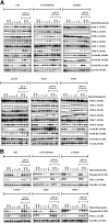
Effect of dinaciclib and ABT-737 on the cell-cycle profile and the expression levels of cell-cycle regulatory proteins. (A and B) Logarithmically growing U87, U87-EGFRviii, LNZ308 (upper panel), LN229, LN18, and T98G (lower panel) cells were treated with dinaciclib (indicated concentrations) or ABT-737 (100 nM) or the combination of both for 24 hours. Control cells received equivalent concentrations of vehicle, DMSO. Whole-cell extracts were prepared, and equal amounts of protein were separated by SDS-PAGE and subjected to Western blotting analysis with the indicated antibodies. β-actin served as loading control (A). Total Rb served as loading control (B). The results of a representative study are shown; two additional experiments produced similar results.
Fig. 5.
Fig. 5.
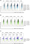
Cotreatment with dinaciclib and ABT-737 induces mitochondrial membrane potential dysfunction and conformational changes of the proapoptotic protein Bax. Logarithmically growing U87, U87-EGFRviii, LNZ308, LN229, LN18, and T98G cells were treated with dinaciclib (indicated concentrations) (A), ABT-737 (indicated concentrations (B), or the combination of both (indicated concentrations (C) for 24 hours. The integrity of the mitochondrial membranes of the cells was examined by DiOC6 staining and flow cytometry. (D) U87, U87-EGFRviii, LNZ308, LN229, LN18, and T98G cells were treated with dinaciclib (50 nM) or ABT-737 (50 nM) or the combination of both for indicated durations. Cytosolic extracts were prepared, and equal amounts of protein were separated by SDS-PAGE and subjected to Western blotting analysis with the indicated antibodies. (E), U87, U87-EGFRviii, LNZ308, LN229, LN18, and T98G cells were treated with dinaciclib (50 nM) or ABT-737 (50 nmol/L) or the combination of both for the indicated duration and lysed with 1% CHAPS buffer. An equal amount of protein (500 µg) was immunoprecipitated with monoclonal anti-Bax (6A7; Sigma-Aldrich) antibody and then subjected to Western blot analysis with polyclonal anti-Bax antibody (Cell Signaling Technology). (F) U87, U87-EGFRviii, LNZ308, LN229, LN18, and T98G cells were treated with dinaciclib (50 nM) or ABT-737 (50 nM) or the combination of both for 12 hours. Control cells received equivalent amounts of DMSO. Membrane fractions were obtained as described in Materials and Methods, and proportional amounts corresponding to total protein were analyzed for Bax oligomerization by Western blotting under nonreducing conditions. Slow-moving Bax oligomers in DSP cross-linked cells were derived from Bax monomers, and the molecular masses of oligomers containing Bax were calculated by plotting their migrations against migrations of molecular mass standards (left panel, mol. wt. marker). The results of a representative study are shown; two additional experiments produced similar results.
Fig. 5.
Fig. 5.
Cotreatment with dinaciclib and ABT-737 induces mitochondrial membrane potential dysfunction and conformational changes of the proapoptotic protein Bax. Logarithmically growing U87, U87-EGFRviii, LNZ308, LN229, LN18, and T98G cells were treated with dinaciclib (indicated concentrations) (A), ABT-737 (indicated concentrations (B), or the combination of both (indicated concentrations (C) for 24 hours. The integrity of the mitochondrial membranes of the cells was examined by DiOC6 staining and flow cytometry. (D) U87, U87-EGFRviii, LNZ308, LN229, LN18, and T98G cells were treated with dinaciclib (50 nM) or ABT-737 (50 nM) or the combination of both for indicated durations. Cytosolic extracts were prepared, and equal amounts of protein were separated by SDS-PAGE and subjected to Western blotting analysis with the indicated antibodies. (E), U87, U87-EGFRviii, LNZ308, LN229, LN18, and T98G cells were treated with dinaciclib (50 nM) or ABT-737 (50 nmol/L) or the combination of both for the indicated duration and lysed with 1% CHAPS buffer. An equal amount of protein (500 µg) was immunoprecipitated with monoclonal anti-Bax (6A7; Sigma-Aldrich) antibody and then subjected to Western blot analysis with polyclonal anti-Bax antibody (Cell Signaling Technology). (F) U87, U87-EGFRviii, LNZ308, LN229, LN18, and T98G cells were treated with dinaciclib (50 nM) or ABT-737 (50 nM) or the combination of both for 12 hours. Control cells received equivalent amounts of DMSO. Membrane fractions were obtained as described in Materials and Methods, and proportional amounts corresponding to total protein were analyzed for Bax oligomerization by Western blotting under nonreducing conditions. Slow-moving Bax oligomers in DSP cross-linked cells were derived from Bax monomers, and the molecular masses of oligomers containing Bax were calculated by plotting their migrations against migrations of molecular mass standards (left panel, mol. wt. marker). The results of a representative study are shown; two additional experiments produced similar results.
Fig. 6.
Fig. 6.
Dinaciclib promotes proteasomal degradation of Mcl-1 and enhances ABT-737-mediated cell death in malignant human glioma cell lines. (A) Logarithmically growing T98G, U87, U87-EGFRviii, and LNZ308 cells were pretreated with 1.0 µM of MG-132 (proteasomal inhibitor) for 2 hours followed by dinaciclib (250 nM) for the indicated duration. Cell extracts were subjected to Western blot analysis with the indicated antibody. β-actin served as loading control. (B) U87, U87-EGFRviii, LNZ308, and T98G cells were transfected with nontarget (NT) or Mcl-1 shRNA as described in Materials and Methods. Forty-eight hours post-transfection, cells were treated with the indicated concentrations of ABT-737 for 24 hours, and viability was assessed by annexin V/PI apoptosis assay (lower panel). In parallel, cell lysates were collected and protein was subjected to Western blot analysis using Mcl-1 antibody. Immunoblots were stripped and reprobed with β-actin. (C) U87 and LNZ308 cells were transfected with vector (pCMV) or Mcl-1 expression vector as described in Materials and Methods. Forty-eight hours post-transfection, cells were treated with dinaciclib (dina, 100 nM) or ABT-737 (ABT, 100 nM) or the combination of both (dina + ABT) for 24 hours, and viability was assessed by annexin V/PI apoptosis assay (lower panel). In parallel, cell lysates were collected, and protein was subjected to Western blot analysis using Mcl-1 antibody. Immunoblots were stripped and reprobed with β-actin. Data are representative of triplicate studies from three independent experiments. *P < 0.005.

Similar articles

Cited by

References

    1. Akhavan D, Cloughesy TF, Mischel PS. (2010) mTOR signaling in glioblastoma: lessons learned from bench to bedside. Neuro-oncol 12:882–889. - PMC - PubMed
    1. Annis MG, Soucie EL, Dlugosz PJ, Cruz-Aguado JA, Penn LZ, Leber B, Andrews DW. (2005) Bax forms multispanning monomers that oligomerize to permeabilize membranes during apoptosis. EMBO J 24:2096–2103. - PMC - PubMed
    1. Asghar U, Witkiewicz AK, Turner NC, Knudsen ES. (2015) The history and future of targeting cyclin-dependent kinases in cancer therapy. Nat Rev Drug Discov 14:130–146. - PMC - PubMed
    1. Bastien JI, McNeill KA, Fine HA. (2015) Molecular characterizations of glioblastoma, targeted therapy, and clinical results to date. Cancer 121:502–516. - PubMed
    1. Bleeker FE, Molenaar RJ, Leenstra S. (2012) Recent advances in the molecular understanding of glioblastoma. J Neurooncol 108:11–27. - PMC - PubMed

Publication types

MeSH terms

LinkOut - more resources