. 2015 Oct 28;90(1):506-20.
doi: 10.1128/JVI.01903-15. Print 2016 Jan 1.

Modulation of Mitochondrial Antiviral Signaling by Human Herpesvirus 8 Interferon Regulatory Factor 1

Affiliations

Modulation of Mitochondrial Antiviral Signaling by Human Herpesvirus 8 Interferon Regulatory Factor 1

Keun Young Hwang et al. J Virol. .

Abstract

Mitochondrial lipid raft-like microdomains, experimentally also termed mitochondrial detergent-resistant membrane fractions (mDRM), play a role as platforms for recruiting signaling molecules involved in antiviral responses such as apoptosis and innate immunity. Viruses can modulate mitochondrial functions for their own survival and replication. However, viral regulation of the antiviral responses via mDRM remains incompletely understood. Here, we report that human herpesvirus 8 (HHV-8) gene product viral interferon regulatory factor 1 (vIRF-1) is targeted to mDRM during virus replication and negatively regulates the mitochondrial antiviral signaling protein (MAVS)-mediated antiviral responses. The N-terminal region of vIRF-1 interacts directly with membrane lipids, including cardiolipin. In addition, a GxRP motif within the N terminus of vIRF-1, conserved in the mDRM-targeting region of mitochondrial proteins, including PTEN-induced putative kinase 1 (PINK1) and MAVS, was found to be important for vIRF-1 association with mitochondria. Furthermore, MAVS, which has the potential to promote vIRF-1 targeting to mDRM possibly by inducing cardiolipin exposure on the outer membrane of mitochondria, interacts with vIRF-1, which, in turn, inhibits MAVS-mediated antiviral signaling. Consistent with these results, vIRF-1 targeting to mDRM contributes to promotion of HHV-8 productive replication and inhibition of associated apoptosis. Combined, our results suggest novel molecular mechanisms for negative-feedback regulation of MAVS by vIRF-1 during virus replication.

Importance: Successful virus replication is in large part achieved by the ability of viruses to counteract apoptosis and innate immune responses elicited by infection of host cells. Recently, mitochondria have emerged to play a central role in antiviral signaling. In particular, mitochondrial lipid raft-like microdomains appear to function as platforms in cell apoptosis signaling. However, viral regulation of antiviral signaling through the mitochondrial microdomains remains incompletely understood. The present study demonstrates that HHV-8-encoded vIRF-1 targets to the mitochondrial detergent-resistant microdomains via direct interaction with cardiolipin and inhibits MAVS protein-mediated apoptosis and type I interferon gene expression in a negative-feedback manner, thus promoting HHV-8 productive replication. These results suggest that vIRF-1 is the first example of a viral protein to inhibit mitochondrial antiviral signaling through lipid raft-like microdomains.


Figures

FIG 1
FIG 1
vIRF-1 targets to mDRM. (A) Detergent solubility of vIRF-1 in mitochondria. The mitochondria isolated from reactivated BCBL-1 cells were incubated in TNE buffer containing the detergents n-dodecyl β-d-maltoside (DDM), digitonin, NP-40, Triton X-100, CHAPS, and SDS, and the supernatant (Sup) and pellet (Pel) fractions separated by centrifugation were immunoblotted with the indicated primary antibodies. (B) Identification of vIRF-1 in mDRM. The Triton X-100-insoluble fraction of the mitochondria derived from reactivated BCBL-1 cells was further fractionated on iodixanol gradients, 5% to 40%, and each fraction was collected and immunoblotted with antibodies to vIRF-1 and mDRM-localized voltage-dependent anion channel 1 (VDAC1).
FIG 2
FIG 2
The proline-rich domain (PD) is required and sufficient for vIRF-1 targeting to mDRM. (A) Mapping of the mDRM targeting regions of vIRF-1. Each of 18 blocks of 25 amino acids (aa) was deleted from the N-terminal end of vIRF-1. The mDRM fractions and total cell extracts derived from 293T cells transfected with Flag-tagged full-length vIRF-1 (FL) or 18 deletion mutants were immunoblotted (IB) with the indicated antibodies. Flotillin-1 (FLOT1) and lactate dehydrogenase (LDH) were used as loading controls for mDRM and total cell extracts, respectively. (B) mDRM targeting of vIRF-1 PD alone. Total cell extracts (T) and mitochondrial detergent-soluble (S) and -insoluble (P) fractions derived from 293T cells transfected with GFP or GFP-fused vIRF-1 PD (PD-GFP) were immunoblotted with primary antibodies to GFP or FLOT1. (C) Membrane integration of vIRF-1 PD. The mitochondria isolated from 293T cells transfected with GFP or PD-GFP were washed in 0.1 M sodium carbonate (Na2CO3, pH 11.5) for 30 min and then centrifuged at 17,000 × g for 10 min. The supernatant (S) and pellet (P) fractions were immunoblotted with anti-GFP antibody. The expression levels of GFP and PD-GFP proteins were examined by anti-GFP immunoblotting of total cell extracts.
FIG 3
FIG 3
vIRF-1 PD has sequence similarity with cellular mDRM-targeting proteins. (A) Two regions (I and II) within vIRF-1 PD have sequence similarity to the latter part of PINK1 mitochondrial targeting sequences (MTS). Identical and homologous amino acids are highlighted with dark blue and pale blue, respectively. TM, transmembrane domain. (B) Identification of the mDRM-targeting region of PINK1. An mDRM targeting assay was performed with the mitochondria isolated from 293T cells transfected with full-length PINK1 (FL) and a mutant (Δ32-60) lacking residues 32 to 60. “ΔN” indicates a natural proteolytic product of PINK1. MAVS was used as a loading control. (C) Functional equivalence of vIRF-1 PD and PINK1 MTS with respect to mDRM targeting. An mDRM targeting assay was performed with 293T cells transfected with vIRF-1 ΔPD or MTS-tagged ΔPD constructs. The indicated MTSs were fused to the N-terminal region of vIRF-1 ΔPD. FLOT1, cytochrome c, and TOM20 were used as loading controls. (D) Alignment of the peptide sequences containing the GxRP motifs of vIRF-1, MAVS, and PINK1. The GxRP sequences among the proteins are highlighted in red. The sequences conserved at the N-terminal region of the GxRP motifs of PINK1 and MAVS are highlighted in purple. (E) The requirement of the GxRP motif for vIRF-1 binding to the mitochondria. In vitro mitochondria pulldown assay was performed with purified recombinant GFP, PD-GFP, and PD (GxRPX)-GFP proteins. ‘GxRPX’ indicates the mutation of the GxRP motif to AxAA. Recombinant proteins (10% of input) and precipitated complexes were resolved by SDS-PAGE and immunoblotted with antibodies to GFP or heat shock protein 60 (HSP60), a mitochondrial protein. T, total cell extracts, S, supernatant, P, pellet; Mito, mitochondria.
FIG 4
FIG 4
vIRF-1 binds to membrane lipids. (A) Colloidal blue staining of purified recombinant GFP and GFP-fused vIRF-1 proteins. (B) Protein lipid overlay assay. Membrane lipid strips were probed with 1 μg/ml of recombinant GFP or GFP-fused vIRF-1 expanded PD (aa 1 to 153)-GFP proteins and immunoblotted with anti-GFP antibody. TG, triacylglycerol; DAG, diacylglycerol; PA, phosphatidic acid; PS, phosphatidylserine; PE, phosphatidylethanolamine; PC, phosphatidylcholine; PG, phosphatidylglycerol; CL, cardiolipin; PI, phosphatidylinositol; CHOL, cholesterol; SM, sphingomyelin; SULF, sulfatide. (C) ELISA-based analysis of CL binding of vIRF-1 PD. Recombinant GFP or GFP-fused vIRF-1 PD or expanded PD (1 to 153) proteins (2 μM) were added to CL immobilized on a 96-well plate, and captured proteins were detected by colorimetric reaction (absorbance at 450 nm) using HRP-conjugated GFP antibody and TMB ELISA substrate. (D) A CL-coated plate was incubated with the indicated concentrations of recombinant vIRF-1–GFP fusion proteins. The relative amount of protein bound to CL was determined by calculating 1 − (BmaxB/BmaxBmin), where B is absorbance at 450 nm, as a percentage and fitted to sigmoidal curve. (E) Involvement of a putative cholesterol-binding domain (CBD) of vIRF-1 in stable targeting to mDRM. A putative CBD motif corresponding to the cholesterol recognition amino acid consensus (CRAC; L/V-(X)(1-5)-Y-(X)(1-5)-R/K) was identified in the expanded PD region, and the consensus sequences were substituted for alanine (CBDx). An mDRM targeting assay was performed with 293T cells transfected with constructs carrying GFP-fused vIRF-1 expanded PD (1 to 151) with intact or mutated CBD. MAVS was used as loading control. T, total cell extracts; S, supernatant; P, pellet; Mito, mitochondria.
FIG 5
FIG 5
MAVS is required for mDRM targeting of vIRF-1. (A) MAVS targeting to mDRM during HHV-8 lytic replication. The mitochondria (Mito) of BCBL-1 TRE:RTA cells incubated with or without 1 μg/ml of doxycycline (DOX) for 2 days were isolated on a self-generating Percoll gradient. Purified mitochondria were incubated in TNE buffer containing 1% Triton X-100 on ice for 30 min, centrifuged into the supernatant (S) and the pellet (P), and immunoblotted with the primary antibodies to vIRF-1, MAVS, and subcellular markers: calregulin for endoplasmic reticulum (ER), acetyl coenzyme A (acetyl-CoA) synthetase long-chain family member 4 (ACSL4) for mitochondria-associated ER membranes (MAM), catalase for peroxisome, and VDAC1 and cytochrome c for mitochondria. Twenty-fold excesses of mitochondrial extracts over total cell extract (T) were loaded onto gels to achieve near normalization. (B) MAVS requirement for HHV-8 replication-induced mDRM targeting of vIRF-1. An mDRM targeting assay was performed with the control and MAVS-depleted BCBL-1 TRE:RTA cells treated with DOX for 24 h. Combined MAVS shRNAs (1 and 2) were lentivirally transduced into the cells for 2 days before DOX treatment. Relative band intensities of vIRF-1 in mDRM were determined by fold intensity compared to control (sh-Cont, no DOX). (C) MAVS requirement for VSV infection-induced mDRM targeting of vIRF-1. An mDRM targeting assay was performed in MAVS+/+ and MAVS−/− 293T cells transfected with vIRF-1 and infected with VSV at an MOI of 1 for 24 h. Relative band intensities of vIRF-1 in mDRM were determined by fold intensity compared to the control (MAVS+/+, no VSV infection). (D) Requirement of the PD domain for MAVS-induced vIRF-1 targeting to mDRM. An mDRM targeting assay was performed with 293T cells transfected with the indicated vIRF-1 constructions, including full-length (FL) and ΔPD versions together with or without MAVS. The relative band intensities of mitochondrial vIRF-1 in the detergent soluble (S) and insoluble (P) fractions normalized by total vIRF-1 (T) are depicted in the stacked-column graph. FLOT1 and HSP60 were used as loading controls.
FIG 6
FIG 6
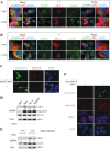
vIRF-1 targeting to mDRM is promoted by mitochondrial reactive oxygen species. (A and B) Specific targeting of vIRF-1 to the mitochondria after virus infection. HeLa cells were transiently transfected with vIRF-1–Flag for 24 h, infected with VSV at an MOI of 5 for 6 h, and immunostained with rat anti-Flag (L5)-Alexa Fluor 647, mouse anti-TOM20-Alexa Fluor 488, and rabbit anti-catalase-Alexa Fluor 555 (A) or ACSL4-Alexa Fluor 555 (B) antibodies. For comparative analysis with TOM20 (green), pseudocolor images of catalase (green) and ACSL4 (green) were generated using Element software. The images in rectangles with white line are enlarged and shown in the next images. (C) MAVS production of mitochondrial reactive oxygen species (mROS). 293T cells were transfected with or without MAVS for 24 h and then incubated with 5 μM MitoSOX red reagent in HBSS buffer for 10 min. After fixation with chilled methanol, the cells were immunostained with anti-Flag antibody (green) and nuclei were counterstained with 4′,6-diamidino-2-phenylindole (DAPI) (blue). (D) Role of mROS in MAVS-induced mDRM targeting of vIRF-1. 293T cells were transfected with vIRF-1 and/or MAVS and 12 h later treated with mROS scavengers, including 1 μM MitoQ and 10 μM MitoTEMPO, for 12 h. The mDRM fractions and total cell extracts were immunoblotted with the indicated antibodies. Relative band intensities of vIRF-1 in mDRM were calculated by normalization with the band intensity of corresponding MAVS and are shown under the top blot. (E) Role of mROS for HHV-8 replication-induced mDRM targeting of vIRF-1. BCBL-1 TRE:RTA cells were incubated with or without DOX in the presence or absence of 1 μM MitoQ for 1 day. The mDRM fractions and total cell extracts were immunoblotted with the indicated antibodies. Relative band intensities of vIRF-1 in mDRM were calculated by normalization with the band intensity of corresponding FLOT1 and noted under the top blot. (F) Absence of effect of vIRF-1 on basal and MAVS-induced mROS production. 293T cells were transfected with vIRF-1 with or without MAVS for 24 h and incubated with MtioSOX red as described above. The cells were immunostained with anti-Flag (green) and vIRF-1 (purple), and nuclei were counterstained with DAPI.
FIG 7
FIG 7
CL externalization is promoted by MAVS or HHV-8 replication. (A) Principle of externalized CL assay using human anti-CL antibody. Mitochondria isolated from the sample cells were incubated with anti-CL antibody (0.5 μl) on ice for 30 min, washed, resolved by SDS-PAGE, and immunoblotted with goat anti-human IgG (α-hIgG). Hc, heavy chain; Lc, light chain. (B) MAVS-induced CL externalization. The externalized CL assay was performed with the mitochondria isolated from 293T cells transfected with or without MAVS for 24 h. One tenth of the input (α-CL) was loaded on the gel. HSP60 was used to examine the level of precipitated mitochondria. (C) MitoQ inhibition of HHV-8 replication-induced CL externalization. The mitochondria isolated from BCBL-1 TRE:RTA cells incubated with or without DOX in the absence or presence of MitoQ for 24 h were subjected to the externalized-CL assay. Normalized band intensities are depicted in the graph. Representative gel images are shown and the data in the graph indicate means ± SD from three independent experiments.
FIG 8
FIG 8
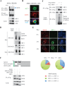
vIRF-1 interacts with MAVS and inhibits MAVS aggregation. (A) HHV-8 replication-induced vIRF-1–MAVS interaction. BCBL-1 TRE:RTA cells were treated with 1 μg/ml of DOX or mock treated for 1 day, and total cell extracts were used for immunoprecipitation with anti-vIRF-1 antibody or normal rabbit immunoglobulin (nIgG). The immunoprecipitates and lysates were immunoblotted with the indicated primary antibodies. The arrow indicates MAVS that coimmunoprecipitated with vIRF-1. The asterisks indicate heavy chain of IgG used for immunoprecipitation. (B) Colocalization of vIRF-1 with MAVS in lytic BCBL-1 cells. BCBL-1 TRE:RTA cells were treated with DOX for 24 h and immunostained with mouse anti-MAVS and rabbit anti-vIRF-1 antibodies along with nuclear counterstaining with DAPI. The arrows indicate regions of colocalization. (C) Intracellular interaction of vIRF-1 with MAVS. The Flag-tagged immunoprecipitates and homogenates of the total DRM fractions derived from 293T cells transfected with Flag-MAVS and/or Myc-vIRF-1 were immunoblotted with anti-Myc or Flag antibodies. (D) vIRF-1 inhibition of MAVS aggregation. The total cell extracts and mitochondrial fractions derived from 293T cells transfected with MAVS alone or together with vIRF-1 were resolved by SDD-AGE and SDS-PAGE and immunoblotted with the indicated antibodies. (E) Altered fluorescence of MAVS in vIRF-1-expressing BCBL-1 cells. BCBL-1 TRE:RTA cells were treated with DOX for 0, 24, and 48 h and immunostained with antibodies to vIRF-1 and MAVS. Nuclei were counterstained with DAPI. Example images showing reduced MAVS intensity in vIRF-1-positive (+) cells (white circle) at 24 and 48 h are shown. The MAVS fluorescent intensity and/or aggregates (big speckles) of 150 vIRF-1 (+) cells from 50 randomly selected microscopic fields (60×) were compared with those of the neighboring vIRF-1 negative (−) cells, and three types of population are grouped and depicted in the pie diagrams: red, stronger MAVS signals and/or intense MAVS aggregates in vIRF-1 (+) cells than in neighbor vIRF-1 (−) cells [vIRF-1 (+) > vIRF-1 (−)]; green, the same as neighbor vIRF-1 (−) cells [vIRF-1 (+) = vIRF-1 (−)]; and blue, less MAVS signal in vIRF-1 (+) cells than in neighbor vIRF-1 (−) cells [vIRF-1 (+) < vIRF-1 (−)]. (F) Mapping of vIRF-1 binding region of MAVS. A coimmunoprecipitation assay was performed with RIPA buffer-soluble extracts derived from 293T MAVS−/− cells transfected with full-length, ΔCARD (lacking aa 10 to 77), or ΔPD (lacking aa 103 to 153) versions of Flag-MAVS together with vIRF-1.
FIG 9
FIG 9
vIRF-1 inhibits MAVS-mediated antiviral signalings. (A) vIRF-1's nuclear localization signal (NLS) and its mutation (NLSX). (B and C) vIRF-1 inhibition of MAVS-induced and a constitutively active RIG-I (ΔRIG-I)-induced IFN-β expression. IFN-β-Luc reporter assays were performed with 293T cells transfected with the indicated constructs for 24 h. Results are presented as means ± standard deviations in triplicate. *, P < 0.05; **P < 0.01. (D) vIRF-1 inhibition of MAVS-induced cell death. The viability of 293 cells was assessed at 24 h and 48 h after transfection with MAVS and/or vIRF-1 using a CellTiter-Glo luminescence assay kit. Representative results are presented as means ± standard deviations. (E) The total cell extracts collected at 24 h for panel D were immunoblotted with the indicated antibodies, including anti-phospho-JNK1/2 (T183/Y185) antibody. (F) vIRF-1 inhibition of ΔRIG-I-induced apoptotic signaling. 293 cells were transfected with GFP or ΔRIG-I-GFP in the presence and absence of vIRF-1 for 24 h and immunoblotted with the indicated antibodies.
FIG 10
FIG 10
vIRF-1 PD is important for HHV-8 replication. (A) Diagram of a single lentiviral vector encoding control or vIRF-1 shRNAs together with vIRF-1R variants resistant to vIRF-1 shRNA. (B) Evaluation of vIRF-1 shRNA and vIRF-1R variants resistant to vIRF-1 shRNA. 293T cells were cotransfected with control or vIRF-1 shRNAs along with Flag-vIRF-1R (full-length or ΔPD) and Myc-vIRF-1, which is sensitive to vIRF-1 shRNA. Total cell extracts were immunoblotted with anti-Flag, Myc, and β-actin antibodies. (C) Contribution of the PD domain to vIRF-1 promotion of HHV-8 productive replication in PEL cells. BCBL-1 TRE:RTA cells were lentivirally transduced with control or vIRF-1 shRNAs together with vIRF-1R variants. Encapsidated HHV-8 virions from the culture media of the transduced BCBL-1 TRE:RTA cells treated or untreated with DOX for 2 days were collected and subjected to qPCR-based viral genome copy number analysis. The data in the graph are means ± SD from triplicate samples. (D) Verification of vIRF-1 depletion and reconstitution in BCBL-1 TRE:RTA cells. Total extracts collected from the cells for panel C were immunoblotted with the indicated antibodies to PARP, vIRF-1, Flag, and LDH.

Similar articles

Cited by

References

    1. Carbone A, Gloghini A. 2008. KSHV/HHV8-associated lymphomas. Br J Haematol 140:13–24. - PubMed
    1. Ganem D. 2006. KSHV infection and the pathogenesis of Kaposi's sarcoma. Annu Rev Pathol 1:273–296. doi:10.1146/annurev.pathol.1.110304.100133. - DOI - PubMed
    1. Cousins E, Nicholas J. 2014. Molecular biology of human herpesvirus 8: novel functions and virus-host interactions implicated in viral pathogenesis and replication. Recent Results Cancer Res 193:227–268. doi:10.1007/978-3-642-38965-8_13. - DOI - PMC - PubMed
    1. Lee H-R, Kim MH, Lee J-S, Liang C, Jung JU. 2009. Viral interferon regulatory factors. J Interferon Cytokine Res 29:621–627. doi:10.1089/jir.2009.0067. - DOI - PMC - PubMed
    1. Jacobs SR, Damania B. 2011. The viral interferon regulatory factors of KSHV: immunosuppressors or oncogenes? Front Immunol 2:19. - PMC - PubMed

Publication types

MeSH terms

Substances