. 2015 Oct 14;90(1):180-8.
doi: 10.1128/JVI.02342-15. Print 2016 Jan 1.

Kaposi's Sarcoma-Associated Herpesvirus Reduces Cellular Myeloid Differentiation Primary-Response Gene 88 (MyD88) Expression via Modulation of Its RNA

Affiliations

Kaposi's Sarcoma-Associated Herpesvirus Reduces Cellular Myeloid Differentiation Primary-Response Gene 88 (MyD88) Expression via Modulation of Its RNA

Amy Lingel et al. J Virol. .

Abstract

Kaposi's sarcoma (KS)-associated herpesvirus (KSHV) is a human gammaherpesvirus associated with several human malignancies. The replication and transcription activator (RTA) is necessary and sufficient for the switch from KSHV latency to lytic replication. Interleukin 1 (IL-1) is a major mediator for inflammation and plays an important role in both innate and adaptive immunity. Myeloid differentiation primary response gene 88 (MyD88) is an essential adaptor molecule for IL-1 as well as most Toll-like receptor signaling. In this study, we identified a novel mechanism by which KSHV interferes with host inflammation and immunity. KSHV RTA specifically reduces the steady-state protein levels of MyD88, and physiological levels of MyD88 are downregulated during KSHV lytic replication when RTA is expressed. The N-terminal region of RTA is required for the reduction of MyD88. Additional studies demonstrated that RTA targets MyD88 expression at the RNA level, inhibits RNA synthesis of MyD88, and may bind MyD88 RNA. Finally, RTA inhibits IL-1-mediated activation of NF-κB. Because IL-1 is abundant in the KS microenvironment and inhibits KSHV replication, this work may expand our understanding of how KSHV evades host inflammation and immunity for its survival in vivo.

Importance: MyD88 is an important molecule for IL-1-mediated inflammation and Toll-like receptor (TLR) signaling. This work shows that KSHV inhibits MyD88 expression through a novel mechanism. KSHV RTA may bind to MyD88 RNA, suppresses RNA synthesis of MyD88, and inhibits IL-1-mediated signaling. This work may expand our understanding of how KSHV evades host inflammation and immunity.


Figures

FIG 1
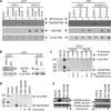
FIG 1
RTA modulates the expression of MyD88 protein. (A) RTA reduces the expression of MyD88 protein. 293T cells were transfected with cDNA3, RTA, EBV-RTA (EBV-R) (0.2 μg), and the MyD88 expression plasmid (0.1 μg) in various combinations, as shown on the top. E-RTA has significant homologies with KSHV RTA. Total DNA for transfection was kept the same with the use of vector DNA. The cell lysates were obtained 1 day later for Western blot analysis. The membrane was stripped and probed with another antibody. Images in the same box are derived from the same membranes. (B) MyD88 is downregulated during KSHV lytic replication. BCBL1 (KSHV+) and P3HR1 (KSHV) cells were treated with sodium butyrate for 24 h. Cell lysates were made, and the expression of endogenous proteins was analyzed by Western blotting. The membrane was stripped and probed with another antibody. The same cell lysates were used, and images in the same box are derived from the same membranes.
FIG 2
FIG 2
Domain analysis of RTA for MyD88 reduction. (A) Schematic diagram of RTA mutant constructs. The numbers denote the amino acid positions. The drawing is not to scale. (B) The N terminus of RTA is required for MyD88 regulation. 293T cells were transfected with vector pcDNA3 and with MyD88 (0.1 μg), RTA (0.2 μg), and RTA mutant expression plasmids (0.2 μg) in various combinations, as shown at the top. The amount of total DNA for transfection was kept the same with the use of vector DNA. Cell lysates were made 1 day later, and Western blot analysis was performed with RTA, FLAG, and tubulin antibodies. (C) Schematic diagram of MyD88 mutant constructs. The numbers denote amino acid positions of MyD88. The drawing is not to scale. (D) Multiple regions of MyD88 are targeted by RTA. 293T cells were transfected with vector pcDNA3 and with MyD88 (0.1 μg), RTA (0.2 μg), or MyD88 deletion mutant plasmids in various combinations, as shown at the top. The amount of total DNA for transfection was kept the same with the use of vector DNA. Cell lysates were made 1 day later, and Western blot analysis was performed with FLAG and GAPDH antibodies.
FIG 3
FIG 3
RTA did not affect the MyD88 protein stability. (A) MyD88 is a relatively stable protein. 293T cells in a 10-cm dish were transfected with MyD88 (0.4 μg) RTA, or MyD88 plus RTA (0.8 μg) expression plasmids. The amount of total DNA for transfection was kept the same with the use of vector DNA. At 6 h after transfection, cells were split into a 6-well plate. Cycloheximide (100 μg/ml) was added after a 12-h incubation. Cell lysates were made at various times, as shown on the top, and Western blot analysis was performed. The membrane was stripped and probed with another antibody. Images in the same box are derived from the same membranes. (B) Detection of IRF1 stability. The lysates used for panel A were used. IRF1 protein stability was measured in MyD88 and MyD88 plus RTA-transected cells.
FIG 4
FIG 4
RTA reduces MyD88 RNA expression. (A) RTA reduces the RNA expression of MyD88 in transfected cells. 293T cells were transfected with a MyD88 or MyD88-plus-RTA expression plasmid. The amount of total DNA for transfection was kept the same with the use of vector DNA. Cell were collected 24 h after transfections, and RNA was isolated. Semiquantitative RT-PCR was carried out with specific primers. (B) RTA reduces endogenous MyD88 RNA expression. 293T cells were transfected with RTA and MyD88 expression plasmids. Total RNA were isolated 24 h after transfections. Semiquantitative RT-PCR was carried out with specific primers. Primers MyD88AF and MyD88BR for MyD88 were used for panels A and B. (C) MyD88 RNA is downregulated during KSHV lytic replication. BCBL1 (KSHV+) cells were treated with sodium butyrate for 24 h. Total RNAs were isolated 24 h after treatments. Semiquantitative RT-PCR was carried out with specific primers at the same time. (D) Lactacystin did not modulate the expression of MyD88 RNA. 293T cells were transfected with various expression plasmids. At 4 to 6 h after transfection, cells were treated with lactacystin (10 μM). On the next day, total RNAs were isolated, and semiquantitative RT-PCR was carried out with specific primers. Primer MyD88BF and MyD88BR were used for panels C and D. (E) Lactacystin did not modulate the expression of MyD88 protein. Cell lysates were obtained from the experiment whose results are shown in panel D, and MyD88, tubulin, and MDM2 expression was examined by Western blotting.
FIG 5
FIG 5
RTA reduces the rate of MyD88 RNA synthesis. (A) RTA reduces the relative RNA synthesis rate for MyD88. 293T cells were transfected with various plasmids, as shown at the top. The cells were labeled with ethynyl uridine (EU) for 30 to 60 min, and total RNAs were isolated. The EU-containing RNA (newly synthesized) was isolated from the total RNA with the use of a Click-iT nascent-RNA capture kit as described in Materials and Methods. RT-PCR were carried out with the appropriate primers. The PCR products were separated in a polyacrylamide gel. The relative MyD88 RNA levels were determined (MyD88 RNA/actin RNA). The relative new synthesis rate was calculated as relative newly synthesized MyD88 RNA (EU-labeled MyD88/EU-actin) versus relative total MyD88 RNA (total MyD88/total actin). (B) Comparison of relative MyD88 synthesis rates. The average relative synthesis rates with and without RTA from five independent experiments are shown. Statistical calculation was done with Microsoft Excel. **, P < 0.01. (C) MyD88 RNA stability is not affected drastically by RTA. RTA and MyD88 plasmids were transfected into 293T cells. More MyD88 plasmid was transfected in the presence of RTA (shown at the top) in order to achieve similar MyD88 RNA levels. One day after transfection, the cells were labeled with actinomycin D to inhibit RNA synthesis. Total RNAs were isolated at the various times (hours posttreatment), and MyD88 RNA levels were detected by semiquantitative RT-PCR. Actin levels were used as a control. Results of one representative experment of three independent experiments are shown.
FIG 6
FIG 6
RTA may bind to MyD88 RNA. (A) Schematic diagram of primers used for detection of MyD88 RNA. The four pairs of primers are used to detect different regions of MyD88 RNA (Table 1). All four products are approximately the same length, around 230 bp. The drawing is not to scale. (B) RTA may bind to MyD88 RNA. 293T cells were transfected with expression plasmids as shown on the top. One day after transfection, cells were fixed with formaldehyde for 30 min and sonicated briefly to avoid extensive damage to RNA. Normal rabbit serum (NRS) and RTA antibody were used for the RNA-ChIP assay. After digestion with DNase, RT-PCR was carried out with the appropriate primers, shown in panel A. Results from four independent experiments are shown. (C) RTA may selectively bind to MyD88 RNA. RNA-ChIP were done as above, and primer pairs A and B were used for detection. Input RNA (1:100) was used as a control. Results of one representative experiment are shown.
FIG 7
FIG 7
RTA blocks IL-1 signaling. (A) RTA blocks IL-1-mediated NF-κB activation. 293T cells were transfected with cDNA3 or RTA along with the NF-κB reporter construct and β-galactosidase expression plasmids. Total DNA for transfection was kept the same with the use of vector DNA. After 4 to 6 h transfection, the cells were washed and then treated (+) with IL-1β (0.5, 1, and 5 ng/ml) or not treated (−). One day later, the cells were collected, and luciferase and β-galactosidase assays were used for detection of reporter activation. The relative activation of NF-κB reporter (with or without IL-1β) is shown. (B) RTAΔC blocks IL-1-mediated NF-κB activation. Various plasmids were transfected into 293T cells as shown on the top. The cells were then treated (+) with IL-1β (5 ng/ml) or not treated (−). The relative activation of NF-κB reporter (with or without IL-1β) is as shown. One set of representative results is shown. (C) RTA blocks MyD88-mediated activation of NF-κB. Various plasmids were transfected into 293T cells as shown on the top. Luciferase and β-galactosidase activities were measured 1 day later. The relative activation of NF-κB reporter is shown. One representative set of results is shown.

Similar articles

Cited by

References

    1. Chang Y, Cesarman E, Pessin MS, Lee F, Culpepper J, Knowles DM, Moore PS. 1994. Identification of herpesvirus-like DNA sequences in AIDS-associated Kaposi's sarcoma. Science 266:1865–1869. doi:10.1126/science.7997879. - DOI - PubMed
    1. Chang Y, Moore PS. 1996. Kaposi's sarcoma (KS)-associated herpesvirus and its role in KS. Infect Agents Dis 5:215–222. - PubMed
    1. Moore P, Chang Y. 2001. Kaposi's sarcoma-associated herpesvirus, p 2803–2833. In Knipe DM, Howley PM, Griffin DE, Lamb RA, Martin MA, Roizman B, Straus SE (ed), Fields virology, 4th ed Lippincott Williams & Wilkins, Philadelphia, PA.
    1. Dourmishev LA, Dourmishev AL, Palmeri D, Schwartz RA, Lukac DM. 2003. Molecular genetics of Kaposi's sarcoma-associated herpesvirus (human herpesvirus-8) epidemiology and pathogenesis. Microbiol Mol Biol Rev 67:175–212. doi:10.1128/MMBR.67.2.175-212.2003. - DOI - PMC - PubMed
    1. West JT, Wood C. 2003. The role of Kaposi's sarcoma-associated herpesvirus/human herpesvirus-8 regulator of transcription activation (RTA) in control of gene expression. Oncogene 22:5150–5163. doi:10.1038/sj.onc.1206555. - DOI - PubMed

Publication types

MeSH terms

LinkOut - more resources