Review
. 2015;390(Pt 1):151-209.
doi: 10.1007/978-3-319-22822-8_8.

EBV Persistence--Introducing the Virus

Affiliations
Review

EBV Persistence--Introducing the Virus

David A Thorley-Lawson. Curr Top Microbiol Immunol. 2015.

Abstract

Persistent infection by EBV is explained by the germinal center model (GCM) which provides a satisfying and currently the only explanation for EBVs disparate biology. Since the GCM touches on every aspect of the virus, this chapter will serve as an introduction to the subsequent chapters. EBV is B lymphotropic, and its biology closely follows that of normal mature B lymphocytes. The virus persists quiescently in resting memory B cells for the lifetime of the host in a non-pathogenic state that is also invisible to the immune response. To access this compartment, the virus infects naïve B cells in the lymphoepithelium of the tonsils and activates these cells using the growth transcription program. These cells migrate to the GC where they switch to a more limited transcription program, the default program, which helps rescue them into the memory compartment where the virus persists. For egress, the infected memory cells return to the lymphoepithelium where they occasionally differentiate into plasma cells activating viral replication. The released virus can either infect more naïve B cells or be amplified in the epithelium for shedding. This cycle of infection and the quiescent state in memory B cells allow for lifetime persistence at a very low level that is remarkably stable over time. Mathematically, this is a stable fixed point where the mechanisms regulating persistence drive the state back to equilibrium when perturbed. This is the GCM of EBV persistence. Other possible sites and mechanisms of persistence will also be discussed.


Figures
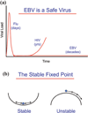
Fig. 1
Fig. 1
EBV establishes a stable, benign, low-level, lifetime persistent infection. a EBV is a safe virus. EBV establishes a persistent, benign infection in virtually every human being for their entire life. This is in comparison with a virus like flu whose infection resolves in a few days or HIV which undergoes an acute infection that resolves into a long-term low-level persistent infection that eventually returns to kill the host. EBV also undergoes acute infection but then enters into a low-level persistent infection which remains stable for the life of the host. b The stable fixed point. The type of equilibrium EBV achieves is referred to mathematically as a stable fixed point. This means that the forces regulating the system act to return it to the same place after perturbation, e.g., a marble in the bottom of a bowl, whereas in an unstable fixed point, small perturbations irrevocably destroy the fixed point, e.g., a marble on top of the bowl. In real-life biology, where there are always perturbations, the only way to achieve long-term stability is through a stable fixed point
Fig. 2
Fig. 2
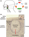
The lymphoepithelium of the tonsil where EBV performs its biology. a Waldeyer’s ring consists of the adenoids and tonsils which form a ring of lymphoid tissue at the back of the throat. b The structure of the lymphoepithelium underlying the saliva. Inset is an expanded view of the marginal zone/epithelium. B cells exit the circulation and enter the lymphoid tissue through the HEV and migrate to the mantle zone of the follicle. Here, they reside for a period of time and then either leave or, if they see antigen, enter the follicle to undergo a GC reaction which produces memory cells that can then enter the peripheral circulation. This is the B cell system that EBV exploits. For more details, see Figs. 3 and 4 and text (Figure provided by Marta Perry)
Fig. 3
Fig. 3
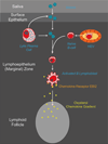
EBV biology mirrors B cell biology. To the left is diagrammed a typical mucosal humoral immune response. Antigen in saliva crosses the epithelial barrier of the tonsil to be sampled by naïve B cells in the underlying lymphoid tissue. When naïve B cells recognize cognate antigen, they become activated blasts and migrate to the follicle to undergo a GC reaction. If they receive signals from antigen and antigen-specific Th cells, they can leave to become resting memory B cells that occasionally undergo division as part of memory B cell homeostasis. To the right is diagrammed how EBV uses the same pathways. EBV is spread through saliva, crosses the epithelial barrier, and infects naïve B cells. These become B cell blasts that enter the GC. Here, the viral latent proteins LMP1 and LMP2 have the capacity to provide surrogate antigen and Th survival signals that allow the latently infected B cells to leave the GC as resting memory cells that also divide through homeostasis. To the right are listed in orange the transcription programs used at each stage. The blue circles represent the viral DNA which is a circular episome
Fig. 4
Fig. 4
The germinal center model (GCM) of EBV persistence. The stages 1–4 follow those in the text from Sect. 3. “EBV Infection in the healthy host—a summary of the GCM.” For details, see the text
Fig. 5
Fig. 5
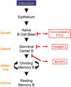
The origin of EBV-positive lymphomas. EBV lymphomas arise from different stages of the infection process. The figure shows diagrammatically the flow of virus from infectious virions to latently infected resting memory B cell as detailed in Figs. 3 and 4 and the text. To the right are shown the 3 EBV-associated lymphomas and their proposed origin and to the left are listed the viral transcription programs expressed in the tumors and at the equivalent stage of infection. IL is proposed to arise from a latently infected blast that is unable to differentiate and so continues to proliferate. HD is derived from a GC B cell, and BL is a GC cell that has left the follicle. Note that a tumor is proposed to arise from each of the three stages of EBV biology that involve proliferation
Fig. 6
Fig. 6
The first steps of EBV infection. Naïve B cells emerge from the HEV and migrate toward the mantle zone of the follicle. On the way, they encounter EBV that either has crossed the epithelial barrier or is derived from lyrically infected plasma cells. The newly infected lymphoblast upregulates the chemokine receptor EBI2 and follows a gradient of oxysterol chemokine into the follicle
Fig. 7
Fig. 7
A summary of the functions of LMP1 and LMP2a demonstrated in vitro or in vivo with transgenic mice that could contribute to the GC processing of a latently infected B cell
Fig. 8
Fig. 8
A model of EBV reactivation and shedding. The known data fit a model where a single latently infected memory B cell in the tonsil occasionally differentiates into a plasma cell and releases virus that infects epithelial cells. The infection spreads exponentially through the epithelium, resulting in the shedding of virus. The plaque is eventually eliminated by the immune response. Meanwhile, another plaque initiates elsewhere in the Waldeyer’s ring. The data are consistent with their being no more than three such plaques in Waldeyer’s ring at any one time. Virus is continuously shed into the mouth where it mingles with saliva for about 2 min before being swallowed. Thus, the mouth is a flow stream of EBV not a static reservoir
Fig. 9
Fig. 9
The cyclic pathogen model (CPM). a CPM is a mathematical description of the GCM. It consists of a cycle of 6 infected stages (blue circles based on the biological GCM illustrated in Fig. 4). These are blast, GC, memory and immediate early, early and late lyrically infected B cells, each of which is potentially controlled by the immune response (red circles). The single lytic stage in the GCM is broken down into three discrete stages which are known to be recognized independently by the immune response. Biologically, there is never a CTL response against the memory stage; however, the model allows analysis of theoretical conditions such as the memory compartment being regulated by CTL. This model can be described by a system of differential equations employing rate constants for the stimulation of CTL (blue arrows), killing of CTL targets (red arrows), and the proliferation and death of each stage (green arrows). For this system, there is one and only one mathematical solution that is stable and biologically credible. This solution accurately describes biologically persistent infection. b Shows the infected populations as circles whose area is proportional to their frequency within all tonsils (1:5:1.5.102:104:104:0.5.104, Late:Early:ImmEarly:Memory:GC:Blast). This highlights the very large range in the sizes of these populations
Fig. 10
Fig. 10
Are there 2 pathways to persistence? The current data suggest the following possible hypothetical model. Infected naive blasts will migrate to the follicle because they express the chemokine receptor EBI2. They express AID and undergo SHM but will not enter the GC because they are bcl-6 negative. If they receive the necessary signals (cytokines/T cell help), they will enter the follicle switch on bcl-6, undergo CSR, and eventually leave as resting memory B cells as described by the GCM (Route 1). If, however, the cells do not receive the necessary signal to turn on bcl-6, they will continue to proliferate as marginal zone memory B cells (Route 2). The ultimate fate of such cells is unclear. For example, to be biologically relevant, they would need to release infectious virus at some point. What is clear is that they appear capable of extensive proliferation despite the presence of CTL

Similar articles

Cited by

References

    1. Adams A, Lindahl T. Epstein-Barr virus genomes with properties of circular DNA molecules in carrier cells. Proc Natl Acad Sci USA. 1975;72:1477–1481. - PMC - PubMed
    1. Allday MJ. How does Epstein-Barr virus (EBV) complement the activation of Myc in the pathogenesis of Burkitt’s lymphoma? Semin Cancer Biol. 2009;19:366–376. - PMC - PubMed
    1. Allday MJ. EBV finds a polycomb-mediated, epigenetic solution to the problem of oncogenic stress responses triggered by infection. Front Genet. 2013;4:212. - PMC - PubMed
    1. Allday MJ, Crawford DH, Griffin BE. Epstein-Barr virus latent gene expression during the initiation of B cell immortalization. J Gen Virol. 1989;70:1755–1764. - PubMed
    1. Allday MJ, Sinclair A, Parker G, Crawford DH, Farrell PJ. Epstein-Barr virus efficiently immortalizes human B cells without neutralizing the function of p53. EMBO J. 1995;14:1382–1391. - PMC - PubMed

Publication types

LinkOut - more resources