. 2015 Dec;89(24):12349-61.
doi: 10.1128/JVI.01365-15. Epub 2015 Sep 30.

Flavivirus Infection Impairs Peroxisome Biogenesis and Early Antiviral Signaling

Affiliations

Flavivirus Infection Impairs Peroxisome Biogenesis and Early Antiviral Signaling

Jaehwan You et al. J Virol. 2015 Dec.

Abstract

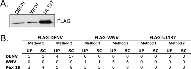
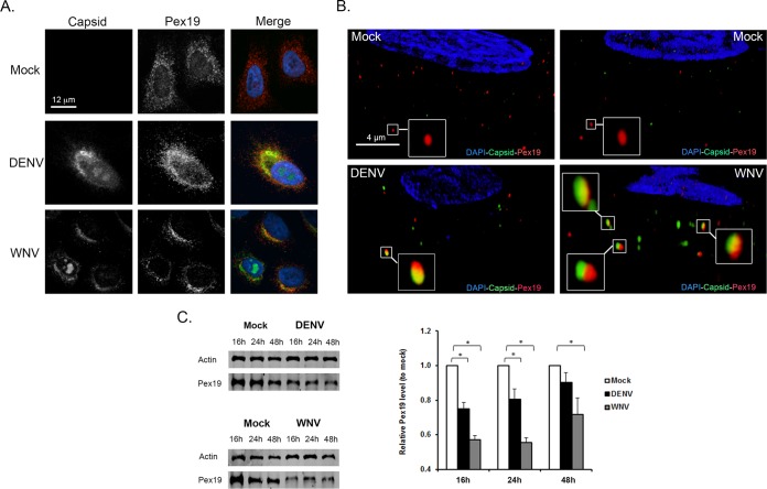
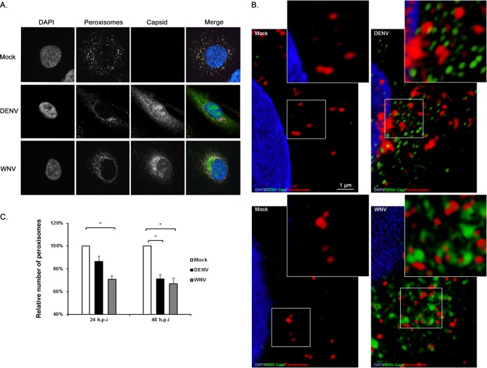
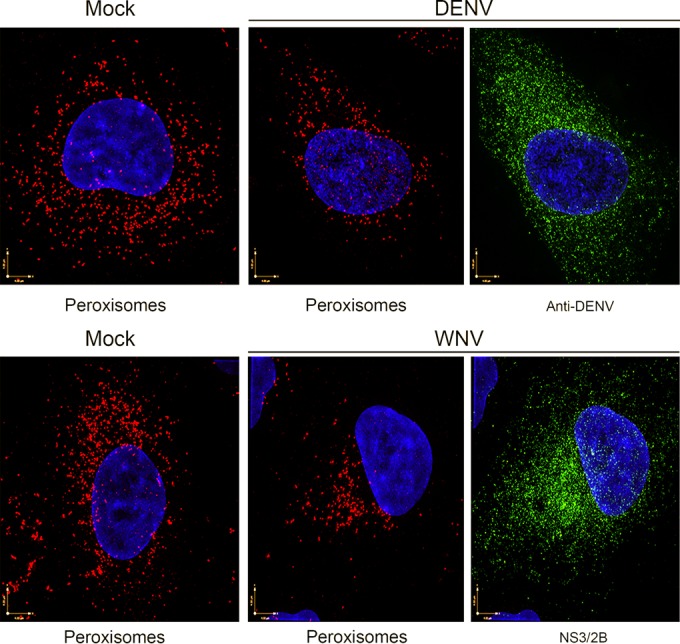
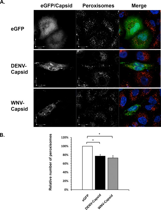
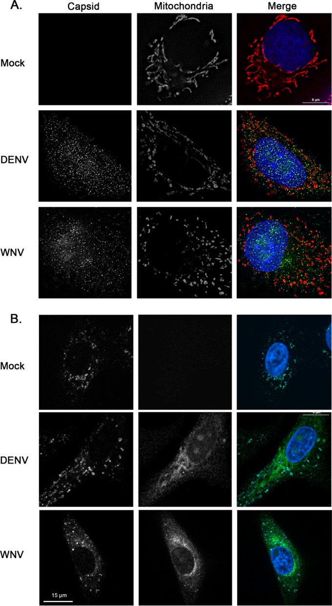
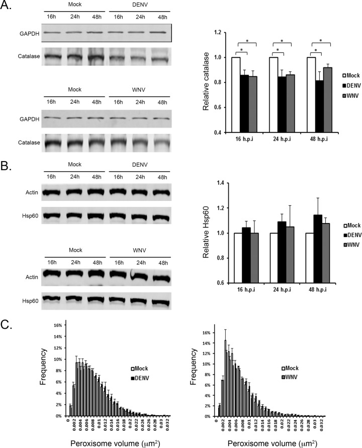
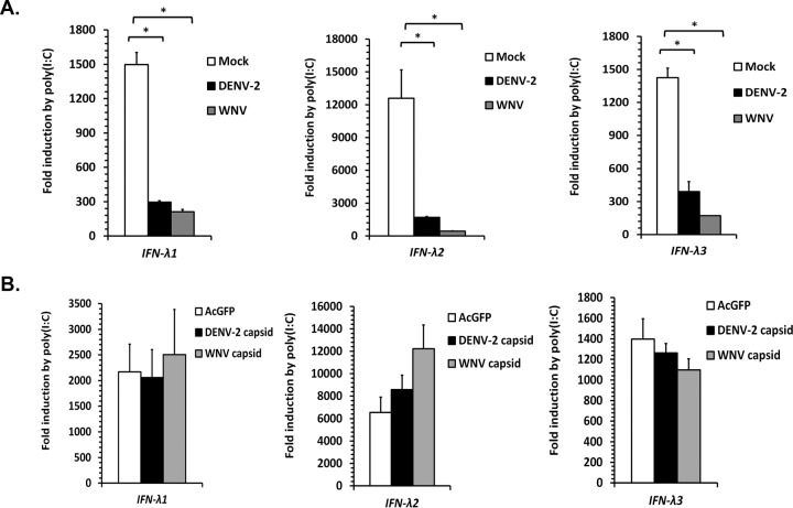
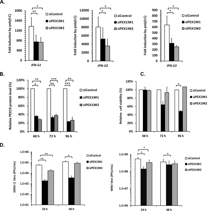
Flaviviruses are significant human pathogens that have an enormous impact on the global health burden. Currently, there are very few vaccines against or therapeutic treatments for flaviviruses, and our understanding of how these viruses cause disease is limited. Evidence suggests that the capsid proteins of flaviviruses play critical nonstructural roles during infection, and therefore, elucidating how these viral proteins affect cellular signaling pathways could lead to novel targets for antiviral therapy. We used affinity purification to identify host cell proteins that interact with the capsid proteins of West Nile and dengue viruses. One of the cellular proteins that formed a stable complex with flavivirus capsid proteins is the peroxisome biogenesis factor Pex19. Intriguingly, flavivirus infection resulted in a significant loss of peroxisomes, an effect that may be due in part to capsid expression. We posited that capsid protein-mediated sequestration and/or degradation of Pex19 results in loss of peroxisomes, a situation that could result in reduced early antiviral signaling. In support of this hypothesis, we observed that induction of the lambda interferon mRNA in response to a viral RNA mimic was reduced by more than 80%. Together, our findings indicate that inhibition of peroxisome biogenesis may be a novel mechanism by which flaviviruses evade the innate immune system during early stages of infection.

Importance: RNA viruses infect hundreds of millions of people each year, causing significant morbidity and mortality. Chief among these pathogens are the flaviviruses, which include dengue virus and West Nile virus. Despite their medical importance, there are very few prophylactic or therapeutic treatments for these viruses. Moreover, the manner in which they subvert the innate immune response in order to establish infection in mammalian cells is not well understood. Recently, peroxisomes were reported to function in early antiviral signaling, but very little is known regarding if or how pathogenic viruses affect these organelles. We report for the first time that flavivirus infection results in significant loss of peroxisomes in mammalian cells, which may indicate that targeting of peroxisomes is a key strategy used by viruses to subvert early antiviral defenses.


Figures

FIG 1
FIG 1
Recovery of FLAG-tagged DENV and WNV capsid proteins from human cells identifies an interaction with Pex19. HEK293T cells were transfected with plasmids encoding FLAG-tagged DENV or WNV capsid protein or with FLAG-tagged UL137 from human cytomegalovirus (negative control). Forty-eight hours later, the cells were lysed in physiological salt buffer (method 1) or high-salt buffer followed by dialysis (method 2). The FLAG-tagged proteins and their interactors were recovered from lysates on anti-FLAG resin. (A) Immunoblot of the proteins (50%) isolated using method 1 (probed with anti-FLAG antibody) showing that all three viral proteins were recovered. (B) The recovered proteins were trypsinized and analyzed by mass spectrometry. The recovery of peptides from Pex19 and the tagged DEN and WNV proteins is shown. UP, number of unique peptides; SC, total spectral counts (total number of peptides).
FIG 2
FIG 2
Pex19 forms stable complexes with capsid proteins during flavivirus infection. A549 cells were infected with WNV or DENV (multiplicity of infection [MOI] = 3). Forty-eight hours later, cell lysates were subjected to immunoprecipitation (IP) with anti-capsid (Cap) and anti-Pex19 antibodies. The levels of capsid proteins and Pex19 were detected in whole-cell lysates (WCL) and immunoprecipitates by immunoblotting (IB) with the indicated antibodies. The secondary antibodies were Dylight 680 donkey anti-rabbit or anti-mouse IgG and Dylight 800 donkey anti-mouse, anti-rabbit, or anti-guinea-pig IgG.
FIG 3
FIG 3
Redistribution and loss of Pex19 during flavivirus infection. (A and B) A549 cells were infected with WNV or DENV (MOI = 1) and processed for indirect immunofluorescence microscopy 16 to 48 h postinfection. WNV and DENV capsid proteins were detected using guinea pig polyclonal antibodies and goat anti-guinea pig IgG conjugated to Alexa Fluor 546. Pex19 was detected with a mouse monoclonal antibody and goat anti-mouse IgG conjugated to Alexa Fluor 488. Nuclei were stained using DAPI. The images were obtained using spinning-disc confocal (A) and structured-illumination (B) microscopes. The enlarged areas in panel B show structures that contain both Pex19 and capsid proteins. (C) Infection by DENV or WNV leads to decreased Pex19. Immunoblot and quantitative analyses of Pex19 levels in mock-infected and flavivirus-infected cells are presented. The error bars represent standard errors of the mean. *, P < 0.05.
FIG 4
FIG 4
Effect of flavivirus infection on peroxisomes. (A) A549 cells were infected with WNV or DENV (MOI = 1) and processed for indirect immunofluorescence microscopy 24 and 48 h postinfection. Cells infected with DENV or WNV were identified using guinea pig antibodies to the respective capsid proteins and goat anti-guinea pig IgG conjugated to Alexa Fluor 546. Peroxisomes were detected with a rabbit polyclonal antibody to the peroxisomal targeting signal SKL and donkey anti-rabbit IgG conjugated to Alexa Fluor 568. Nuclei were stained using DAPI. (B) Superresolution microscopy images of mock-infected and DENV- or WNV-infected cells showing lack of colocalization between capsid proteins and peroxisomes. The insets show enlargements of the boxed areas. (C) The average numbers of SKL-positive structures in mock-infected and flavivirus-infected cells were determined from three independent experiments. The error bars represent standard errors of the mean. *, P < 0.05.
FIG 5
FIG 5
Loss of peroxisomes in flavivirus-infected cells as revealed by superresolution microscopy. A549 cells were infected with WNV or DENV and then processed for superresolution microscopy after 48 h. Cells infected with DENV or WNV were identified using human anti-DENV E antibodies or a mouse monoclonal antibody to the WNV NS2B-NS3 complex. Primary antibodies were detected with goat anti-human IgG conjugated to Alexa Fluor 488 or donkey anti-mouse IgG conjugated to Alexa Fluor 488. Peroxisomes were detected with rabbit polyclonal antibodies to SKL and donkey anti-rabbit IgG conjugated to Alexa Fluor 568. Nuclei were stained using DAPI. The images were acquired and reconstructed using a DeltaVision OMX structured-illumination microscope.
FIG 6
FIG 6
Expression of flavivirus capsid proteins causes loss of peroxisomes. A549 cells were transfected with plasmids encoding DENV or WNV capsid proteins or eGFP (negative control). The cells were processed for indirect immunofluorescence microscopy 24 h posttransfection. (A) Peroxisomes were detected with rabbit anti-SKL antibodies and donkey anti-rabbit IgG conjugated to Alexa Fluor 546. Capsid proteins were detected with guinea antibodies to DENV or WNV capsids and goat anti-guinea pig IgG conjugated to Alexa Fluor 647. Nuclei were stained with DAPI. The cells were viewed by confocal microscopy. (B) The average numbers of SKL-positive structures in cells expressing eGFP, DENV capsid, and WNV capsid were determined from three independent experiments (minimum of 15 cells). Peroxisome numbers were determined using Volocity image analysis software. The error bars represent standard errors of the mean. *, P < 0.05.
FIG 7
FIG 7
Mitochondrial morphology is not dramatically affected by flavivirus infection or expression of capsid proteins. A549 cells were infected with DENV or WNV (MOI = 1). Forty-eight hours later, samples were processed for indirect immunofluorescence microscopy. Infected cells were identified using guinea pig polyclonal antibodies to capsid proteins and goat anti-guinea pig IgG conjugated to Alexa Fluor 488. (A) Mitochondria were detected using a monoclonal antibody to the matrix protein p32 and donkey anti-mouse IgG conjugated to Alexa Fluor 546. (B) Lysosomes were detected using a mouse monoclonal antibody to the lysosome membrane protein, LAMP-1, and donkey anti-mouse IgG conjugated to Alexa Fluor 647. Nuclei were stained using DAPI. Cells were viewed by confocal microscopy.
FIG 8
FIG 8
Flavivirus infection results in loss of the peroxisomal matrix enzyme, catalase. (A and B) A549 cells were infected with DENV or WNV (MOI = 1); cell lysates were harvested 16, 24, and 48 h postinfection; and the levels of the peroxisomal and mitochondrial matrix proteins, catalase (A) and Hsp60 (B), respectively, were determined by immunoblotting (left) and quantified (right). The error bars represent standard errors of the mean. *, P < 0.05. (C) Peroxisome size is not affected by flavivirus infection. A549 cells were infected (MOI = 1) with either DENV or WNV. At 48 h postinfection, the cells were processed for indirect immunofluorescence microscopy. Peroxisomes were detected with rabbit polyclonal antibodies to SKL and donkey anti-rabbit IgG conjugated to Alexa Fluor 568, and infected cells were identified using human anti-DENV E antibodies or a mouse monoclonal antibody to the WNV NS2B-NS3 complex. Primary antibodies were detected with goat anti-human IgG conjugated to Alexa Fluor 488 or donkey anti-mouse IgG conjugated to Alexa Fluor 488. The images were acquired and reconstructed using a DeltaVision OMX structured-illumination microscope. Volocity software was used to determine the sizes and numbers of peroxisomes in mock-infected and flavivirus-infected cells.
FIG 9
FIG 9
Flavivirus infection inhibits poly(I·C)-induced expression of type III interferon. (A) A549 cells were mock treated or infected with WNV or DENV-2 (MOI = 2) for 10 to 12 h. The cells were then transfected with 4 μg of poly(I·C) or pCMV5, an empty-vector negative control, for 12 h to induce expression of IFN-λ genes. The cell lysates were collected and processed for RNA extraction and subsequent qRT-PCR. (B) A549 cells were transduced with a lentivirus expressing GFP derived from Aequorea coerulescens (AcGFP) alone, AcGFP/myc-DENV-2 capsid or AcGFP/myc-WNV capsid. 48 h postransduction, the cells were transfected with poly(I·C) for 12 h. The cell lysates were collected and processed for RNA extraction and subsequent qRT-PCR. The data are averaged from the results of 3 independent experiments. The error bars represent standard errors of the mean. *, P < 0.05.
FIG 10
FIG 10
Knockdown of Pex19 reduces poly(I·C)-induced IFN-λ expression and flavivirus titers. (A) A549 cells were treated with a nontargeting siRNA (siControl) or Pex19-specific siRNAs for 48 h. The cells were then transfected with 4 μg of poly(I·C) for 12 h to induce IFN-λ expression or with an empty plasmid vector, pCMV5. The cell lysates were processed for RNA extraction and subsequent qRT-PCR. (B) To determine knockdown efficiency, lysates from siRNA-transfected cells were subjected to immunoblotting with anti-Pex19 antibodies. Quantification was performed using Li-COR software. ***, P < 0.001. (C) The effect of Pex19 knockdown on cell viability assay was determined by counting viable cells at different time points posttransfection. (D) A549 cells transfected with siRNAs for 48 h were infected with DENV-2 or WNV at an MOI of 0.5. At 24 and 48 h postinfection, the viral titers in the cell supernatants were determined. *, P < 0.05; **, P < 0.01 (n = 4).

Similar articles

Cited by

References

    1. Pierson TC, Diamond MS. 2013. Flaviviruses, p 747–794. In Knipe DM, Howley PM (ed), Fields virology, 6th ed, vol 1 Lippincott Williams & Wilkins, Philadelphia, PA.
    1. Urbanowski MD, Hobman TC. 2013. The West Nile virus capsid protein blocks apoptosis through a phosphatidylinositol 3-kinase-dependent mechanism. J Virol 87:872–881. doi:10.1128/JVI.02030-12. - DOI - PMC - PubMed
    1. Urbanowski MD, Ilkow CS, Hobman TC. 2008. Modulation of signaling pathways by RNA virus capsid proteins. Cell Signal 20:1227–1236. doi:10.1016/j.cellsig.2007.12.018. - DOI - PMC - PubMed
    1. Moriya K, Fujie H, Shintani Y, Yotsuyanagi H, Tsutsumi T, Ishibashi K, Matsuura Y, Kimura S, Miyamura T, Koike K. 1998. The core protein of hepatitis C virus induces hepatocellular carcinoma in transgenic mice. Nat Med 4:1065–1067. doi:10.1038/2053. - DOI - PubMed
    1. Xu Z, Hobman TC. 2012. The helicase activity of DDX56 is required for its role in assembly of infectious West Nile virus particles. Virology 433:226–235. doi:10.1016/j.virol.2012.08.011. - DOI - PMC - PubMed

Publication types