. 2015 Sep 24;11(9):e1005171.
doi: 10.1371/journal.ppat.1005171. eCollection 2015 Sep.

A KSHV microRNA Directly Targets G Protein-Coupled Receptor Kinase 2 to Promote the Migration and Invasion of Endothelial Cells by Inducing CXCR2 and Activating AKT Signaling

Affiliations

A KSHV microRNA Directly Targets G Protein-Coupled Receptor Kinase 2 to Promote the Migration and Invasion of Endothelial Cells by Inducing CXCR2 and Activating AKT Signaling

Minmin Hu et al. PLoS Pathog. .

Abstract

Kaposi's sarcoma (KS) is a highly disseminated angiogenic tumor of endothelial cells linked to infection by Kaposi's sarcoma-associated herpesvirus (KSHV). KSHV encodes more than two dozens of miRNAs but their roles in KSHV-induced tumor dissemination and metastasis remain unknown. Here, we found that ectopic expression of miR-K12-3 (miR-K3) promoted endothelial cell migration and invasion. Bioinformatics and luciferase reporter analyses showed that miR-K3 directly targeted G protein-coupled receptor (GPCR) kinase 2 (GRK2, official gene symbol ADRBK1). Importantly, overexpression of GRK2 reversed miR-K3 induction of cell migration and invasion. Furthermore, the chemokine receptor CXCR2, which was negatively regulated by GRK2, was upregulated in miR-K3-transduced endothelial cells. Knock down of CXCR2 abolished miR-K3-induced cell migration and invasion. Moreover, miR-K3 downregulation of GRK2 relieved its direct inhibitory effect on AKT. Both CXCR2 induction and the release of AKT from GRK2 were required for miR-K3 maximum activation of AKT and induction of cell migration and invasion. Finally, deletion of miR-K3 from the KSHV genome abrogated its effect on the GRK2/CXCR2/AKT pathway and KSHV-induced migration and invasion. Our data provide the first-line evidence that, by repressing GRK2, miR-K3 facilitates cell migration and invasion via activation of CXCR2/AKT signaling, which likely contribute to the dissemination of KSHV-induced tumors.


Conflict of interest statement

The authors have declared that no competing interests exist.

Figures
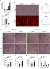
Fig 1
Fig 1. Ectopic expression of miR-K3 promotes endothelial cell migration and invasion.
(A). KSHV miR-K3 expression in HUVEC infected with KSHV BAC16 virus induced from iSLK-BAC16 cells or transduced by the different MOI of lentiviral miR-K3 were determined by qPCR. The miR-K3 level in KSHV group was set as ‘‘1” for comparison. (B). HUVEC were transduced with 2 MOI of lentivirus empty vector (mpCDH; left) and lentivirus-miR-K3 (miR-K3; right), and representative images were taken under light microscope (Phase; top) and fluorescent microscope (RFP; bottom) (Original magnification, ×100). (C). Cells treated as in (B) were analyzed for RFP expression by flow cytometry to determine transduction efficiency. y axis units are numbers of cells. (D). Luciferase activity was detected in 2 MOI of lentivirus empty vector (mpCDH) or lentivirus-miR-K3 (miR-K3) transduced HUVEC transfected by the pGL3-Control (Control) or the pGL3-miR-K3 sensor reporter (miR-K3-Sensor). *** P < 0.001 for Student’s t-test. n.s., not significant. (E). Transwell migration (left panel) and Matrigel invasion (right panel) assays for HUVEC transduced with lentivirus empty vector (mpCDH) or lentivirus-miR-K3 (miR-K3). The representative images were captured at 6 and 12 h post seeding (original magnification, ×100). (F). The quantification results of Transwell migration assay in (E). (G). The quantification results of Matrigel invasion assay in (E). (H). The quantification results of wound healing assay in S1 Fig. (I). The mRNA expression of MMP1, 9, 10 and IL-6, 8 in HUVEC treated as in (B) were determined by qPCR.
Fig 2
Fig 2. GRK2 expression is reduced in miR-K3-expressing HUVEC and KS lesion samples.
(A). The mRNA level of GRK2 in HUVEC transduced with lentivirus empty vector (mpCDH) and lentivirus-miR-K3 (miR-K3) were examined by qPCR. (B). The expression of GRK2 proteins in HUVEC transduced with lentivirus empty vector (mpCDH) and lentivirus-miR-K3 (miR-K3) were detected by Western blotting analysis. Results shown were from a representative experiment of three independent experiments with similar results. The values of density of protein bands after normalization to housekeeping were shown; same for all of the following Western blotting figures. (C). HUVEC were infected with KSHV, and image captured under light microscope (Phase) and fluorescent microscope (GFP) (Original magnification, ×100). (D). qPCR analysis for GRK2 mRNA in KSHV-infected HUVEC as described in (C) (KSHV) or in HUVEC treated with PBS as the negative control (PBS). (E). Western blotting analysis of the expression of GRK2 protein in KSHV-infected HUVEC as described in (C) (KSHV) or in HUVEC treated with PBS as the negative control (PBS). (F). Hematoxylin and eosin (H&E) staining of KS lesion (right) and normal (left) tissue sections to show histologic features (left panel; original magnification, ×100) and immunohistochemical staining (IHC) of KSHV LANA and GRK2 (middle and left panels; original magnification, ×200). (G). Quantification of results in (F). *** P < 0.001 for Student’s t-test.
Fig 3
Fig 3. GRK2 is directly targeted by miR-K3.
(A). Luciferase activity was detected in HEK 293T cells co-transfected by a mimic of miR-K3 (miR-K3) or a negative control nucleotide of miRNA (Neg. Ctrl.) together with pGL3-Control or pGL3-GRK2 3’UTR luciferase reporter (pGL3-GRK2 3’UTR). ** P < 0.01 for Student’s t-test. n.s., not significant. (B). Luciferase assay of 293T cells co-transfected by pGL3-GRK2 3′UTR together with increasing amounts (10, 20, and 50 nM) of miR-K3. (C). Schematic illustration of the putative seed sequences of miR-K3 complementary with GRK2 3’UTR and mutagenesis of binding sites in the 3’UTR of GRK2. (D). The luciferase activity was assayed in 293T cells co-transfected by GRK2 wild type 3’UTR (WT GRK2) or the mutant GRK2 3’UTR construct (mut GRK2) together with miR-K3 or mutant miR-K3 mimic (mut miR-K3). * P < 0.05 and ** P < 0.01 for Student’s t-test. (E). miR-K3 inhibited the expression of exogenous GRK2 protein by targeting its native 3’UTR. Western blotting was performed in HEK 293T cells co-transfected by pcDNA3.1–3×Flag-GRK2-3’UTR together with pEGFP and increasing amounts (10 and 20 nM) mimic of miR-K3. (F). miR-K3 inhibited the expression of endogenous GRK2 protein in HUVEC transfected with increasing amounts (10 and 20 nM) mimic of miR-K3. (G). Mutant miR-K3 failed to target endogenous GRK2. Western blotting was performed in HUVEC transfected by Neg. Ctrl., miR-K3 mimic (20 nM) or mut miR-K3 lacking the seed sequences. (H). Transfection of miR-K3 mimic (20 nM) has the same inhibition level on GRK2 expression as that of KSHV infection.
Fig 4
Fig 4. Ectopic expression of GRK2 inhibits miR-K3-induced endothelial cell migration and invasion.
(A). Transwell migration (top) and Matrigel invasion (bottom) assays for HUVEC transduced with lentivirus-mediated empty vector (mpCDH) or miR-K3 (miR-K3), which were subsequently co-transduced with lentivirus-mediated empty vector (pHAGE) and lentivirus-GRK2 (GRK2), respectively. The representative images were captured at 6 and 12 h post seeding (original magnification, ×100). (B). The quantification results of Transwell migration assay in (A). * P < 0.05, ** P < 0.01 and *** P < 0.001 for Student’s t-test. (C). The quantification results of Matrigel invasion assay in (A). ** P < 0.01 and *** P < 0.001 for Student’s t-test. (D). Western blotting was performed in HUVEC treated as in (A) with the indicated antibodies. The antibody against His-tag was used to detect the exogenous expression of GRK2. (E). Western blotting was performed in normal HUVEC transduced with lentivirus-GRK2 (GRK2) and its control (pHAGE), or KSHV-infected HUVEC transduced with lentivirus-GRK2 (GRK2) and its control (pHAGE) with the indicated antibodies. The antibody against His-tag was used to examine the exogenous expression of GRK2. (F). Transwell migration assay for HUVEC treated as in (E) at 6 and 12 h post seeding. * P < 0.05, ** P < 0.01 and *** P < 0.001 for Student’s t-test. (G). Matrigel invasion assay for HUVEC treated as in (E) at 6 and 12 h post seeding. * P < 0.05, ** P < 0.01 and *** P < 0.001 for Student’s t-test.
Fig 5
Fig 5. KSHV infection promotes endothelial cell migration and invasion through miR-K3 by targeting GRK2.
(A). Western blotting was performed in KSHV-infected HUVEC (KSHV + HUVEC) transduced with lentivirus empty vector (mpCDH) or lentivirus-miR-K3 (miR-K3) with the indicated antibodies. (B). MiR-K3 sponge was functional. HEK 293T cells were co-transfected with miR-K3 sensor reporter and miR-K3 mimic, and subsequently transduced with increasing MOI of lentivirus-mediated miR-K3 sponge (miR-K3 sponge) or its control (pCDH). The cells were collected at 48 h post-transduction for luciferase assays. *** P < 0.001 for Student’s t-test. n.s., not significant. (C). Western blotting was performed in KSHV-infected HUVEC (KSHV + HUVEC) transduced with miR-K3 sponge (miR-K3 sponge) or its control (pCDH) with the indicated antibodies. (D). Transwell migration (Left panel) and Matrigel invasion (Right panel) assays for cells treated as in (C) at 6 and 12 h post seeding. (E). Western blotting was performed in normal HUVEC transduced with lentivirus-mediated a mixture of short hairpin RNAs targeting GRK2 (shGRK2) or the control (mpCDH) with the indicated antibodies. (F). Transwell migration (Left panel) and Matrigel invasion (Right panel) assays for cells treated as in (E) at 6 and 12 h post seeding.
Fig 6
Fig 6. Activation of CXCR2, which was negatively regulated by GRK2, contributes to miR-K3-induced endothelial cell migration and invasion.
(A). The mRNA level of CXCR2 in HUVEC transduced with lentivirus empty vector (mpCDH) and lentivirus-miR-K3 (miR-K3) or HUVEC treated with PBS (PBS) and infected with KSHV (KSHV) were examined by qPCR. (B). The expressions of CXCR2 protein in HUVEC treated as in (A). (C). Confocal microscopy of HUVEC treated as in (A), then stained for red fluorescence protein (refers to CXCR2; red). 4’, 6’-diamidino-2-phenylindole (DAPI) (blue) stains nuclei. (D). Representative flow cytometry histograms for CXCR2 expression on the surface of HUVEC treated as in (A). Cells were stained with anti-CXCR2 MAb and fluorescein isothiocyanate-labeled IgG was used as an isotype control antibody. (E). Immunohistochemical (IHC) staining of CXCR2 in normal skin (Normal Skin; top) and KS lesions (Skin KS; bottom). (F). Quantification of the results in (E). *** P < 0.001 for Student’s t-test. (G). Western blotting was performed using HUVEC transduced with lentivirus-mediated miR-K3 (miR-K3) or empty vector (mpCDH), and co-transduced with GRK2 (GRK2) or its control (pHAGE), respectively, with the indicated antibodies. The antibody against His-tag was used to detect the exogenous GRK2. (H). Western blotting was performed in HUVEC transduced with miR-K3 (miR-K3) or empty vector (mpCDH), which were further transduced with a mixture of short hairpin RNAs targeting CXCR2 (shCXCR2) or its control (mpCDH), respectively. (I). Transwell migration (left) and Matrigel invasion (right) assays were performed in HUVEC treated as in (E). * P < 0.05, ** P < 0.01 and *** P < 0.001 for Student’s t-test.
Fig 7
Fig 7. MiR-K3 enhances the activation of AKT in HUVEC by targeting GRK2.
(A). Western blotting analysis of phosphorylated and total AKT in HUVEC transduced with mpCDH or miR-K3, and HUVEC treated with PBS (PBS) or infected with KSHV (KSHV), respectively. (B). Western blotting analysis of phosphorylated AKT in HUVEC transduced with mpCDH or miR-K3, which were further transduced with lentivirus-GRK2 (GRK2) or its control (pHAGE). (C). Western blotting analysis for CXCR2 and phosphorylation levels of AKT in normal HUVEC or KSHV-infected HUVEC transduced with lentivirus-GRK2 (GRK2) or its control (pHAGE). (D). Western blotting analysis of CXCR2 and the phosphorylation levels of AKT in KSHV-infected HUVEC transduced with lentivirus-mediated miR-K3 (miR-K3) and its control (mpCDH) or lentivirus-mediated miR-K3 sponge (miR-K3 sponge) and its control (pCDH), respectively. (E). Western blotting analysis of phosphorylation levels of AKT in normal HUVEC transduced with lentivirus-mediated a mixture of short hairpin RNAs targeting GRK2 (shGRK2) or the control (mpCDH). (F). Western blotting analysis of phosphorylation levels of AKT in HUVEC transduced with lentivirus-mediated miR-K3 (miR-K3) or empty vector (mpCDH), and further with lentivirus-mediated a mixture of short hairpin RNAs targeting CXCR2 (shCXCR2). (G). Western blotting analysis of phosphorylated AKT in normal HUVEC or KSHV-infected HUVEC transduced with lentivirus-mediated a mixture of short hairpin RNAs targeting CXCR2 (shCXCR2) or its control (mpCDH). (H). GRK2 physiologically hijacked AKT in GRK2-expressing HUVEC. HUVEC were transduced with lentivirus-GRK2 (GRK2) or its control (pHAGE) and subjected to co-immunoprecipitation with the antibody against GRK2 (IP: Anti GRK2) or AKT (IP: Anti AKT) followed by Western blotting using indicated antibodies.
Fig 8
Fig 8. Activation of AKT is necessary to miR-K3-induced endothelial cell migration and invasion.
(A). Transwell migration (Left panel) and Matrigel invasion (Right panel) assays for HUVEC which were transduced with lentivirus-mediated empty vector (mpCDH) or miR-K3 (miR-K3) expression and further with lentivirus-AKT-DN (AKT-DN) or its control (pCDH). * P < 0.05, ** P < 0.01 and *** P < 0.001 for Student’s t-test. (B). Western blotting analysis of phosphorylated AKT in HUVEC treated as in (A). The antibody against HA-tag was used to detect the transduction of AKT-DN. (C). Transwell migration (Left panel) and Matrigel invasion (Right panel) assays for KSHV-infected HUVEC transduced with lentivirus-mediated a mixture of short hairpin RNAs targeting AKT (shAKT) or its control (pCDH). * P < 0.05 and *** P < 0.001 for Student’s t-test. (D). Western blotting analysis of phosphorylated AKT levels in HUVEC treated as in (C). (E). The mRNA expression of MMP1, 9, 10 and IL-6, 8 in HUVEC, which were transduced with lentivirus-mediated empty vector (mpCDH) or miR-K3 (miR-K3) expression and further treated with the AKT inhibitor, MK-2206 (MK-2206) or its control (DMSO), were determined by qPCR. * P < 0.05, ** P < 0.01 and *** P < 0.001 for Student’s t-test. (F). The mRNA expression of MMP1, 9, 10 and IL-6, 8 in KSHV-infected HUVEC treated with the AKT inhibitor, MK-2206 (MK-2206) or its control (DMSO) were determined by qPCR. * P < 0.05, ** P < 0.01 and *** P < 0.001 for Student’s t-test.
Fig 9
Fig 9. Deletion of miR-K3 from the KSHV genome attenuates KSHV induction of endothelial cell migration and invasion.
(A). Transwell migration assay for HUVEC infected with BAC16 KSHV wide type virus (KSHV_WT) or BAC16 KSHV miR-K3 deletion mutant virus (miR-K3_Mut). * P < 0.05, ** P < 0.01 and *** P < 0.001 for Student’s t-test. (B). Matrigel invasion assay for HUVEC treated as in (A). * P < 0.05, ** P < 0.01 and *** P < 0.001 for Student’s t-test. (C). The mRNA expression of MMP1, 9, 10 and IL-6, 8 in HUVEC treated as in (A) were determined by RT-qPCR. (D). Western blotting analysis of expression of GRK2, CXCR2, phosphorylated AKT, and KSHV RTA in HUVEC treated as in (A) with the indicated antibodies. (E). Schematic representation of the mechanism by which miR-K3 facilitates endothelial cell migration and invasion.

Similar articles

Cited by

References

    1. Chang Y, Cesarman E, Pessin MS, Lee F, Culpepper J, et al. (1994) Identification of herpesvirus-like DNA sequences in AIDS-associated Kaposi's sarcoma. Science 266: 1865–1869. - PubMed
    1. Cesarman E, Chang Y, Moore PS, Said JW, Knowles DM (1995) Kaposi's sarcoma-associated herpesvirus-like DNA sequences in AIDS-related body-cavity-based lymphomas. N Engl J Med 332: 1186–1191. - PubMed
    1. Soulier J, Grollet L, Oksenhendler E, Cacoub P, Cazals-Hatem D, et al. (1995) Kaposi's sarcoma-associated herpesvirus-like DNA sequences in multicentric Castleman's disease. Blood 86: 1276–1280. - PubMed
    1. Mesri EA, Cesarman E, Boshoff C (2010) Kaposi's sarcoma and its associated herpesvirus. Nat Rev Cancer 10: 707–719. 10.1038/nrc2888 - DOI - PMC - PubMed
    1. Ganem D (1997) KSHV and Kaposi's sarcoma: the end of the beginning? Cell 91: 157–160. - PubMed

Publication types

MeSH terms

LinkOut - more resources