. 2015 Sep 14;10(9):e0137965.
doi: 10.1371/journal.pone.0137965. eCollection 2015.

Membrane Curvature Sensing by Amphipathic Helices Is Modulated by the Surrounding Protein Backbone

Affiliations

Membrane Curvature Sensing by Amphipathic Helices Is Modulated by the Surrounding Protein Backbone

Christine M Doucet et al. PLoS One. .

Abstract

Membrane curvature is involved in numerous biological pathways like vesicle trafficking, endocytosis or nuclear pore complex assembly. In addition to its topological role, membrane curvature is sensed by specific proteins, enabling the coordination of biological processes in space and time. Amongst membrane curvature sensors are the ALPS (Amphipathic Lipid Packing Sensors). ALPS motifs are short peptides with peculiar amphipathic properties. They are found in proteins targeted to distinct curved membranes, mostly in the early secretory pathway. For instance, the ALPS motif of the golgin GMAP210 binds trafficking vesicles, while the ALPS motif of Nup133 targets nuclear pores. It is not clear if, besides curvature sensitivity, ALPS motifs also provide target specificity, or if other domains in the surrounding protein backbone are involved. To elucidate this aspect, we studied the subcellular localization of ALPS motifs outside their natural protein context. The ALPS motifs of GMAP210 or Nup133 were grafted on artificial fluorescent probes. Importantly, ALPS motifs are held in different positions and these contrasting architectures were mimicked by the fluorescent probes. The resulting chimeras recapitulated the original proteins localization, indicating that ALPS motifs are sufficient to specifically localize proteins. Modulating the electrostatic or hydrophobic content of Nup133 ALPS motif modified its avidity for cellular membranes but did not change its organelle targeting properties. In contrast, the structure of the backbone surrounding the helix strongly influenced targeting. In particular, introducing an artificial coiled-coil between ALPS and the fluorescent protein increased membrane curvature sensitivity. This coiled-coil domain also provided membrane curvature sensitivity to the amphipathic helix of Sar1. The degree of curvature sensitivity within the coiled-coil context remains correlated to the natural curvature sensitivity of the helices. This suggests that the chemistry of ALPS motifs is a key parameter for membrane curvature sensitivity, which can be further modulated by the surrounding protein backbone.


Conflict of interest statement

Competing Interests: The authors have declared that no competing interests exist.

Figures

Fig 1
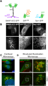
Fig 1. ALPS motifs confer organelle selectivity to a naïve protein.
(A) Schematic of the GMAP-ACC-GFP, GFP133 and Sar1-GFP constructs. (B) Confocal images of live U2OS cells expressing GMAP-ACC-GFP, GFP133 or Sar1-GFP. (C-D) U2OS cells were transfected with GMAP-ACC-GFP, fixed and stained with the cis-Golgi marker anti-GM130. Cells were then imaged by confocal microscopy (C) or structured illumination (D). (D) Upper panels show a maximum projection of a Z-stack, lower panels are single cross-sections. Scale bars are 10μm unless otherwise stated.
Fig 2
Fig 2. GFP133 and Sar1-GFP recapitulate membrane curvature sensitivity of native proteins.
(A) Confocal image of a cell co-expressing GFP133 and the ER marker Sec61-mCherry. Arrowheads indicate flat cisternae. (B) Confocal image of a cell co-expressing Sec61-GFP and Sec61-mCherry. Arrowheads indicate flat cisternae. (C) Fluorescence intensities of GFP133 and Sec61-mCherry (right panel) along a line spanning an ER tubule and an ER flat sheet (in yellow in the left panel). (D) Several plots as in (C) were analysed in cells co-expressing GFP133 and Sec61-mCherry or Sec61-GFP and Sec61-mCherry. For each plot, paired Student t-tests were run to assess if the GFP and mCherry intensities are significantly different. This analysis was done on plot portions spanning cytoplasm (base line), cisternae or tubules. The dashed red line indicates a p-value of 0.05. Black lines represent median values. (E) Cross-section (left) and nuclear surface (right) of a cell co-expressing GFP133 and Sec61-mCherry. The cyan line delineates the nuclear surface. (F) Cross-section (left) and nuclear surface (right) of a cell expressing Sar1-GFP. (G) Representative intensity plots of GFP133 (upper panel) or Sar1-GFP (lower panel) along a line spanning a portion of nuclear envelope and a portion of cytoplasm. (H) Fluorescence intensity of Sar1-GFP (right panel) along a line spanning ER tubules and an ER flat sheets (in cyan in the left panel). Scale bars are 10μm unless otherwise stated.
Fig 3
Fig 3. Reducing its hydrophobicity does not increase membrane curvature sensitivity of Nup133 ALPS motif.
(A) Amino-acid sequence of the ALPS motif of Nup133. The strongest hydrophobic region is highlighted in yellow, and the secondary region is framed in red. The hydrophobic residues mutated in this study are indicated by arrows. (B) L252 in GFP133 was mutated to less hydrophobic residues, namely Valine, Alanine and Glycine. (C) F263 in GFP133 was mutated to less hydrophobic residues, namely Valine, Alanine and Glycine. Scale bars are 10μm. (D) Normalized GLCM contrasts, as an indication of membrane-bound fractions, were measured in peripheral regions of cells transfected with the indicated constructs. Horizontal bars represent median values.
Fig 4
Fig 4. Increasing its hydrophobicity does not impair membrane curvature sensitivity of Nup133 ALPS motif.
(A) V259 and I265 of GFP133 were mutated to bulkier hydrophobic residues. (B) Intensity plots of GFP133 / Sec61-mCherry along a line spanning an ER tubule and an ER flat sheet. (C) Cross-section and nuclear surface of a cell co-expressing GFP133 and Sec61-mCherry. All scale bars are 10μm. (D) Normalized GLCM contrasts measured in peripheral regions of cells transfected with the indicated constructs. Horizontal bars represent median values. (E) For each mutant, we calculated the side chain volume ratio of the mutated to wt residues, and the average of the corresponding normalized contrast ratios. Contrast ratios were then plotted as a function of volume ratios. Error bars are standard deviations.
Fig 5
Fig 5. Altering the charges of ALPS Nup133 does not change its specificity.
(A) Helical projection of Nup133 ALPS motif, showing the position of the two basic residues. The projection was generated by the Heliquest software (http://heliquest.ipmc.cnrs.fr). (B) Confocal images of live cells expressing GFP133 and mutants of lysine K258. (C) Confocal images of live cells expressing GFP133 and mutants where the R257 residue has been mutated to uncharged residues. (D) Neutralizing or adding a negative charge at the interface between the polar and hydrophobic faces of the helix reduces its binding to membranes. Scale bars = 10μm. (E) Normalized GLCM contrasts of mutated constructs, as indicated. Bars are median values.
Fig 6
Fig 6. Geometry of the ALPS motif, imposed by the backbone, influences Nup133 ALPS motif localization.
(A) Confocal images of live cells transfected with GFP133, 133-GFP, GFP-133 or 133-ACC-GFP. (B) Confocal images of cells transfected with GFP133, 133-ACC-GFP or GMAP-ACC-GFP, fixed and stained with anti-GM130 (red). Note that fixation alters ER morphology. (C) Confocal images of live cells transfected with GMAP-ACC-GFP, GFP-GMAP or GFP-GMAP-GMAP. (D) Confocal images of live cells co-transfected with Sec61-mCherry and GMAP-ACC-GFP or GFP-GMAP-GMAP. Scale bars = 10μm.
Fig 7
Fig 7. The coiled-coil domain provides membrane curvature sensitivity to Sar1 AH.
(A) Confocal images of live cells transfected with Sar1-GFP or Sar1-ACC-GFP. (B) Confocal images of cells transfected with Sar1-GFP or Sar1-ACC-GFP, fixed and stained with anti-GM130 (red). (C) Confocal image of a cell co-transfected with Sar1-ACC-GFP and Sec61-mCherry. Arrowheads indicate ER cisternae. (D) Relative intensities of Sar1-ACC-GFP and Sec61-mCherry along a line spanning ER tubules and a cisternae (shown in cyan in left panel). (E) Confocal images of the nuclear surface of cells expressing Sar1-GFP or Sar1-ACC-GFP. On the right are shown representative intensity plots along a segment spanning portions of nuclear envelope and cytoplasm. Scale bars = 10μm.
Fig 8
Fig 8. Schematic cellular localization of Sar1 AH and Nup133 ALPS motif within various backbones.
This diagram illustrates that addition of the ACC domain increases membrane curvature sensitivity of the considered helices. But importantly, this gain remains correlated to their initial degree of membrane curvature sensitivity. This supports that the AH physico-chemical properties are determinant for membrane curvature sensitivity even when they are modulated by the surrounding backbone.

Similar articles

Cited by

References

    1. Van Meer G, Voelker DR, Feigenson GW. Membrane lipids : where they are and how they behave. Nat Rev Mol Cell Biol. 2008;9: 112–124. 10.1038/nrm2330.Membrane - DOI - PMC - PubMed
    1. Antonny B. Membrane deformation by protein coats. Curr Opin Cell Biol. 2006;18: 386–94. 10.1016/j.ceb.2006.06.003 - DOI - PubMed
    1. Stachowiak JC, Brodsky FM, Miller E a. A cost–benefit analysis of the physical mechanisms of membrane curvature. Nat Cell Biol. Nature Publishing Group; 2013;15: 1019–1027. 10.1038/ncb2832 - DOI - PMC - PubMed
    1. Shibata Y, Hu J, Kozlov MM, Rapoport T a. Mechanisms shaping the membranes of cellular organelles. Annu Rev Cell Dev Biol. 2009;25: 329–54. 10.1146/annurev.cellbio.042308.113324 - DOI - PubMed
    1. Farsad K. Mechanisms of membrane deformation. Curr Opin Cell Biol. 2003;15: 372–381. 10.1016/S0955-0674(03)00073-5 - DOI - PubMed

Publication types

MeSH terms

Grants and funding

CMD is supported by the Centre National de la Recherche Scientifique and by a Marie Curie grant (IRG 277018) from the 7th Framework Programme for Research. BA is supported by an advanced grant from the European Research Council (ERC 268888). The funders had no role in study design, data collection and analysis, decision to publish or preparation of the manuscript.

LinkOut - more resources