. 2015 Nov;89(22):11572-83.
doi: 10.1128/JVI.01482-15. Epub 2015 Sep 9.

Kaposi's Sarcoma-Associated Herpesvirus Viral Interferon Regulatory Factor 1 Interacts with a Member of the Interferon-Stimulated Gene 15 Pathway

Affiliations

Kaposi's Sarcoma-Associated Herpesvirus Viral Interferon Regulatory Factor 1 Interacts with a Member of the Interferon-Stimulated Gene 15 Pathway

Sarah R Jacobs et al. J Virol. 2015 Nov.

Abstract

Kaposi's sarcoma-associated herpesvirus (KSHV) is a gammaherpesvirus known to establish lifelong latency in the human host. We and others have previously shown that three KSHV homologs of cellular interferon regulatory factors (IRFs), known as viral IRFs (vIRFs), participate in evasion of the host interferon (IFN) response. We report that vIRF1 interacts with the cellular interferon-stimulated gene 15 (ISG15) E3 ligase, HERC5, in the context of Toll-like receptor 3 (TLR3) activation and IFN induction. The ISG15 protein is covalently conjugated to target proteins upon activation of the interferon response. Interaction between vIRF1 and HERC5 was confirmed by immunoprecipitation, and the region between amino acids 224 and 349 of vIRF1 was required for interaction with HERC5. We further report that expression of vIRF1 in the context of TLR3 activation results in decreased ISG15 conjugation of proteins. Specifically, TLR3-induced ISG15 conjugation and protein levels of cellular IRF3, a known ISG15 target, were decreased in the presence of vIRF1 compared to the control. vIRF1 itself was also identified as a target of ISG15 conjugation. KSHV-infected cells exhibited increased ISG15 conjugation upon reactivation from latency in coordination with increased IFN. Furthermore, knockdown of ISG15 in latently infected cells resulted in a higher level of KSHV reactivation and an increase in infectious virus. These data suggest that the KSHV vIRF1 protein affects ISG15 conjugation and interferon responses and may contribute to effective KSHV replication.

Importance: The KSHV vIRF1 protein can inhibit interferon activation in response to viral infection. We identified a cellular protein named HERC5, which is the major ligase for ISG15, as a vIRF1 binding partner. vIRF1 association with HERC5 altered ISG15 modification of cellular proteins, and knockdown of ISG15 augmented reactivation of KSHV from latency.


Figures

FIG 1
FIG 1
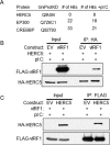
KSHV vIRF1 binds to cellular HERC5 in the context of TLR3 activation. (A) 293-TLR3 stable expressing cells were transfected for 36 h with vIRF1-FLAG and stimulated for 24 h with poly(I·C) (pI:C). After stimulation, FLAG affinity purification was performed, and the products were separated via PAGE and silver stained. Bands of interest were excised and subjected to mass spectrometry. Selected results for proteins interacting with KSHV vIRF1 with or without treatment with poly(I·C) are shown. (B) 293-TLR3 cells were transfected with HA-HERC5 and control EV or FLAG-vIRF1 plasmids. Twenty-four hours after transfection, the cells were treated with poly(I·C) and harvested 24 h after treatment. The cell lysates were immunoprecipitated (IP) with anti-HA antibody-conjugated beads; resolved by SDS-PAGE, along with input control lysates; and probed with antibodies as indicated. (C) 293-TLR3 cells were transfected with FLAG-vIRF1 and control EV or HA-HERC5 plasmid. Twenty-four hours after transfection, the cells were treated with poly(I·C) and harvested 24 h after treatment. The cell lysates were immunoprecipitated with anti-FLAG antibody-conjugated beads and were subjected to SDS-PAGE along with a fraction of the input lysates, and immunoblotting was performed with various antibodies as indicated. The data are representative of the results of more than four independent experiments.
FIG 2
FIG 2
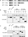
vIRF1 interacts with HERC5 between amino acids 224 and 349. (A) Two vIRF1 truncation mutants and full-length (FL) vIRF1. The black squares indicate the location of the FLAG tag. DBD, DNA-binding domain; IAD, IRF association domain. (B) 293-TLR3 cells were transfected with HA-HERC5 or control vector (−) and FL or aa 1 to 349 or aa 1 to 224 mutant FLAG-vIRF1 constructs. Twenty-four hours after transfection, the cells were treated with poly(I·C) and harvested 24 h after treatment. The cell lysates were immunoprecipitated with FLAG antibody-conjugated beads. Eluate and input samples were subjected to SDS-PAGE and immunoblotted as indicated. (C) 293-TLR3 cells were transfected with FL or aa 1 to 349 or aa 1 to 224 mutant FLAG-vIRF1 constructs or control vector (−) and HA-HERC5. Twenty-four hours after transfection, the cells were treated with poly(I·C) and harvested 24 h posttreatment. The cell lysates were immunoprecipitated with anti-HA antibody-conjugated beads prior to resolution with SDS-PAGE and immunoblotting with the indicated antibodies. The data are representative of the results of three independent experiments.
FIG 3
FIG 3
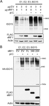
vIRF1 decreases conjugation of ISG15 to cellular proteins in the context of TLR3 activation. (A) 293-TLR3 cells were transfected with 0, 1, or 4 μg of FLAG-vIRF1 and control EV to a total of 4 μg DNA. The cells were also transfected with the E1, E2, and E3 ligases for ISG15, as well an ISG15 construct. Twenty-four hours after transfection, the cells were treated with poly(I·C) as indicated and harvested 24 h after treatment. The cell lysates were subjected to SDS-PAGE and immunoblotting and probed with antibodies as indicated. (B) 293-TLR3 cells were transfected with FL or aa 1 to 349 or aa 1 to 224 mutant FLAG-vIRF1 constructs and the E1, E2, and E3 ligases for ISG15 and HA-ISG15. Twenty-four hours after transfection, the cells were treated with poly(I·C) and harvested 24 h posttreatment. The cell lysates were subjected to SDS-PAGE, followed by immunoblotting with the antibodies indicated. The data are representative of the results of three independent experiments.
FIG 4
FIG 4
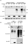
vIRF1 expression decreases protein levels of IRF3 following TLR3 activation. (A) 293-TLR3 cells were transfected with expression plasmids for E1, E2, and E3 ligases for ISG15 and ISG15 where indicated. The cells were also transfected with constructs of FLAG-vIRF1 or control EV. Twenty-four hours after transfection, the cells were treated with poly(I·C) as indicated and harvested 24 h after treatment. The cells were lysed, resolved via SDS-PAGE, and subjected to immunoblotting with the antibodies indicated. (B) 293-TLR3 cells were transfected with the E1, E2, and E3 ligases for ISG15 and FLAG-ISG15 and Myc-vIRF1 or control EV. Twenty-four hours after transfection, the cells were treated with poly(I·C) and harvested 24 h after treatment. The cells were lysed and immunoprecipitated with anti-FLAG antibody-conjugated beads prior to SDS-PAGE and immunoblotting with the indicated antibodies. The data are representative of the results of more than five independent experiments.
FIG 5
FIG 5
KSHV vIRF1 is ISG15 conjugated. (A) 293-TLR3 cells were transfected with the E1, E2, and E3 ligases for ISG15, FLAG-ISG15, and Myc-vIRF1 or control empty vector (−). Twenty-four hours after transfection, the cells were treated with poly(I·C) and harvested 24 h after treatment. The cell lysates were immunoprecipitated with anti-FLAG antibody-conjugated beads, followed by elution and resolution of eluate and input lysates via SDS-PAGE and immunoblotting with the indicated antibodies. (B) 293-TLR3 cells were transfected with the E1, E2, and E3 ligases for ISG15, HA-ISG15, and FLAG-vIRF1 or control empty vector (−). Twenty-four hours after transfection, the cells were treated with poly(I·C) and harvested 24 h posttreatment. The cell lysates were immunoprecipitated with anti-FLAG antibody-conjugated beads, followed by elution and resolution of eluate and input lysates via SDS-PAGE and immunoblotting with the indicated antibodies. The data are representative of the results of three independent experiments.
FIG 6
FIG 6
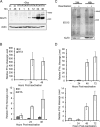
Reactivation of KSHV increases ISG15 conjugation and IFN message. TREx BCBL1 cells expressing empty-vector control (pc) or RTA under a doxycycline (Dox)-inducible promoter were treated with doxycycline for the number of hours indicated. (A) Cells were harvested and lysed prior to separation with SDS-PAGE and immunoblotting with the antibodies indicated. (B) Doxycycline-treated TREx BCBL1 cells were harvested, RNA was extracted, and cDNA was generated prior to quantification of IFN-α and IFN-β message levels by real-time (RT) PCR. (C) BCBL1 PEL cells were reactivated with sodium butyrate treatment or vehicle control (−) for 24 or 48 h. The cells were harvested, and the lysates were resolved via SDS-PAGE, which was followed by immunoblotting with the indicated antibodies. (D) BCBL1 PEL cells reactivated with sodium butyrate for 0 (−), 24, 48, or 72 h were harvested. RNA was extracted, and cDNA was generated prior to quantification of IFN-α and IFN-β message levels by RT-PCR. The data are representative of the results of three independent experiments, and the values represent the means ± the standard deviations of the means from triplicate biological replicates.
FIG 7
FIG 7
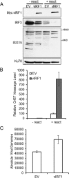
Expression of vIRF1 in PEL cells decreases ISG15 conjugation and increases KSHV reactivation. BCBL1 cells were infected with lentivirus containing EV control or Myc-vIRF1 for 48 h prior to reactivation with TPA and sodium butyrate. (A) Cells were harvested 48 h after reactivation, lysed, and resolved via SDS-PAGE, followed by immunoblotting with the indicated antibodies. − react, no reactivation; + react, with reactivation. (B) Lentivirus-transduced BCBL-1 cells were harvested 48 h after reactivation, RNA was extracted, and cDNA was generated prior to quantification of KSHV Orf57 message levels by real-time quantitative PCR (qPCR). (C) BCBL-1 cell supernatants were harvested 48 h after reactivation and used to infect naive 293 cells, and infected cells were harvested 48 h postinfection; DNA was isolated, and Orf57 DNA levels were compared to a standard curve to assess the absolute number of viral genomes by real-time qPCR. The data are representative of the results of two independent experiments, and the values represent the means ± the standard deviations of the means from triplicate biological replicates.
FIG 8
FIG 8
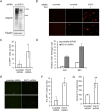
Knockdown of ISG15 increases KSHV replication upon reactivation. (A) iSLK.219 cells were infected with scrambled control (scr) or ISG15 shRNA lentivirus for 72 h. Cells were then lysed and subjected to SDS-PAGE and immunoblotting for the detection of ISG15 knockdown. (B) Seventy-two hours after lentiviral infection with scrambled control or ISG15 shRNA lentivirus, iSLK.219 cells were reactivated for 48 h with doxycycline. Lytic virus was detected by RFP expression via fluorescence microscopy. (C) RFP-expressing iSLK.219 cells were quantified by counting the cells in 4 random fields. (D) iSLK.219 cells were infected with scrambled control or ISG15 shRNA lentivirus for 72 h prior to reactivation with doxycycline. Cells were harvested 24 or 48 h after reactivation, and the RNA was isolated, reverse transcribed, and subjected to real-time qPCR to quantitate KSHV Orf57 message levels. (E) iSLK.219 cells were infected with scrambled control or ISG15 shRNA lentivirus and reactivated for 48 h. The cell supernatants were harvested and used to infect naive 293 cells, and GFP expression was quantitated 72 h postinfection. (F) GFP-expressing 293 cells were quantified by counting the cells in 5 randomly selected fields. (G) iSLK.219 cells were infected with scrambled control or ISG15 shRNA lentivirus and reactivated for 48 h. The cell supernatants were then used to infect naive 293 cells. The infected cells were harvested 48 h postinfection; DNA was isolated, and Orf57 DNA levels were compared to a standard curve to assess the absolute number of viral genomes by real-time qPCR. The data are representative of the results of four independent experiments, and the values represent the means ± the standard deviations of the means from triplicate biological replicates. *, P < 0.005; **, P < 0.05 (Student's t test).
FIG 9
FIG 9
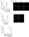
Knockdown of HERC5 increases KSHV replication upon reactivation. (A) iSLK.219 cells were infected with scrambled control or HERC5 shRNA lentivirus for 72 h prior to harvesting for detection of HERC5 knockdown through real-time qPCR. (B) Seventy-two hours after lentiviral infection with scrambled control or HERC5 shRNA lentivirus, iSLK.219 cells were reactivated for 48 h with doxycycline. Lytic virus was detected by RFP expression via fluorescence microscopy. (C) RFP-expressing iSLK.219 cells were quantified by counting the cells in 6 randomly selected fields. (D) iSLK.219 cells were infected with scrambled control or ISG15 shRNA lentivirus and reactivated for 48 h. The cell supernatants were harvested and used to infect naive 293 cells, and infection was detected by GFP expression via fluorescence microscopy 48 h postinfection. (E) iSLK.219 cells were infected with scrambled control or ISG15 shRNA lentivirus and reactivated for 48 h. The cell supernatants were then used to infect naive 293 cells. The infected cells were harvested 48 h postinfection; DNA was isolated, and Orf57 DNA levels were compared to a standard curve to assess the absolute number of viral genomes by real-time qPCR. The data are representative of the results of three independent experiments, and the values represent the means ± the standard deviations of the means from triplicate biological replicates. *, P < 0.01 (Student's t test).

Similar articles

Cited by

References

    1. Potter JL, Narasimhan J, Mende-Mueller L, Haas AL. 1999. Precursor processing of pro-ISG15/UCRP, an interferon-beta-induced ubiquitin-like protein. J Biol Chem 274:25061–25068. doi:10.1074/jbc.274.35.25061. - DOI - PubMed
    1. Der SD, Zhou A, Williams BR, Silverman RH. 1998. Identification of genes differentially regulated by interferon alpha, beta, or gamma using oligonucleotide arrays. Proc Natl Acad Sci U S A 95:15623–15628. doi:10.1073/pnas.95.26.15623. - DOI - PMC - PubMed
    1. Loeb KR, Haas AL. 1992. The interferon-inducible 15-kDa ubiquitin homolog conjugates to intracellular proteins. J Biol Chem 267:7806–7813. - PubMed
    1. Narasimhan J, Potter JL, Haas AL. 1996. Conjugation of the 15-kDa interferon-induced ubiquitin homolog is distinct from that of ubiquitin. J Biol Chem 271:324–330. doi:10.1074/jbc.271.1.324. - DOI - PubMed
    1. Hemelaar J, Borodovsky A, Kessler BM, Reverter D, Cook J, Kolli N, Gan-Erdene T, Wilkinson KD, Gill G, Lima CD, Ploegh HL, Ovaa H. 2004. Specific and covalent targeting of conjugating and deconjugating enzymes of ubiquitin-like proteins. Mol Cell Biol 24:84–95. doi:10.1128/MCB.24.1.84-95.2004. - DOI - PMC - PubMed

Publication types

MeSH terms

LinkOut - more resources