. 2015 Sep 8;12(10):1555-1563.
doi: 10.1016/j.celrep.2015.08.011. Epub 2015 Aug 28.

Cell-to-Cell Transmission of HIV-1 Is Required to Trigger Pyroptotic Death of Lymphoid-Tissue-Derived CD4 T Cells

Affiliations

Cell-to-Cell Transmission of HIV-1 Is Required to Trigger Pyroptotic Death of Lymphoid-Tissue-Derived CD4 T Cells

Nicole Lk Galloway et al. Cell Rep. .

Abstract

The progressive depletion of CD4 T cells underlies clinical progression to AIDS in untreated HIV-infected subjects. Most dying CD4 T cells correspond to resting nonpermissive cells residing in lymphoid tissues. Death is due to an innate immune response against the incomplete cytosolic viral DNA intermediates accumulating in these cells. The viral DNA is detected by the IFI16 sensor, leading to inflammasome assembly, caspase-1 activation, and the induction of pyroptosis, a highly inflammatory form of programmed cell death. We now show that cell-to-cell transmission of HIV is obligatorily required for activation of this death pathway. Cell-free HIV-1 virions, even when added in large quantities, fail to activate pyroptosis. These findings underscore the infected CD4 T cells as the major killing units promoting progression to AIDS and highlight a previously unappreciated role for the virological synapse in HIV pathogenesis.


Figures

Figure 1
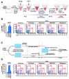
Figure 1. The mode of HIV-1 transfer generates disparate death responses of target lymphoid CD4 T cells
The death of lymphoid CD4 T cells was examined by spinoculation of target cells with large quantities of cell-free virions to target cells (A, B), as previously described (Doitsh et al., 2014; Geng et al., 2014; O’Doherty et al., 2000), or in co-cultures of CFSE-labeled target CD4 T with productively infected HLACs (Doitsh et al., 2010; Jekle et al., 2003) (C, D). All samples were infected with a multiple-round X4-tropic NL4-3 strain of HIV-1. NL4-3 was selected because tonsillar tissue contains a high percentage of resting CD4 T cells that express CXCR4 (90–100%). Target cells were treated with the same concentrations of drugs prior to co-culture with productively infected HLACs or spinoculation with free virions. Inhibitors blocking HIV entry (AMD3100) or early steps of reverse transcription (efavirenz) prevented death of target CD4 T cells. In sharp contrast, inhibiting later events in the viral life cycle (raltegravir) did not prevent cell death in co-cultures with productively infected cells (B), but abrogated the death response of target cells spinoculated with cell-free HIV-particles (D). Bar graph represents summary of flow data presented. Error bars represent SD/√n (SEM) of three independent donors. FACS plots are representative of six independent experiments performed with tonsils from different donors. See also Figure S1.
Figure 2
Figure 2. Free HIV-1 particles do not induce cell death of abortively infected lymphoid CD4 T cells
(A) HLACs were spinoculated with a multiple-round (NLENG1I), a single-round (NLENG1-ES-IRES, pseudotyped with HIV-1 Env), or integrase deficient (NLENG1I-D116N) viral clones containing a GFP reporter. An IRES upstream of the nef gene preserves Nef expression and supports LTR-driven GFP expression in productively infected target cells (Levy et al., 2004). No drugs were added to the spinoculated cultures. The levels of productive infection and CD4 T-cell depletion in the cultures were analyzed by flow cytometry four days after spinoculation. (B) Treatment with the entry inhibitor AMD3100 four hours after spinoculation with multiple-round NLENG1I HIV-1 particles does not prevent productive infection, but efficiently blocks the killing of target resting CD4 T cells. Thus, death of CD4 T cells occurs after establishment of productive infection, but not during initial spinoculation of cell-free viruses. Of note, spinoculation of supernatant from infected HLACs failed to induce death of target CD4 T cells (See Figure S3). (C) A method to assess death of CD4 T cells with non-infectious HIV-1 clones. The single-round and integrase-deficient HIV-1 clones are not competent for multiple rounds of viral replication. Instead, we modified the experimental system by overlaying HLAC cells on a monolayer of 293T cells transfected with these proviral clones, as previously described (Doitsh et al., 2014). As illustrated, fresh human tonsil is processed into HLAC and cells are cultured in suspension. After 12 hours, transfected 293T cells in a 24-well plate are washed and overlaid with 4×10⁶ HLAC cells in RPMI. Virus-producing 293T cells directly interact with target overlaying HLAC cells. After 24–72 hours, HLAC suspensions were collected from wells and analyzed by flow cytometry. (D) Single-round and integrase-deficient HIV-1 clones kill target CD4 T cells as efficiently as multiple-round HIV-1 clones when transmitted via virus-producing cells. FACS plots are representative of three independent experiments performed with tonsils from different donors. Error bars, SEM; *** P < 0.001 (Student’s t test); n.s., not significant, P > 0.05. See also Figure S2.
Figure 3
Figure 3. Death of lymphoid CD4 T cells requires close interaction with productively infected cells
(A) Increasing the cell culture surface area in HIV-infected cultures decreases cell-to-cell interactions, and reduces the kinetics of CD4 T-cell depletion. (B) Inverse correlation between culture surface area and CD4 T-cell death. Death of target CD4 T cells in each vessel was examined after four days of co-culturing with HIV-infected cells. Note that cell death decreases even in vessels where the volume of culture medium remained constant. (C, D) Blocking antibodies against either ICAM-1 or CD11a prevent death of target CD4 T cells (E) Exclusive expression of ICAM-1 on activated (permissive) lymphoid CD4 T cells. ICAM-1 expression is also high in antigen presenting B cells, but not CD8 T cells. (F) High expression of LFA-1 on lymphoid CD4 and CD8 T cells, but not on B cells. In contrast to ICAM-1, LFA-1 expression is not increased on activated CD4 T cells. Error bars, SEM; *P < 0.05, ***P < 0.001 (Student’s t test). EFV, Efavirenz. See also Figures S3, S4, and S5.
Figure 4
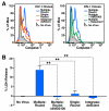
Figure 4. Cell-to-Cell transmission of HIV-1 is required to trigger innate recognition and caspase 1-dependent pyroptosis of lymphoid CD4 T Cells
(A) HLACs were spinoculated with multiple- or single-round NLRX-IRES reporter clones as indicated. Saquinavir was added to the culture before spinoculation. AMD3100 was added to the culture four hours after spinoculation. Cells were analyzed by flow cytometry using cell permeable fluorogenic substrates that contain amino acids sequences specifically cleaved by active caspase 1 (CaspaLux1). Abundant caspase 1 activity is exclusively observed in cultures spinoculated with multiple-round HIV-1 clones. Essentially no caspase 1 activity is observed in target CD4 T cells where cell-to-cell spread of HIV-1 is blocked or does not occur. Histograms show one experiment, a representative of three independent experiments performed with tonsils from different donors (B) Supernatants from spinoculated cultures were analyzed for levels of released cytoplasmic LDH enzyme, an indicator of pyroptosis (Decker and Lohmann-Matthes, 1988) FACS plots are representative of three independent experiments performed with tonsils from different donors. Error bars, SEM; **P < 0.01 (Student’s t test). See also Figure S5.
Figure 5
Figure 5. The mode of HIV-1 spread determines the outcome form of cell death
Infection of free HIV-1 particles produces productive infection and caspase 3-apoptosis in a small fraction of permissive lymphoid CD4 T cells. Next, cell-to-cell spread of HIV-1 is required to deplete the non-permissive lymphoid CD4 T cells, which represent 95% or more of the target cells in lymphoid tissues, via caspase 1-dependent pyroptosis. Free HIV-1 particles, even at high quantities, cannot trigger innate immune recognition and produce this form of cell death. Thus, in contrast to the previous view indicating productive and abortive HIV-1 infection as independent cytopathic events causing CD4 T-cell death (Cummins and Badley, 2010; Doitsh et al., 2010; Fevrier et al., 2011; Garg et al., 2012), these events are essentially linked in a single pathogenic cascade. Therefore, antiretroviral drugs such as AZT or raltergravir that do not directly prevent the death of abortively infected CD4 T cells (Doitsh et al., 2010), effectively inhibit HIV pathogenesis in vivo by blocking upstream events of productive HIV-1 infection. Caspase 1 inhibitors do not inhibit productive HIV-1 infection but block pyroptosis of abortively infected CD4 T cells (Doitsh et al., 2014). See also Supplemental Experimental Procedures.

Similar articles

Cited by

References

    1. Agosto LM, Uchil PD, Mothes W. HIV cell-to-cell transmission: effects on pathogenesis and antiretroviral therapy. Trends in microbiology. 2015;23:289–295. - PMC - PubMed
    1. Bounou S, Leclerc JE, Tremblay MJ. Presence of host ICAM-1 in laboratory and clinical strains of human immunodeficiency virus type 1 increases virus infectivity and CD4(+)-T-cell depletion in human lymphoid tissue, a major site of replication in vivo. J Virol. 2002;76:1004–1014. - PMC - PubMed
    1. Cooper A, Garcia M, Petrovas C, Yamamoto T, Koup RA, Nabel GJ. HIV-1 causes CD4 cell death through DNA-dependent protein kinase during viral integration. Nature. 2013 - PubMed
    1. Cummins NW, Badley AD. Mechanisms of HIV-associated lymphocyte apoptosis: 2010. Cell death & disease. 2010;1:e99. - PMC - PubMed
    1. Decker T, Lohmann-Matthes ML. A quick and simple method for the quantitation of lactate dehydrogenase release in measurements of cellular cytotoxicity and tumor necrosis factor (TNF) activity. Journal of immunological methods. 1988;115:61–69. - PubMed

Publication types