Mice Deficient in NF-κB p50 and p52 or RANK Have Defective Growth Plate Formation and Post-natal Dwarfism
- PMID: 26273511
- PMCID: PMC4175530
- DOI: 10.4248/BR201304004
Mice Deficient in NF-κB p50 and p52 or RANK Have Defective Growth Plate Formation and Post-natal Dwarfism
Abstract
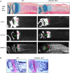
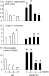
NF-κBp50/p52 double knockout (dKO) and RANK KO mice have no osteoclasts and develop severe osteopetrosis associated with dwarfism. In contrast, Op/Op mice, which form few osteoclasts, and Src KO mice, which have osteoclasts with defective resorptive function, are osteopetrotic, but they are not dwarfed. Here, we compared the morphologic features of long bones from p50/p52 dKO, RANK KO, Op/Op and Src KO mice to attempt to explain the differences in their long bone lengths. We found that growth plates in p50/p52 dKO and RANK KO mice are significantly thicker than those in WT mice due to a 2-3-fold increase in the hypertrophic chondrocyte zone associated with normal a proliferative chondrocyte zone. This growth plate abnormality disappears when animals become older, but their dwarfism persists. Op/Op or Src KO mice have relatively normal growth plate morphology. In-situ hybridization study of long bones from p50/p52 dKO mice showed marked thickening of the growth plate region containing type 10 collagen-expressing chondrocytes. Treatment of micro-mass chondrocyte cultures with RANKL did not affect expression levels of type 2 collagen and Sox9, markers for proliferative chondrocytes, but RANKL reduced the number of type 10 collagen-expressing hypertrophic chondrocytes. Thus, RANK/NF-κB signaling plays a regulatory role in post-natal endochondral ossification that maintains hypertrophic conversion and prevents dwarfism in normal mice.
Keywords: NF-κB; RANK; chondrocytes; dwarfism; growth plate.
Figures


Similar articles
-
NF-kappaB p50 and p52 expression is not required for RANK-expressing osteoclast progenitor formation but is essential for RANK- and cytokine-mediated osteoclastogenesis.J Bone Miner Res. 2002 Jul;17(7):1200-10. doi: 10.1359/jbmr.2002.17.7.1200. J Bone Miner Res. 2002. PMID: 12096833
-
Expression of either NF-kappaB p50 or p52 in osteoclast precursors is required for IL-1-induced bone resorption.J Bone Miner Res. 2003 Feb;18(2):260-9. doi: 10.1359/jbmr.2003.18.2.260. J Bone Miner Res. 2003. PMID: 12568403
-
Constitutive activation of the alternative NF-κB pathway disturbs endochondral ossification.Bone. 2019 Apr;121:29-41. doi: 10.1016/j.bone.2019.01.002. Epub 2019 Jan 3. Bone. 2019. PMID: 30611922
-
NF-kappaB functions in osteoclasts.Biochem Biophys Res Commun. 2009 Jan 2;378(1):1-5. doi: 10.1016/j.bbrc.2008.10.146. Epub 2008 Nov 6. Biochem Biophys Res Commun. 2009. PMID: 18992710 Review.
-
Perspective. Osteoclastogenesis and growth plate chondrocyte differentiation: emergence of convergence.Crit Rev Eukaryot Gene Expr. 2003;13(2-4):181-93. Crit Rev Eukaryot Gene Expr. 2003. PMID: 14696966 Review.
Cited by
-
CHIP regulates skeletal development and postnatal bone growth.J Cell Physiol. 2020 Jun;235(6):5378-5385. doi: 10.1002/jcp.29424. Epub 2020 Jan 3. J Cell Physiol. 2020. PMID: 31898815 Free PMC article.
-
Nicorandil Inhibits Osteoclast Formation Base on NF-κB and p-38 MAPK Signaling Pathways and Relieves Ovariectomy-Induced Bone Loss.Front Pharmacol. 2021 Sep 8;12:726361. doi: 10.3389/fphar.2021.726361. eCollection 2021. Front Pharmacol. 2021. PMID: 34566650 Free PMC article.
-
Physiological role of receptor activator nuclear factor-kB (RANK) in denervation-induced muscle atrophy and dysfunction.Receptors Clin Investig. 2016 May 30;3(2):e13231-e13236. doi: 10.14800/rci.1323. Receptors Clin Investig. 2016. PMID: 27547781 Free PMC article.
-
Therapeutic potentials and modulatory mechanisms of fatty acids in bone.Cell Prolif. 2020 Feb;53(2):e12735. doi: 10.1111/cpr.12735. Epub 2019 Dec 4. Cell Prolif. 2020. PMID: 31797479 Free PMC article. Review.
-
Maternal RANKL Reduces the Osteopetrotic Phenotype of Null Mutant Mouse Pups.J Clin Med. 2018 Nov 8;7(11):426. doi: 10.3390/jcm7110426. J Clin Med. 2018. PMID: 30413057 Free PMC article.
References
-
- Wagner EF, Karsenty G. Genetic control of skeletal development. Curr Opin Genet Dev. 2001;11:527–532. - PubMed
-
- Karsenty G, Wagner EF. Reaching a genetic and molecular understanding of skeletal development. Dev Cell. 2002;2:389–406. - PubMed
-
- Holmbeck K, Bianco P, Caterina J, Yamada S, Kromer M, Kuznetsov SA, Mankani M, Robey PG, Poole AR, Pidoux I, Ward JM, Birkedal-Hansen H. MT1-MMP-deficient mice develop dwarfism, osteopenia, arthritis, and connective tissue disease due to inadequate collagen turnover. Cell. 1999;99:81–92. - PubMed
-
- Takuma S. Electron microscopy of cartilage resorption by chondroclasts. J Dent Res. 1962;41:883–889. - PubMed
Grants and funding
LinkOut - more resources
Full Text Sources
Other Literature Sources
Research Materials
Miscellaneous

